Introduction
In the process of spermatogenesis, Sertoli cells (SC) provide structural support and cytokines for sperm cells, regulate nutrition and development, and play a crucial role in maintaining a stable micro-environment for spermatogenesis (Lee and Cheng, 2004a). The microtubules of Sertoli cells are involved in spermatogenesis through the regulation of cell morphology, intracellular transport, organelle positioning, cell motility, cell division and other physiological processes. If the function of a microtubule is damaged, spermatogenesis will be impaired, causing male infertility (Zhu et al., Reference Zhu, Emanuele and Van2004).
Nitric oxide (NO) has a dual role as a signal molecular element. A low concentration of NO (<1 μmol/l) has a protective effect on cells, but high concentrations of NO (>1 μmol/l) can react with molecular oxygen or superoxide anions and result in cytoskeleton protein nitro-solution, nitration and oxidative damage, leading to impaired cell function, or even apoptosis (Davis et al., Reference Davis, Martin, Turko and Murad2001; Hanafy et al., Reference Hanafy, Krumenacker and Murad2001; Lee and Cheng, 2004a). Landino and colleagues (Reference Landino, Koumas, Mason and Alston2007) found that cysteine residues of tubulin were more easily oxidized by reactive nitrogen oxide. The dimerization of α- and β-tubulin subunits was therefore inhibited, leading to damage of the cytoskeleton of normal cells.
SCs are the first target for many drugs acting on testicular cells. A high concentration of NO, produced by a variety of drugs in testes, could impair the activity of SCs, particularly by causing damage to the SC cytoskeleton. Such changes in the SC cytoskeleton would have a negative impact on the blood–testis barrier. Furthermore, the concentrations of anti-oxidant enzymes in spermatogenic cells are so low that high concentrations of NO could disrupt spermatogenesis and even result in male infertility. As the development model of the pig's gonad is very similar to that of a human being, pigs have become important experimental animals in developmental biology and reproductive medicine. In order to further understand the effects of NO on SCs, we used piglets and sodium nitroprusside (SNP) (NO donor) to study the effects of NO on the microtubules of SCs.
Materials and methods
Materials
Testes were obtained in sterile conditions from immature pigs (2–3 weeks old) from a local pig farm. All animal treatment procedures were approved by the Animal Care Committee of Southwest University. Dulbecco's modified eagle medium (DMEM)/F-12 Ham's, fetal bovine serum and collagenase type VI were purchased from Gibco, Inc (California, USA). Trypsin and SB203580 were obtained from Sigma-Aldrich, Inc. (St Louis, MO, USA), SNP, the nitric oxide assay kit, the N-nitro-l-arginine-methyl ester (L-NAME), and glutathione (GSH) assay kit were purchased from Beyotime Institute of Biotechnology (Shanghai, China). The cell counting 8 Kit came from Dojindo Molecular Technologies, Inc. (Kumamoto, Japan). The superoxide dismutase (SOD) assay kit and total anti-oxygen capability (T-AOC) assay kit were purchased from Nanjing Jiancheng Biotechnology Co., Ltd. (Nanjing, China). The antibodies used in this study, β-actin, p38MAPK and phosphorylated (p)-p38MAPK came from Santa Cruz (USA). The antibodies to α-tubulin and transferrin were purchased from Invitrogen (California, USA) and Bethy (Montgomery, USA), respectively.
Methods
Effect of SNP on the level of NO in SCs
SCs were isolated from testes using the protocol described by Maccarrone et al. (Reference Maccarrone, Cecconi, Rossi, Battista, Pauselli and Finazzi-Agro2003) . After collagenase dissociation (3% collagenase, 40 min at 32°C) and trypsin dissociation (0.25% trypsin, 20 min at 32°C), cells were washed by centrifugation (200 g, 10 min). After sedimentation, the tubules were collected and washed 3–4 times at unit gravity in DMEM/F-12 Ham's. Freshly isolated SCs were suspended in DMEM/F-12 Ham's containing 10% fetal bovine serum, then plated at 1×10⁸ cells/cm2 in culture flasks with 3 ml DMEM/F-12 per well and cultured in a humidified atmosphere of 5%, CO2 at 32°C. After 4–8 h, cells were treated with 20 mM Tris-HCl, pH 7.4, to lyse residual germ cells, and allowed to grow for another 24 h. The SCs were then incubated for 24 h in 1 μmol/l, 10 μmol/l, 100 μmol/l and 1 mmol/l SNP as described by Tepperman et al. (Reference Tepperman, Abrahamson and Soper1998) . Meanwhile, L-NAME was used to inhibit the production of endogenous NO. Cells were then washed twice with pre-heated phosphate buffer saline (PBS; 34°C) and the protein extracted as described by Wang et al. (Reference Wang, Sun, Pan, Wu and Zhang2006), and the levels of NO detected using the NO assay kit.
Effect of SNP on SC viability
Cells were treated with different concentrations of SNP (0–1 mmol/l) for 24 h in a 96-well plate, adding 10 μl of the CCK-8 solution (WST-8 solution) to each well 4 h before the end of treatment. Absorbance was measured at 450 nm using a microplate reader (Shanghai shenggong, Shanghai, China). Cell viability was expressed as a percentage of the control culture value.
Effect of NO on the activity of p38MAPK in SCs
Cells were incubated with SNP for 24 h, then proteins were isolated from the cultured cells as described. The protein content of the supernatant after centrifugation was determined by spectrophotometer using bovine serum albumin (BSA) as a standard. Approximately 40 μg total protein per lane was separated using 12% sodium dodecyl sulphate-polyacrylamide gel electrophoresis (SDS-PAGE; 5% concentration gel, 20 mA, 1.5 h; 12% separation gel, 25 mA, 3 or 4 h) and transferred to nitrocellulose membranes (90 v, 2 h, Bio-Rad, Hercules, CA, USA). The primary antibodies, anti-p38MAPK and anti-phospho-p38MAPK, were diluted to 1:500. Band intensities were determined by Quantity one software (Bio-Rad).
Effect of NO on the distribution of α-tubulin in SCs
After culturing SC in vitro for 24 h, 1 μmol/l, 10 μmol/l, 100 μmol/l, 1 mmol/l SNP and 1 mmol/l SNP plus SB203580 (20 μmol/l, adding SB203580 30 min in advance) were used for incubation of SCs for another 24 h. Cells were then washed twice in pre-heated PBS (34°C, 5 min each wash). Cells were fixed with 2% methanol for 10 min, then washed twice with pre-heated PBS (34°C, 5 min each wash). Cells were incubated with 0.1% Triton-X 100 (prepared with PBS containing 10% FBS) for 3 or 5 min, then washed twice with pre-heated PBS (34°C, 5 min each wash). After blocking in 1% BSA for 30 min, cells were incubated in a 1:500 dilution of anti-α-tubulin antibody at 4°C overnight. Cells were washed twice with PBS (5 min each time) and then incubated with FITC secondary antibodies (1:200) at room temperature for 2 h. Cells were washed with PBS to remove unbound secondary antibodies, then observed using an inverted microscope and photos taken.
The effect of NO on transferrin in SCs
After cells were incubated with SNP for 24 h as described, they were washed by centrifugation (13 000 rpm, 5 min). Proteins of cells were collected, and electrophoresis and transmembrane carried out for those proteins. The primary antibodies, anti-transferrin and anti-β-actin, were diluted to 1:1000 and 1:500, respectively. Band intensities were determined by Quantity one software (Bio-Rad).
The effect of NO on anti-oxidant activity
After cells were incubated with SNP for 24 h as described, cells were homogenized in 5 ml of ice-cold Tris-HCl buffer (0.01 mol/l, pH 7.4) containing 0.01% ethylenediaminetetraacetic acid disodium (EDTA-2Na), 0.01 mol/l saccharose and 0.8% NaCl. The homogenization procedure was carried out for 2 min at 13 000 rpm. All procedures were performed at 4°C. Homogenate, supernatant and extracted samples were prepared to determine the activities of SOD, T-GSH, T-AOC as described by Bauche and colleagues (Reference Bauche, Fouchard and Jegou1994) .
Statistical analysis
Every test was carried out in triplicate, and all data were reported as means±SEM. Statistical analyses were performed using SPSS (version 16.0; SPSS Inc, Chicago, IL, USA). Data were analysed by one-way ANOVA and Tukey's post hoc test (Tukey, Reference Tukey1965) to determine treatment differences. A probability of P<0.05 was considered to be statistically significant.
Results
Effect of SNP dose on the levels of NO in SCs
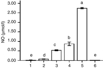
The levels of NO in SCs varied between 0.07±0.01 μmol/l and 2.74±0.03 μmol/l when SCs were incubated with different concentrations of SNP for 24 h. When SCs were incubated with L-NAME, the level of NO in the SCs was 0.008±0.003 μmol/l, indicating endogenous NO had no harmful effect on SCs in this study (P>0.05; Fig. 1).
Effect of SNP dose on the level of NO in Sertoli cells. Bars with different letters are significantly different from each other (P<0.05). 1, control; 2, 1 μmol/l SNP; 3, 10 μmol/l SNP; 4, 100 μmol/l SNP; 5, 1 mmol/l SNP; 6, 1 mmol/l SNP+100 μmol/l L-NAME. Error bars indicate relative accuracy between samples.

The effect of SNP dose on cell viability of SCs
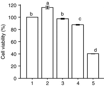
When the concentration of SNP was 1 μmol/l, cell viability increased by ~15.90% above control (P<0.05). At an SNP concentration of 10 μmol/l, there was no significant difference in cell viability when compared with control (P>0.05). With a further increase in concentration of SNP, cell viability decreased significantly (P<0.05; Fig. 2).
Effect of SNP dose on cell viability of Sertoli cells. Bars with different letters are significantly different from each other (P<0.05). 1, control; 2, 1 μmol/l SNP; 3, 10 μmol/l SNP; 4, 100 μmol/l SNP; 5, 1 mmol/l SNP. Error bars indicate relative accuracy between samples.

The effect of NO on the activity of p38MAPK in SCs
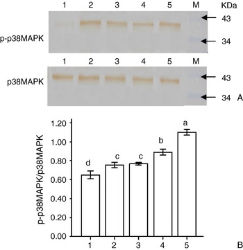
All doses of SNP used in this study increased the activity of p38MAPK. With an increase in the concentration of SNP, the activity of p38MAPK increased significantly (P<0.05; Fig. 3).
Effect of SNP dose on activity of p38MAPK in Sertoli cells. Bars with different letters are significantly different from each other (P<0.05). Lanes: 1, control; 2, 1 μmol/l SNP; 3, 10 μmol/l SNP; 4, 100 μmol/l SNP; 5, 1 mmol/l SNP; M, protein marker. (A) Western blot analysis. (B) Statistical analysis. (See online for a colour version of this figure.)

Effect of NO on the distribution of α-tubulin in SCs
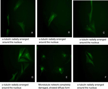
When the SCs were incubated in 1 μmol/l, 10 μmol/l and 100 μmol/l SNP with SB203580, α-tubulin radially arranged around the nucleus as in controls (Fig. 4A–E). However, when the concentration of SNP increased to 1 mmol/l, the microtubule network was completely damaged, showing a dispersed form (Fig. 4F).
Effect of SNP dose on the distribution of α-tubulin in Sertoli cells (×400): (A) control; (B) 1 μmol/l SNP; (C) 10 μmol/l SNP; (D) 100 μmol/l SNP; (E) 1 mmol/l SNP; (F) 1 mmol/l SNP+20 μmol/l SB203580. (See online for a colour version of this figure.)

Effect of NO on transferrin in SCs
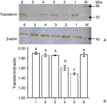
The expression of transferrin was 1.90±0.02 (the ratio of band intensities, transferrin/β-actin) in the control group. When the concentration of SNP increased, the expression of transferrin decreased. With a concentration of SNP of 1 mmol/l, the expression of transferrin decreased significantly to 1.48±0.01 (P>0.05; Fig. 5). When SCs were incubated with 1 mmol/l SNP plus SB203580, the expression of transferrin increased obviously compared to incubation with 1 mmol/l SNP only (P<0.05; Fig. 5).
Effect of SNP dose on transferrin in Sertoli cells. Bars with different letters are significantly different from each other (P<0.05). Lanes: 1, control; 2, 1 μmol/l SNP; 3, 10 μmol/l SNP; 4, 100 μmol/l SNP; 5, 1 mmol/l SNP; 6, 1 mol/l SNP+20 μmol/l SB203580; M, protein marker. (A) Western blot analysis. (B) Statistical analysis. (See online for a colour version of this figure.) Error bars indicate relative accuracy between samples.

Effect of NO on anti-oxidant activity
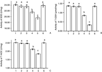
At SNP concentrations of 1 and 10 μmol/l, there were no effects on SOD, T-GSH and T-AOC activity when compared with controls (P>0.05; Fig. 6). When the concentration of SNP was increased, the activities of SOD, T-GSH and T-AOC decreased significantly. At concentrations of 100 μmol/l and 1 mmol/l SNP, the activities of SOD, T-GSH and T-AOC reduced (P<0.05 compared to controls; Fig. 6). Adding SB203580 could reduce the inhibition effect of SNP on these enzymes (P<0.05 compared to the 1 mmol/l group; Fig. 6).
Effect of SNP dose on anti-oxidant activity. Bars with different letters are significantly different from each other (P<0.05). 1, control; 2, 1 μmol/l SNP; 3, 10 μmol/l SNP; 4, 100 μmol/l SNP; 5, 1 mol/l SNP; 6, 1 mmol/l SNP+20 μmol/l SB203580. (A) Activity of SOD. (B) Activity of T-GSH. (C) Activity of T-AOC. Error bars indicate the relative accuracy between samples.

Discussion
SNP is a NO donor, and can generate NO in cells and play an important role in regulating the function of cells. A number of studies have shown that a low concentration of NO is helpful in spermatogenesis, and a high concentration of NO has a negative impact on spermatogenesis (He et al., Reference He, Tian, Li, Liao and Zhu2002; Kang et al., Reference Kang, Zhang, Li and Duan2002). In this study, concentrations of SNP from 0 to 1 mmol/l were used for incubation of the SCs. After 24 h, as the concentrations of SNP increased, the concentration of NO within the SCs increased significantly (between 0.07±0.01 and 2.74±0.03 μmol/l). The results show that a low concentration of NO could lead to the survival of cells, while a high concentration of NO has a harmful effect on cells. This illustrates that this study model could be used for researching the effect of NO on SCs.
Landino and colleagues (Reference Landino, Hasan and McGaw2002, Reference Landino, Koumas, Mason and Alston2007) indicated that cysteine residues of tubulin are more easily oxidized by NO or its derivatives, including ONOO−. The sulphhydryls of cysteine residues are oxidized to disulphides, which inhibit the polymerization of α and β subunits of tubulin, thus destroying the skeleton of the cells. We found that tubulin was distributed with a clear reticular formation. Low concentrations of NO could maintain the SC microtubules, but high concentrations of NO destroy the structure and distribution of the microtubule, reducing cell viability, consistent with the study of Landino and colleagues (Reference Landino, Koumas, Mason and Alston2007) . Many studies have found that NO as a free radical could produce NO2+, OH−, NO2−, NO3− and other materials that are toxic to cells. We found that low concentrations of NO retain the activity of SOD, T-GSH, T-AOC, and keep microtubule and cell viability normal. However, high concentrations of NO significantly decrease the activity of SOD, T-GSH and T-AOC and also cell viability, and destroy the microtubules. This suggests that NO could play a role, using the thiol of the polypeptide or protein to form an S-nitrosyl complex, causing anti-oxidases to lose their activity, problems in clearing oxygen free radicals and abnormal formation of the cytoskeletal structure (Hogg et al., Reference Hogg, Singh and Kalyanaraman1996; Vaananen et al., Reference Vaananen, Kankuri and Rauhala2005). In vivo, damage to the SC frame may disrupt the blood–testis barrier, which has an impact on the quantity and quality of germ cells. In vitro, it can destroy the secretion of transferrin, which is an important indicator of SC function and is related to spermatogenesis. Yang and colleagues (Reference Yang, Hao and Yang2004) indicated that high concentrations of NO could inhibit the secretion of transferrin in SCs. We also found that the concentration of transferrin and NO were negatively correlated. The reason for this could be that the destruction of microtubular structure has an impact on the secretion of protein in the cells.
The role of NO in cells is related to activation of the signalling pathway. Many studies have demonstrated that MAPK is involved in the signal transduction of NO in cells (Carini et al., Reference Carini, de Grazia and Splendore2003; Endo and Launey, Reference Endo and Launey2003). In this study, as the concentrations of NO increased, the levels of p-p38MAPK increased. After adding SB203580, an inhibitor of p38MAPK, to cells incubated with 1 mmol/l SNP, the structure and distribution of the microtubules returned to normal, and the level of transferrin and activities of SOD, T-GSH and T-AOC increased significantly. The results suggest that the high level of NO destroyed the cells' microtubules and affected the secretory function of cells by activating p38MAPK, consistent with Yamamoto and colleagues' (Reference Yamamoto, Yuyama and Yamamoto2006) study. Intracellular signal transduction forms a very complex network, and whether other MAPK cascades are involved, and how they are involved, remains unclear. To address this question, further investigations will be necessary.
Acknowledgements
This study was supported by the grant from the National Natural Science Foundation of China (no. 30270955).