Communications between right pulmonary artery and left atrium cause cyanosis with minimal clinical findings, sometimes presenting in older patients with polycythaemia, brain abscess, or hypoxia. Reference Chowdhury, Kothari, Airan, Subramaniam and Venugopal1–Reference Monge, Russess, Popescu and Robinson3 These malformations are classified according to their site of origin from the right pulmonary artery, pulmonary venous drainage, number of lobes of the right lung to presence of an aneurysmal segment. Reference deSouza eSilva, Giuliani, Ritter, Davis and Pluth4,Reference Ohara, Ito and Kohguchi5 An uncommon association with scimitar syndrome is described, highlighting the differences in anatomy and management between the present case and the previously described types.
Case report
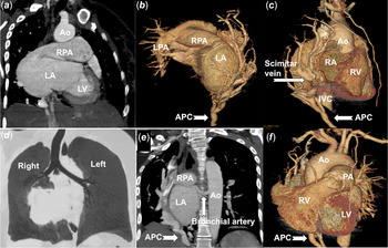
A 28-year-old female with breathlessness for 3 years was identified to have cyanosis and polycythaemia during investigations for infertility. After five venesections for hyperviscosity symptoms, haematocrit was 66%. Physical examination revealed cyanosis with oxygen saturations of 77%, right-sided cardiac pulsations, normal second sound, no precordial murmurs but a soft right infrascapular continuous murmur in the back. Chest X-ray showed mild cardiomegaly with dextroposition and hypoplastic right lung. Even though echocardiogram was uninformative due to dextroposition, agitated saline contrast study confirmed delayed left atrial filling. Computed tomography showed a well-formed right pulmonary artery directly draining into the left atrium without any branches to right lung, anomalous right pulmonary venous drainage to the inferior caval vein, and arterial supply to right lung from a bronchial as well as an abdominal aortic collateral artery (Fig 1). Normal air bronchogram was noted, without any evidence of sequestration. Right lung volumes were around 25% of the left lung volumes. Perfusion scans and magnetic resonance imaging were not done as computed tomography showed absence of pulmonary artery branches to right lung.
Computed tomogram (a) shows direct communication from hilar right pulmonary artery (RPA) to left atrium (LA). Volume rendered image viewed from behind (b) show normal branching of the left pulmonary artery (LPA), absence of RPA branching and its direct drainage into the LA. A 12-mm broad scimitar vein is seen (c) draining to the inferior caval vein (IVC). Normal air bronchogram (d) shows the absence of sequestration in the hypoplastic right lung. Large bronchial artery from thoracic aorta provides nutrition to hypoplastic right lung (e). Frontal surface of heart (f) shows both the ventricles due to the dextroposition.

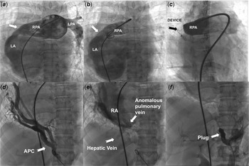
The right pulmonary angiogram showed no branches before its direct drainage to the left atrium (Fig 2, Videos 1, 2). Angiogram of an infradiaphragmatic aortic collateral to the right lung showed anomalous right pulmonary venous drainage through a scimitar vein measuring 12 mm behind the cardiac silhouette to the inferior caval vein above the diaphragm and the hepatic veins. As there was no atrial septal communication, the fistula was crossed from the pulmonary arterial end. After an informed consent, closure of the fistulous communication using a 20-mm Amplatzer post-infarct muscular ventricular septal defect occluder (Abbott, Plymouth, MN, United States of America) with 30 mm retention skirts on either side increased the oxygen saturations to 97%. The pressure in pulmonary trunk remained at 25/10 mmHg after closure, despite having a single-lung perfusion confined to the left lung, indicating a normal vascular resistance in the left lung. Even though a prior balloon interrogation could have given additional information about the size of fistula from the balloon waist, improvement in oxygen saturation, and change in pressure in the pulmonary trunk, it was not done to avoid multiple catheter exchanges that might result in embolism into the left heart. As the oxygenation normalized, the left-to-right shunt through the aortic collateral was also closed with a 14-mm Amplatzer vascular plug (Abbott, Plymouth, MN, United States of America) after confirming an adequate bronchial arterial supply on the tomographic images. She was asymptomatic with good effort tolerance and normal oxygen saturations at 6 months.
Pulmonary angiogram (a and b) shows normal branching of the left pulmonary artery (LPA), absence of branching of the right pulmonary artery (RPA) that directly drains to the left atrium (LA). A muscular occluder (c) closes this communication. The abnormal hypoplastic right lung (d) is perfused by aortic collateral (APC) from abdominal aorta, and venous drainage is anomalous to the inferior caval vein (e). A plug (f) closes the aortic collateral.

Discussion
Abnormal right pulmonary artery-to-left atrial communication, a rare and frequently missed diagnosis, is classified into four morphological types. Reference deSouza eSilva, Giuliani, Ritter, Davis and Pluth4,Reference Ohara, Ito and Kohguchi5 The commonest type I connects the mediastinal right pulmonary artery to left atrium with normal right pulmonary artery branches and pulmonary venous drainage. Reference Zeebregts, Nijveld, Lam, van Oort and Lacquet6 Right lung is single or bilobed in type II, where the lower lobe branch of the right pulmonary artery connects directly to the left atrium and the lower pulmonary vein is absent. Reference Lucas, Lund and Edwards7 These two types are amenable for device closures. Reference Francis, Sivakumar and Kumar8 In the rarer type III, the fistulous tract from the right pulmonary artery receives all the pulmonary veins before entering the left atrium. Reference Tuncali and Aytac9 The fistula originates from the proximal right pulmonary artery in type IV to form an aneurysmal pouch before drainage into the left atrium. The individual right pulmonary veins drain separately into this pouch. Reference Ohara, Ito and Kohguchi5 Surgery is preferred in the last two types due to the proximity of the pulmonary veins to the fistulous tract.
The peculiarity of our patient was a total absence of post hilar right pulmonary artery branching to the right lung associated with scimitar syndrome. Unlike type II, where right lower pulmonary veins and right lower lobe of lung were absent, our patient had hypoplasia of right lung, complete lack of branching of the right pulmonary artery to the right lung, and total absence of pulmonary venous drainage to the left atrium. Even though we could not find any embryological association between scimitar syndrome and such a fistula from any previous reports, this combination possibly represents a gross abnormality in the right lung morphogenesis. There was no effective pulmonary blood flow from the right pulmonary artery to the right lung before the procedure, leading to a single lung situation, and the intervention carried out almost amounted to functional pneumonectomy of the non-functional lung.
The disease has bimodal clinical presentation in neonates and older patients. Persisting fetal pulmonary vascular resistance and patent arterial duct increase pulmonary artery pressure, create gradients, and give audible murmurs. Reference Jiminez, Fournier and Choussat2,Reference Monge, Russess, Popescu and Robinson3 Fetal diagnosis of such turbulent flows lead to early postnatal interventions. Reference Sean and Sivakumar10 Large fistula in older patients equalize the pressure between the chambers and murmurs are absent.
Transcatheter interventions are described in types I and II. Reference Francis, Sivakumar and Kumar8,Reference Sean and Sivakumar10 Surgery may be preferred when the right pulmonary veins drain within the aneurysmal fistula in types III and IV. Difficult surgical access after sternotomy due to marked dextroposition and risk of bleeding after thoracotomy from the anomalous arterial collaterals favoured catheter closure in our patient. The added interventional advantage is the ability to close the aortic collaterals to the abnormal hypoplastic lung, which contributes to a left-to-right shunt.
Supplementary Material
To view supplementary material for this article, please visit https://doi.org/10.1017/S1047951121000287
Acknowledgements
None.
Financial support
This research received no specific grant from any funding agency, commercial, or not-for-profit sectors.
Conflicts of interest
None.
Ethical standards
The authors assert that all procedures contributing to this work comply with the ethical standards of the Indian council of medical research and with the Helsinki Declaration of 1975, as revised in 2008, and has been approved by the institutional committee of Madras Medical Mission, Chennai, India.