Introduction
The earliest known Atrypida emerged during the upper Darriwilian (Middle Ordovician) (Copper, Reference Copper and Kaesler2002), corresponding to the most significant pulse of the Great Ordovician Biodiversification Event (Harper et al., Reference Harper, Rasmussen, Liljeroth, Blodgett, Candela, Jin, Percival, Rong, Villas, Zhan, Harper and Servais2013). Later in the Silurian and Devonian periods this order would become dominated by larger forms such as its namesake Atrypa that became a major component of the global brachiopod fauna, but atrypides were much smaller and of limited biodiversity during the Ordovician.
These early, smaller atrypides are a common component of the Late Ordovician brachiopod fauna from the plates that now make up central Asia and China. A total of 12 genera have been assigned to the order from China, although several have since been synonymized (see Rong et al., Reference Rong, Zhan, Huang, Xu, Fu, Li, Rong, Jin, Shen and Zhan2017 for an extensive summary in English of the Ordovician brachiopods reported from China). The Upper Ordovician rocks of the Kazakh terranes host a remarkably similar suite of atrypides with several additional endemic forms (see Popov et al., Reference Popov, Nikitin and Sokiran1999 for an English-language summary of these forms that includes information from earlier Russian references). Schachriomonia is common to both regions.
Schachriomonia was first described by Nikiforova (Reference Nikiforova1978) from the Upper Ordovician of Uzbekistan. Several other species have since been attributed to the genus from the Upper Ordovician of the Kazakh terranes and North and South China, often initially assigned to other genera such as Zygospira, Kuzgunia, and Ovalospira that have been reclassified or synonymized (see discussion that follows; see also Zhan and Cocks, Reference Zhan and Cocks1998; Popov et al., Reference Popov, Nikitin and Sokiran1999; Rong et al., Reference Rong, Zhan, Huang, Xu, Fu, Li, Rong, Jin, Shen and Zhan2017).
A large brachiopod collection was made from a remote locality in the northeastern part of the Taklamakan Desert in Xinjiang, Northwest China, from the exposed rocks that comprise the Kuruktag Platform on the edge of the Tarim Basin. The shells are from the strongly weathered argillaceous limestones of the upper Katian Hadabulaktag Formation and comprise a low diversity fauna dominated by atrypides and rhynchonellides.
The main goal of this study is to systematically document Schachriomonia pentagonia (Fang in Liu and Fang, Reference Liu and Fang1990) from this locality, but the size and diversity of the collection also provides an opportunity to discuss several important aspects of this species from Tarim. Along with the previously reported rhynchonellide Altaethyrella tarimensis (Sproat and Zhan, Reference Sproat and Zhan2018), finding Schachriomonia on the Tarim paleoplate has paleogeographic implications as the precise position of the plate during the Late Ordovician remains uncertain. The unusually high abundance of these shells at a single sampling horizon may indicate a short-term disruption of the benthic environment that allowed this species to proliferate as an opportunistic colonizer of the seafloor or may reflect a substrate selectivity during the larval stage of these brachiopods. The wide range of shell sizes in the collection was analyzed to examine shell growth trends in Schachriomonia that may reflect the ontogeny of other related early atrypides.
Geographical and geological setting
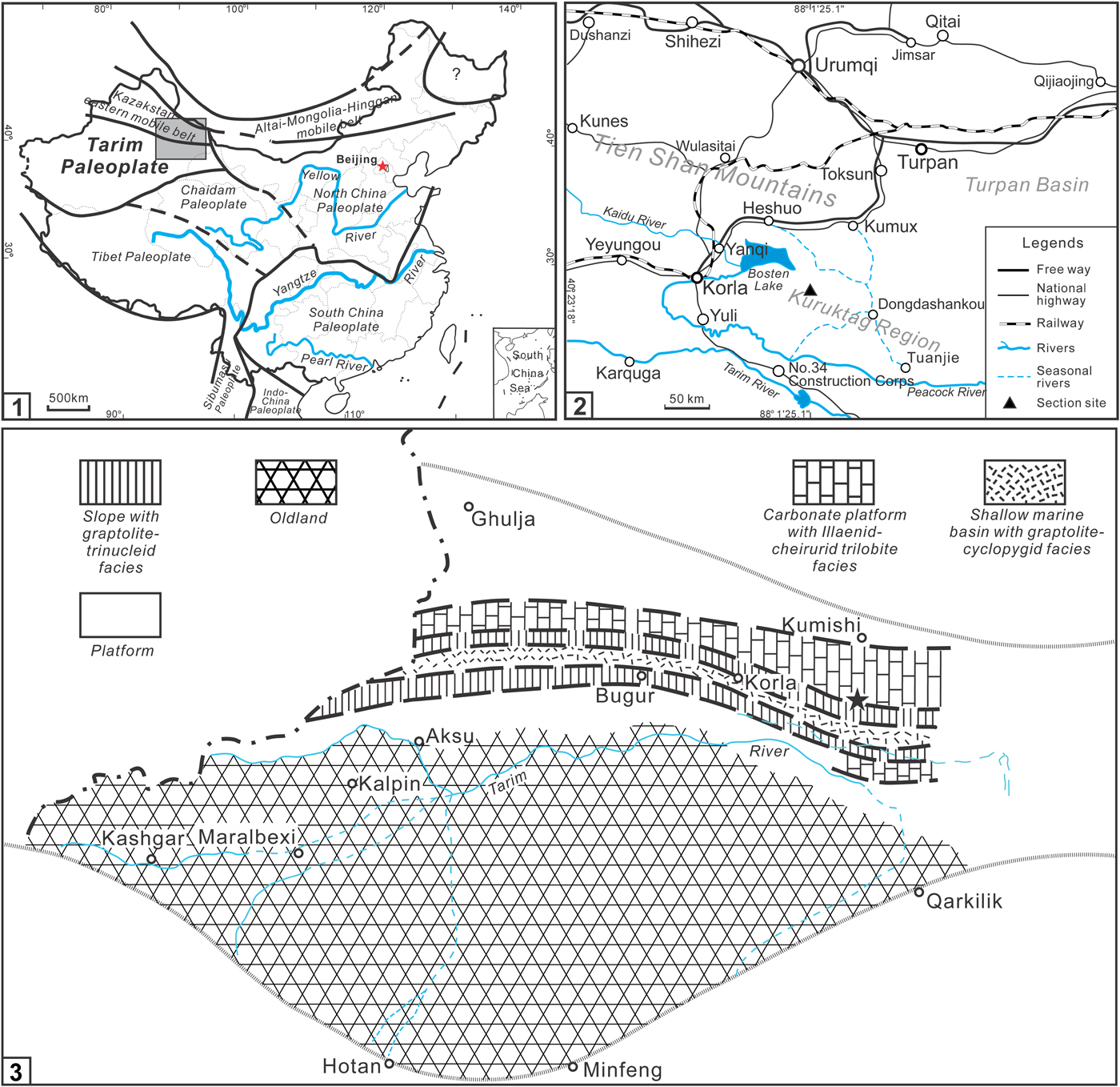
These brachiopods were collected from a remote locality on the Kuruktag Platform that forms the northeastern margin of the Tarim Basin (Fig. 1) and consists of a rocky desert landscape mostly denude of vegetation. The remote nature of the locality and dry, windy environment make access to the site difficult, and the abrasive desert winds have negatively affected the preservation of the shells.
Map of the study area. (1) Map of China with study area indicated by rectangle; (2) magnified view of the study section in the Kuruktag region of the northeastern part of the Tarim Basin, southern Xinjiang, Northwest China. (3) Paleogeographic reconstruction of the Tarim paleoplate during the Katian (Late Ordovician) showing the distribution of different sedimentological and biotic facies. The star shows the exact location of our studied section.

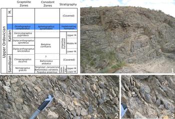
The Ordovician sequence in this region represents a typical platform facies of the narrow and elongate Kuruktag Platform (Zhan et al., Reference Zhan, Rong, Jin, Liang, Yuan, Zhang and Wang2014) that developed during the Ordovician along the northeastern margin of the Tarim paleoplate, separated from the larger Western Tarim-Taklamakan Platform by the Southern Tien Shan Basin (Fig. 1.3). The sedimentary succession in the area includes the Jinlonggou, Xianguletag, Sailikedaban, Uligezitag, and Hadabulaktag formations in ascending stratigraphic order (Fig. 2; see Zhang and Munnecke, Reference Zhang and Munnecke2016).
Upper Ordovician stratigraphy in the study region (adapted from Zhang and Munnecke, Reference Zhang and Munnecke2016) and photos of the outcrop. Modified from Sproat and Zhan (Reference Sproat and Zhan2018). (1) Outcrop of the Hadabulaktag Formation showing typical lithology. Outcrop measures approximately 40 meters in height. (2) The formation is highly fossiliferous in the study area, with (3) numerous brachiopods (albeit poorly preserved) on the surface for collection. Pen cap is approximately 5 cm in length.

The atrypides were collected from thin- to medium-bedded intercalated argillaceous, bioclastic, and micritic limestones and interbedded calcareous mudstone of the upper Hadabulaktag Formation (Fig. 2). Corals, bryozoans, bivalves, and gastropods are common, while brachiopods are the most abundant component of the assemblage. Trilobites and crinoids are present but less common. Conodonts from this formation at other localities indicate a late Katian age in the Aphelognathus pyramidalis Biozone, corresponding to the Dicellograptus complanatus graptolite biozone (Zhang and Munnecke, Reference Zhang and Munnecke2016).
Materials and methods
Collection
A random collection of 2,108 specimens was made during a field excursion in 2007. Most are conjoined shells, with only 12 disarticulated dorsal valves and 17 disarticulated ventral valves, perhaps reflecting the strong joint formed by the cyrtomatodont tooth and socket that had already evolved in atrypides by this time.
Although abundant, these shells are generally poorly preserved. Many are not complete enough to photograph or capture a complete set of diagnostic measurements, and even in relatively well-preserved specimens much of the original shell material usually has been destroyed. The surface ornamentation of shells is commonly partially or completely abraded, and projecting features such as the umbonal regions of shells are often damaged—sometimes completely missing or at least truncated.
Nonetheless, the distinctive ‘tongue’ formed by the dorsal fold and ventral sulcus at the anterior with a single prominent rib in the sulcus and a median furrow flanked by two ribs on the dorsal fold allows for easy identification of these specimens as Schachriomonia. Some specimens can be mistaken for the similar Altaethyrella tarimensis Sproat and Zhan, Reference Sproat and Zhan2018, but the rhynchonellides are generally larger and more globose and always feature several prominent costae in the sulcus. The other atrypides from Central Asia and China have different ribbing patterns and shell shapes except for the very similar Sulcatospira, which may yet be proven to be congeneric with this genus (see discussion in Systematic paleontology).
Measurements
The total number of specimens from each collection was tabulated, and a random subsample of relatively well-preserved specimens was selected for measurement (see Fig. 3 for a diagram showing the measurements made of each shell; see Table 1 for summary statistics). Basic length, width, and thickness in addition to the dimensions of the anterior tongue formed by the deflection of the anterior commissure were recorded, including the width of the tongue at the commissural plane, the width of the tongue at the top of the ventral sulcus where it meets the dorsal fold, and the depth of the tongue perpendicular to these two measurements (see Fig. 3).
Dimensions of each shell measured in dorsal (upper) and anterior (lower) views. L = length of shell from anterior to posterior; W = lateral width; T = thickness of the shell from the most dorsal point to the most ventral point; Ws = width of the sulcus at the commissural plane; Wf = width of the fold at the top of the tongue; Ts = depth of the tongue from the commissural plane to the most dorsal point on the fold at the anterior commissure. Note the prominent tongue formed by the dorsal projection of the commissure at the anterior of the shell.

Summary statistics for the measurements of 244 conjoined Schachriomonia pentagonia shells from the Hadabulaktag Formation in Northwest China. L = length of shell from anterior to posterior; W = lateral width; T = thickness of the shell from the most dorsal point to the most ventral point; Rv = total number of ribs on the ventral valve; Rs = ribs in the sulcus; Ws = width of the sulcus at the commissural plane; Wf = width of the fold at the top of the tongue; Ts = depth of the tongue from the commissural plane to the most dorsal point on the fold at the anterior commissure. Note that ‘0’ in the minimum values associated with the measurements of the sulcus reflects that some small specimens did not have an identifiable sulcus.

These measurements were compiled to create bivariate plots to aid in interpreting the data using the PAST software package (v. 3.20) freely available at https://folk.uio.no/ohammer/past/ (Hammer et al., Reference Hammer, Harper and Ryan2001). The linear regressions applied to select shell dimensions used the reduced major axis method; the linear and power models employed the nonlinear fit module and were compared with other models based on the Akaike information criterion (AIC; the model with the lowest AIC value best fits a given data set). The 95% confidence intervals for these models are based on 1,999 bootstrap replicates.
Serial sectioning
A total of eight shells were serially sectioned, and two were selected for tracing the internal features. A manual parallel grinder was used to grind the shells approximately perpendicular to the commissural plane from the posterior to anterior. The interval between each sectioned surface varied between 0.05 mm and 0.5 mm as small delicate structures near the posterior margin require a finer interval to be captured while larger structures such as the spiralia can be recorded using wider sectioning intervals.
Repository and institutional abbreviation
All specimens figured in this study are catalogued and deposited at the Nanjing Institute of Geology and Palaeontology (NIGP), Chinese Academy of Sciences, in Nanjing, China.
Systematic paleontology
Order Atrypida Rzhonsnitskaya, Reference Rzhonsnitskaya and Orlov1960
Suborder Atrypidina Moore, Reference Moore, Moore, Lalicker and Fischer1952
Superfamily Atrypoidea Gill, Reference Gill1871
Family Atrypinidae McEwan, Reference McEwan1939
Subfamily Spirigerinidae Rzhonsnitskaya, Reference Rzhonsnitskaya1974
Genus Schachriomonia Nikiforova, Reference Nikiforova1978
- Reference Nikiforova1978
Schachriomonia Nikiforova, p. 118.
- Reference Klenina and Bandaletov1984
Zygospira (Kuzgunia) Klenina, p. 113.
- Reference Fu1982
Ovalospira Fu, p. 157.
- Reference Fu1982
Taoqupospira Fu, p. 159.
- Reference Zhan and Cocks1998
Ovalospira; Fu in Zhan and Cocks, p. 60.
- Reference Popov, Nikitin and Sokiran1999
Schachriomonia; Nikiforova in Popov et al., p. 634.
Remarks
Nikiforova (Reference Nikiforova1978) established Schachriomonia based on the type species Schachriomonia schachriomonica from the Archalyk Beds (upper Katian) of Uzbekistan, but several other genera from other regions are now considered junior synonyms (Zhan and Cocks, Reference Zhan and Cocks1998; Popov et al., Reference Popov, Nikitin and Sokiran1999; Rong et al., Reference Rong, Zhan, Huang, Xu, Fu, Li, Rong, Jin, Shen and Zhan2017).
In China, these genera include Ovalospira Fu, Reference Fu1982 and Taoqupospira Fu, Reference Fu1982 (see Rong et al., Reference Rong, Zhan, Huang, Xu, Fu, Li, Rong, Jin, Shen and Zhan2017 for a description and discussion of these genera in English). In their remarks on Ovalospira, Zhan and Cocks (Reference Zhan and Cocks1998) synonymized Taoqupospira from the Upper Ordovician of the western margin of the North China Platform along with Zygospira (Kuzgunia) Klenina, Reference Klenina and Bandaletov1984 from the Ashgillian (middle Katian) Dulankara Formation of Kazakhstan based on similarities to their material. They tentatively considered Ovalospira part of the Zygospiridae (a family now synonymized under the Anazygidae; see Copper, Reference Copper and Kaesler2002) based on its primitive atrypide characteristics and the presence of ribs that would preclude it from the Cyclospiridae but noted that it was somewhat problematic to classify.
Popov et al. (Reference Popov, Nikitin and Sokiran1999) suggested that Ovalospira should be considered a junior synonym of Schachriomonia based on similarities in their costate radial ornamentation, the weakly uniplicate anterior commissure with shallow ventral sulcus originating near the anterior of the shell, and a weak dorsal median fold composed of two slightly accentuated ribs that is reversed from a sulcus near the posterior. They also suggested that the genus would be better classified under the subfamily Spirigerinidae alongside the similar early atrypides Eospirigerina, Boucot and Johnson, Reference Boucot and Johnson1967; Qilianotryma Xu, Reference Xu, Jin, Ye, Xu and Sun1979; and Sulcatospira Xu, Reference Xu, Jin, Ye, Xu and Sun1979. This is probably more appropriate, as Schachriomonia has separate jugal processes while Anazygidae (to which the Zygospiridae have since been assigned) possesses a dorsally to centrally located fused jugum (Copper, Reference Copper1977; Copper, Reference Copper and Kaesler2002). Popov et al. (Reference Popov, Nikitin and Sokiran1999) also provided a detailed discussion of Kuzgunia Klenina, Reference Klenina and Bandaletov1984 in English, summarizing the Russian references and correcting the authorship of the genus.
Copper (Reference Copper and Kaesler2002) considered Ovalospira, Kuzgunia, Taoqupospira, and the contemporaneous atrypide Antizygospira all junior synonyms of Sulcatospira. Rong et al. (Reference Rong, Zhan, Huang, Xu, Fu, Li, Rong, Jin, Shen and Zhan2017), however, noted that the evident V-shaped and well-defined sulcus and corresponding dorsal fold separate Sulcatospira from the broad and shallow U-shaped sulcus and low fold of Ovalospira. In addition, a median dorsal furrow, where the dorsal ribs fuse to become a single continuous ridge on the crest of the dorsal fold, present in Ovalospira is absent in Sulcatospira.
We tentatively agree with the opinions of Popov et al. (Reference Popov, Nikitin and Sokiran1999) and Rong et al., (Reference Rong, Zhan, Huang, Xu, Fu, Li, Rong, Jin, Shen and Zhan2017) in considering Ovalospira along with Taoqupospira and Kuzgunia to be synonyms of Schachriomonia but separate from Antizygospira and Sulcatospira. Rong et al. (Reference Rong, Zhan, Huang, Xu, Fu, Li, Rong, Jin, Shen and Zhan2017) considered Ovalospira Fu, Reference Fu1982 to be the senior synonym, but Schachriomonia Nikiforova, Reference Nikiforova1978 has priority given that it was established four years earlier.
Schachriomonia is very similar to Sulcatospira and could well be congeneric, but a study of additional collections that include well-preserved shells with intact brachidia from both genera is necessary to confirm this. According to Rong et al. (Reference Rong, Zhan, Huang, Xu, Fu, Li, Rong, Jin, Shen and Zhan2017), Sulcatospira differs from Schachriomonia in having a V-shaped, rather than a U-shaped, sulcus, but the description of Schachriomonia by Popov et al. (Reference Popov, Nikitin and Sokiran1999) includes mention of a V-shaped sulcus, so the division is apparently at least somewhat ambiguous. The fused ribs that form the fold in Sulcatospira could be a consequence of poor shell preservation, with abrasion and/or dissolution of the shell surface causing the apparent disappearance of the medial furrow of Schachriomonia. If these genera prove not to be congeneric, they are very closely related, as they are apparently similar in almost every other way.
Antizygospira can still be differentiated from the preceding genera largely on the basis of its ribbing and overall smaller size: while the preceding genera have simple costae with only rare bifurcations, Antizygospira exhibits prominent bifurcation and intercalation of ribs. In the shell interior, Antizygospira lacks true dental plates (Rong et al., Reference Rong, Zhan, Huang, Xu, Fu, Li, Rong, Jin, Shen and Zhan2017) while Schachriomonia has thin dental plates. Antizygospira is usually considerably smaller than the other atrypide genera reported from China.
Schachriomonia pentagonia (Fang in Liu and Fang, Reference Liu and Fang1990)
Figures 4–7
- Reference Liu and Fang1990
Rhynchotrema pentagonia Fang in Liu and Fang, p. 141 (English translation on p. 224), pl. 21, figs. 7, 8, text-fig. 9.
Type specimens
No holotype was selected by Fang in the original description of the species, and the original type material cannot be located. We select NIGP 168782 from our collection as the neotype from the upper Katian Hadabulaktag Formation. The original collection locality of Liu and Fang (Reference Liu and Fang1990) is uncertain, but the current collection is from the same region near Uligezhitag and is at least equivalent to the original type material if not from the exact same locality.
Diagnosis
Small to medium-sized shell with subpentagonal to subcircular outline, ventribiconvex to biconvex, unisulcate anterior in smallest shells becoming rectimarginate and uniplicate in larger shells forming prominent tongue at anterior, prominent median furrow on dorsal fold, and prominent single median costae in dorsal sulcus, spiralia with seven to eight whorls, short jugal process.
Occurrence
Upper Hadabulaktag Formation (upper Katian), in the Kuruktag region of Xinjiang, Northwest China.
Description
(Emended from Liu and Fang, Reference Liu and Fang1990.) Shell small to medium-sized, with length and width approximately 5–15 mm and thickness approximately 2–10 mm; pentagonal to subcircular in outline, lateral profile ventribiconvex to biconvex. Shell equally as long as wide in small shells, becoming approximately 75% as long as wide in larger shells; 30% to 82% as thick as wide. Astrophic hinge line measuring approximately one-third of shell width. Anterior commissure unisulcate in small shells becoming rectimarginate, parauniplicate, and uniplicate in progressively larger shells. Ribs strong, simple, and rounded, mostly originating at umbo with rare bifurcation; one prominent rib in ventral umbo; two prominent ribs on dorsal fold; four to seven on shell flanks.
Ventral umbo large and prominent; ventral interarea anacline and with narrow delthyrium partially covered by elongate deltidial plates. U-shaped ventral fold becoming flattened about one-third of length from umbo and thereafter becoming U-shaped sulcus with one prominent median plication projecting upward at anterior commissure to form prominent tongue.
Dorsal umbo minute and incurved, covered by ventral umbo. U-shaped dorsal sulcus becoming flattened about one-third of length from umbo and thereafter becoming U-shaped fold with flattened top bounded by two prominent plications at each side forming a median groove.
Teeth relatively large with median groove in some shells supported by thin dental plates. Dental cavities triangular, becoming partially infilled by secondary shell material in some shells.
Dorsal median ridge in shell interior very small, extending to midlength of valve, not connected to hinge plates. Sockets relatively deep, with hinge plates separated medially, lacking septalium. Crura projecting anteriorly from crural bases embedded in shell material medial to hinge plates. Spiralia consisting of seven to eight whorls with short jugal process at posterior.
Materials
A total of 2,108 shells (2,079 conjoined valves, 12 dorsal valves, and 17 ventral valves). In addition to the neotype (NIGP 168782), specimens NIGP 168779, 168780, 168781, and 168783 are figured in Figure 4, and NIGP 168784, 168785, and 168786 are shown in Figure 7. NIGP 168787 and 168788 were sectioned and are illustrated in Figures 5 and 6, respectively.
Schachriomonia pentagonia (Fang in Liu and Fang, Reference Liu and Fang1990) from collection AFT-X 398. Note the distinctive tongue at the anterior commissure formed by the dorsal fold with two ribs that form a median furrow and a ventral sulcus that usually contains a single rib. (1–5) NIGP 168779: (1) dorsal, (2) ventral, (3) lateral, (4) anterior, and (5) posterior views. (6–10) NIGP 168780: (6) dorsal, (7) ventral, (8) lateral, (9) anterior, and (10) posterior views. (11–15) NIGP 168781, (11) dorsal, (12) ventral, (13) lateral, (14) anterior, and (15) posterior views. (16–20) NIGP 168782, neotype, (16) dorsal, (17) ventral, (18) anterior, (19) posterior, and (20) lateral views. (21–25) NIGP 168783, (21) dorsal, (22) ventral, (23) lateral, (24) anterior, and (25) posterior views. All shells are photographed at the same scale.

Serial sections of a small Schachriomonia pentagonia (Fang in Liu and Fang, Reference Liu and Fang1990) shell (collection horizon AFT-X 398, specimen no. NIGP 168787). Numbers represent distance (in millimeters) of each section from the most posterior point on the ventral umbo. Dorsal valve uppermost. Speckled region indicates coarse calcite crystals that often fill the mantle cavity in shells from this collection horizon. Despite sectioning several small shells, no spiralia were preserved. The spiralia in these small shells were likely thin and fragile and may have been more easily damaged or dissolved during burial or diagenesis. Most of the outermost layers of the shell are missing, and in some cases, much of the exterior shell surface has been abraded.

Serial sections of a large Schachriomonia pentagonia (Fang in Liu and Fang, Reference Liu and Fang1990) shell (collection horizon AFT-X 398, specimen no. NIGP 168788). Numbers represent distance (in millimeters) of each section from the most posterior point on the ventral umbo. Dorsal valve uppermost. Dotted region indicates coarse calcite crystals that often fill the mantle cavity in shells from this collection horizon. Despite this, the spiralia are well preserved in this specimen although they are slightly offset toward the left side of the shell, perhaps due to damage to the brachidial supports after death of the brachiopod.

Damaged Schachriomonia pentagonia (Fang in Liu and Fang, Reference Liu and Fang1990) shells with spiralia exposed (collection horizon AFT-X 398). (1) NIGP 168784, dorsal view showing seven whorls on the left side of the mantle cavity. (2) NIGP 168785, dorsal view showing seven whorls of the spiralium on the left and four whorls of the incompletely exposed spiralium on the right. (3) NIGP 168786, a smaller shell showing six whorls of the right spiralium in an obliquely oriented cross section of the shell; posterior is on left.

Remarks
These specimens are conspecific with Rhynchotrema pentagonia Fang in Liu and Fang, Reference Liu and Fang1990, also collected from the Hadabulaktag Formation in the Kuruktag region near Uligezhitag. In the description, Fang noted a prominent dorsal groove on the fold and a sulcus with a single prominent rib, indicating that those shells are not Rhynchotrema, which almost always has four ribs on the dorsal fold and three in the ventral sulcus (Sproat et al., Reference Sproat, Sohrabi and Jin2014). Many species that were described as Rhynchotrema in China are now being recognized as belonging to one of the atrypide genera (as in here) or the externally similar rhynchonellide Altaethyrella (Zhan and Li, Reference Zhan and Li1998; Sproat and Zhan, Reference Sproat and Zhan2018). The dimensions of the shells in the collection of Liu and Fang (Reference Liu and Fang1990) are within the range of the specimens we describe here (see Table 1 for summary statistics of each measurement), although our collection additionally includes shells that are much smaller than what they reported. Liu and Fang (Reference Liu and Fang1990) also mention the presence of narrow and elongate deltidial plates characteristic of this genus that partially cover the delthyrium, a diagnostic characteristic of Schachriomonia.
Liu and Fang (Reference Liu and Fang1990) included only four illustrations of sectioned material that are lacking interior details. Note that their first section, labeled ‘12’ on p. 142, is likely reversed and the apparent base of the ventral valve is actually the fused deltidial plates near the posteriormost part of the delthyrium, characteristic of the genus. In our sectioned specimens, six to eight whorls of the spiralia are preserved, but not in the smaller shell (Fig. 5). The larger shell figured here (Fig. 6) clearly shows at least seven whorls, and three incomplete shells in our collection have similar exposed spiralia (Fig. 7). The jugal process is also visible in the larger sectioned shell extending ventromedially from the left crura in the larger shell, although it is not preserved on the right side. The larger shell illustrated here seems to have a prominent midtooth furrow on the exterior-oriented side of the tooth that has not yet been described elsewhere, but this is apparently absent in the smaller specimen, indicating that it may develop over ontogeny through resorption of primary shell material.
Schachriomonia pentagonia is most similar to Schachriomonia parva described by Popov et al. (Reference Popov, Nikitin and Sokiran1999) from the Taldyboi Formation (lower Katian), River Taldyboi, Chingiz Range, including those synonymized by Popov et al. (Reference Popov, Nikitin and Sokiran1999) originally described by Klenina (Reference Klenina and Bandaletov1984). The shell outline is identical (0.97 mean shell length:width ratio in both species), but those S. pentagonia shells are slightly more convex overall than S. parva (0.62 mean shell thickness:length ratio here versus 0.55 in their collection) although the range of convexity is similar between these species. These shells also have slightly fewer ribs (11 to 17 here versus 14 to 21 in their collection).
The type species Schachriomonia schachriomonica Nikiforova (Reference Nikiforova1978) from the upper Katian Archalyk Beds in south Uzbekistan have more numerous ribs and less-pronounced ventral sulcus and dorsal fold. Serial sections for the species show much more substantial dental plates in that species, but the shell also appears to be somewhat distorted, possibly affecting the size and proportions of internal shell structures illustrated there (see also Copper, Reference Copper and Kaesler2002, p. 1433, fig. 4d).
Schachriomonia qinghaiensis (Xu, Reference Xu, Jin, Ye, Xu and Sun1979) from the Qaidam terrane in China is also very similar to S. pentagonia in terms of external morphology. Although some of the internal shell structures are shown in text figures accompanying the description of the genus, the brachidial structure is missing in their figured serial sections. Basic measurements to compare the dimensions and shape of the shell to other species are also lacking. Further study of additional material may show that it is conspecific with either S. pentagonia or S. parva.
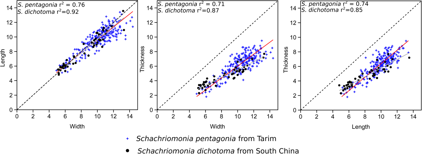
Schachriomonia pentagonia differs from Schachriomonia dichotoma (Fu, Reference Fu1982) from the Xiazhen Formation (upper Katian) in the Zhejiang–Jiangxi border area of South China and central Shaanxi in North China in generally having a more prominent fold and sulcus at the anterior of the shell. S. pentagonia is generally larger in size and exhibits a slightly wider range in shell dimensions (Fig. 8).
Length/width, thickness/width, and thickness/length linear regression plots of Schachriomonia pentagonia from Tarim in Northwest China and Schachriomonia dichotoma from the Zhejiang–Jiangxi border region in South China for comparison. Note that the shell outlines are very similar in these species, but S. pentagonia is generally more convex in profile than S. dichotoma from South China.

Many of the smaller shells in our collection are superficially similar to Antizygospira Fu, Reference Fu1982, especially in outline and the shape of the anterior commissure. However, these shells have the mostly simple costae of Schachriomonia rather than the bifurcating and intercalating costae of Antizygospira, and the smaller shells that have been sectioned all show definite, albeit relatively thin, dental plates that are often lacking in Antizygospira (see Rong et al., Reference Rong, Zhan, Huang, Xu, Fu, Li, Rong, Jin, Shen and Zhan2017).
Paleobiogeographical significance
Locating the Tarim paleoplate in the Late Ordovician
The precise location of the Tarim paleoplate during the Ordovician remains uncertain largely due to the lack of paleomagnetic data from the plate thus far, but it was probably located in the vicinity of the Kazakh terranes and North and South China paleoplates near the equator (Popov and Cocks, Reference Popov and Cocks2017; Torsvik and Cocks, Reference Torsvik and Cocks2017). A full comparison of the brachiopod faunas will be possible once the fauna from Tarim has been completely described, but a few preliminary remarks on the paleogeography of Tarim can be made based on commonalities in the atrypides of these plates.
Tarim, North and South China, and the Kazakh terranes were all parts of a low-latitude brachiopod paleobiogeographic province along with Laurentia, Siberia, and Australia (then part of northern Gondwana) during the Late Ordovician (Harper et al., Reference Harper, Rasmussen, Liljeroth, Blodgett, Candela, Jin, Percival, Rong, Villas, Zhan, Harper and Servais2013). Although there are many broad similarities across this province, there are significant differences within individual orders (e.g., dominance of rhynchotrematides in Laurentia in comparison with the dominance of ancistrorhynchides in the Kazakh terranes and North and South China and the evolution of a distinct epicontinental fauna in the intracontinental seas of Laurentia during the late Katian dominated by taxa such as Hiscobeccus and Hypsiptycha that do not occur elsewhere; see Jin, Reference Jin2001; Jin et al., Reference Jin, Sohrabi and Sproat2013, Sohrabi and Jin, Reference Sohrabi and Jin2013a, Reference Sohrabi and Jin2013b; Sproat et al., Reference Sproat, Sohrabi and Jin2014; Candela, Reference Candela2015) that can be used to distinguish two subprovinces within the larger low-latitude province (e.g., Harper et al., Reference Harper, Rasmussen, Liljeroth, Blodgett, Candela, Jin, Percival, Rong, Villas, Zhan, Harper and Servais2013, fig. 11.10).
Although Schachriomonia is known from the Upper Ordovician of the Kazakh terranes and North and South China, the Kazakh species Schachriomonia parva has thus far only been documented from the Chu Ili and Chingiz terranes (Popov et al., Reference Popov, Nikitin and Sokiran1999; Popov and Cocks, Reference Popov and Cocks2006, Reference Popov and Cocks2014). The similarity of this species to S. pentagonia in Tarim signals a close association between Tarim and these terranes during the Late Ordovician. In fact, S. parva may be a closely related ancestor of S. pentagonia given that S. parva is known from the lower to middle Katian strata in the Kazakh terranes and S. pentagonia is from the upper Katian of Tarim.
Such a relationship agrees with the findings from the rhynchonellides already published. The ancistrorhynchide Altaethyrella tarimensis Sproat and Zhan, Reference Sproat and Zhan2018 occurs alongside S. pentagonia at the collection locality in Tarim and is very similar in terms of shell shape and structures to Altaethyrella otarica (Rukavishnikova, Reference Rukavishnikova1956) found in the Chu-Ili Range, further solidifying the paleogeographic ties between these blocks.
Popov and Cocks (Reference Popov and Cocks2017, text-fig. 13) proposed a Late Ordovician paleogeographic reconstruction, based on similarities in fossil faunas, of the plates and terranes that now make up Central Asia and China. Given that the brachiopod fauna of Tarim is only poorly known, their treatment of Tarim was largely based on similarities of the trilobite faunas. The range of Schachriomonia across North and South China and the Kazakh terranes in addition to Tarim supports their notion that equatorial ocean currents could have enabled the dispersal of the brachiopod fauna across the plates that now comprise China and Central Asia. They projected Tarim to be located adjacent to the Chu-Ili terrane at 455 Ma (equivalent to the Sandbian, slightly earlier in age than the late Katian fauna being studied here), and thus far, the evidence from our work on the brachiopods of Tarim supports this arrangement. However, how the same species would have dispersed to or from the Chingiz terrane that they have plotted on the opposite side of their map closer to Gondwana is difficult to explain.
Central and East Asian atrypides in comparison to Laurentia
Schachriomonia and the other atrypides from Central Asia and China differ from those that evolved during the Sandbian and Katian in Laurentia. In Laurentia, the costate forms Anazyga, Catazyga, and Zygospira were common components of Katian brachiopod assemblages, with smooth-shelled Manespira, Protozyga, Idiospira, and Cyclospira being less common in the shallow marine platforms and basins (Copper, Reference Copper1977, Reference Copper1982, Reference Copper1986). Of these, only Cyclospira is known from the Upper Ordovician of South China (Zhan and Cocks, Reference Zhan and Cocks1998).
Many atrypides in China and Central Asia were initially classified under Zygospira, including those now considered Schachriomonia, and indeed these genera are similar in shell shape and ornamentation. However, the medial external shell structures are reversed—Zygospira has a dorsal sulcus and ventral fold while Schachriomonia has a dorsal fold and ventral sulcus. In the shell interior, the brachidial supports also differ significantly among these groups although these structures are often undocumented in older references. These Asian species are now mostly classified under the family Spirigerinidae, a group that would not reach Laurentia until the Hirnantian (Copper, Reference Copper1986). Thus, two different atrypide lineages occupied these regions during the Late Ordovician but apparently remained mostly isolated from one another until after the onset of the Late Ordovician Mass Extinction in the Hirnantian.
In the Late Ordovician, Australia was located at the northern tip of Gondwana between Laurentia and the plates that now comprise central and eastern Asia (Torsvik and Cocks, Reference Torsvik and Cocks2017). The Late Ordovician atrypides of Australia (Laurie, Reference Laurie1991; Percival, Reference Percival1991) seem to share closer affinities with those of Laurentia rather than with those of the Kazakh terranes, Tarim, and North and South China. In New South Wales, Zygospira carinata Percival, Reference Percival1991 shares the coarse, simple costae and overall shell shape of both Schachriomonia and Zygospira, but its dorsal sulcus and ventral fold suggest affinities to the latter. Protozyga, also found in Laurentia, was reported alongside the endemic forms Webbyspira Percival, Reference Percival1991 and Australispira Percival, Reference Percival1991. Laurie (Reference Laurie1991) reported a species of Protozyga from the Benjamin Limestone of Tasmania as well. This faunal similarity between Laurentia and Australia not only is apparent in the brachiopod fauna but was also shown to occur in Late Ordovician stromatoporoids (Webby, Reference Webby1991).
Paleoecological significance
S. pentagonia is not widespread throughout the studied section, being found in only three collections (AFT-X 398, 399, and 400). The majority (2,047 of 2,108 shells) were collected from AFT-X 398, where they are extremely abundant and significantly outnumber other species, with only 35 shells from AFT-X 389 and 26 from AFT-X 400.
The reason for this high concentration of shells of S. pentagonia in this single horizon is uncertain. The horizon features a high degree of calcite crystallization, but it is not clear how this could be connected to the abundance of these atrypides in relation to other brachiopods. There does not appear to be any taphonomic reason to favor the preservation of the atrypides over other species, as there is no unusual abundance of shell fragments that might suggest other species were present in the same living environment but damaged or destroyed before lithification of the sediments. Winnowing by currents prior to burial could be a factor, but shells from other taxa of similar size should be concentrated in the same layer if that were the case, whereas here the specimens of Altaethyrella are often larger and the orthides that occur at this horizon are significantly larger. No sorting for different shell sizes, shell shapes, or different taxa was noted in the field, and no consistent orientation of the brachiopod shells or other fossils was found.
If geological and taphonomic processes cannot explain this occurrence, biological and ecological factors may be at play. High-dominance, low-diversity assemblages are often interpreted as a response to instability in the environment, with the dominant taxa being rapid-reproducing or high-fecundity generalists that take advantage of the disruption to quickly colonize the seafloor before other taxa can become established (Levinton, Reference Levinton1970). However, there is little evidence of storm activity, debris flows, or periods of anoxia or fluctuating salinity in the Upper Ordovician of this region that would have regularly disrupted the benthic ecosystem. Perhaps Schachriomonia formed dense clusters on the seafloor due to substrate selectivity by the planktonic larvae, much like oysters form dense beds on the seafloor today. If so, collection AFT-X 398 may have been the only horizon that sampled one of these clusters in our collection. These types of associations have not been commonly reported for this lineage, and further detailed study at other localities and regions would be necessary to confirm this hypothesis.
Popov et al. (Reference Popov, Nikitin and Sokiran1999) noted that S. parva in the lower part of the Akdombak Formation (middle Katian) near Akdombak Mountain in Kazakhstan is dominant in a low-diversity assemblage together with the brachiopod Sowerbyella akdombakensis Klenina, Reference Klenina and Bandaletov1984 and tabulate corals. In Tarim, however, Altaethyrella tarimensis and orthides are the second- and third-most abundant components of the assemblage, respectively, so these probably represent different associations.
Growth trends
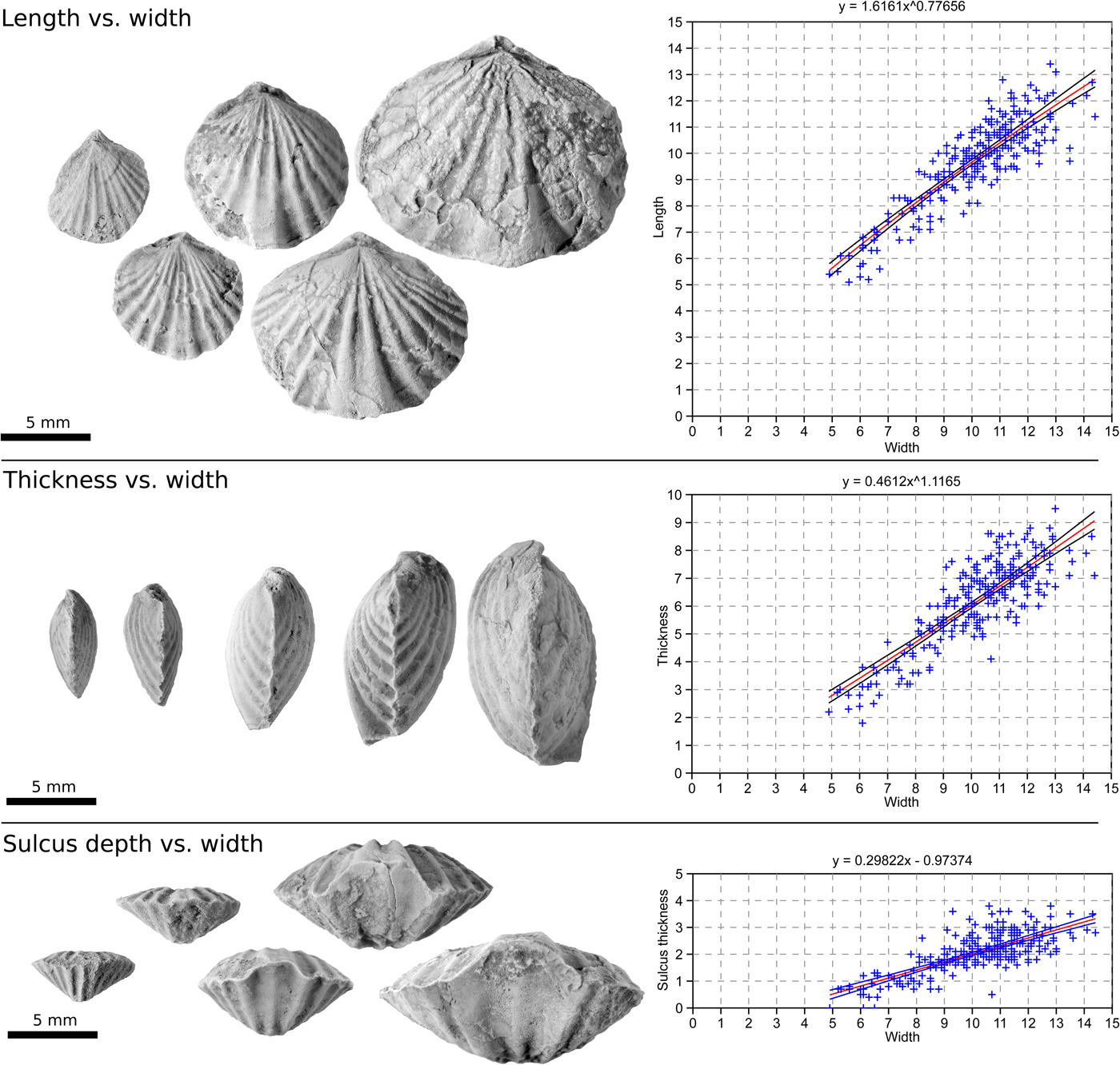
Variation in the rate of shell growth along different shell axes has been identified by comparing the measurements of 244 shells selected across the spectrum of shell sizes (Fig. 9). The most obvious change in shell shape is a complete reversal in the form of the dorsal and ventral valves, but variation in shell outline is also notable between small and large shells.
Figure showing patterns of growth over a sequence of Schachriomonia pentagonia shells at different stages of growth. Scale is constant for all specimen photos to highlight differences. (Note: these shells are also shown in full in Fig. 5.) Dorsal, lateral, and anterior views are shown in conjunction with plots of length/width, thickness/width, and sulcus depth/width. Length, width, and thickness are in millimeters. Plots include a power law trend line of best fit with x and y intercept values of zero (except sulcus versus width, which have nonzero x and y intercepts) with a 95% confidence interval. The equations for each trend line are provided above each graph.

Variation in shell outline and convexity over ontogeny
The ratio of length (L) to width (W) corresponds to the outline of a shell—a larger L/W ratio indicates a more elongate shell while a smaller value indicates a wider shell. Most of the shells in our collection have a value near 1 (see Table 1), but the larger shells deviate from this trend, becoming significantly wider than long, with an L/W ratio as low as 0.73. A power function (Equation 1) best fits a regression line through the data points with an x and y intercept of zero (or c value).
 $${\rm Y}{\rm} = {\rm} {\rm a}{\rm x}^{\rm b} + {\rm} {\rm c}$$
A zero intercept is necessary assuming that the shell started growing from a single point (or 0, 0). Without this stipulation, a linear trend can be fitted to the data, but it is impossible to start growth from an initial stage that is 0 mm long but 1 mm wide, for example.
A growth trend defined by a power law may indicate a degree of allometry, or differential growth in terms of the shell outline. If both measurements increase linearly with regard to each other, the value of x in the power law equation should be equal to one, indicating isometric growth. A value larger or smaller than one indicates a differential change in one dimension relative to the other over ontogeny. The paleontological application of the power law and to allometric growth was thoroughly reviewed and discussed at length by Gould (Reference Gould1966). Literature is sparse on allometry in brachiopods, although Parkinson (Reference Parkinson1952) investigated the growth of Dielasma hasta Sowerby, Reference Sowerby1825 from the lower Carboniferous near Derbyshire, finding not only allometry in terms of length and thickness but also an unusual interruption in length and thickness growth trends midway through growth within the population.
To truly show allometry, the data should include specimens of all sizes, and although we have a wide range of shell dimensions here, the smallest specimens are missing. Nonetheless, the bivariate plots show the power law relationship between length and width, with an exponential (b) value of 0.777, confirming that large shells grow wider at a greater rate than they grow longer.
A similar power function can be fitted to the plot of shell thickness (i.e., shell depth, not thickness of the shell layers) versus shell width as a proxy for shell convexity. This trend is only weakly allometric, with an exponential value of 1.165, indicating that the shell became relatively more convex as it became larger, but not markedly so, as shown in Figure 9.
The widening and slight increase in convexity of the shell over its growth would laterally (and, to a far lesser extent, dorsoventrally) expand the mantle cavity as the shell became larger. Two main structures fill the mantle cavity; one is the spiralia that support the lophophore. As the brachiopod grew in size, its metabolic requirements also grew, necessitating the development of a larger lophophore. Given that the shells did not become significantly more convex over ontogeny, a wider shell would have allowed the lophophore to expand laterally as the energy requirements of the brachiopod grew, allowing for more efficient filter feeding as a greater volume of water would pass through the mantle cavity.
The other structures that would have occupied the mantle cavity are the gonads. These are soft tissues, and thus they are not amenable to preservation in fossil material; there is no direct evidence of their presence in these shells. However, given that only larger shells seem to have been strongly allometric in growth, this could be a signal of sexual maturity in the brachiopod. The spiralia appear to fill most of the mantle cavity even in large shells, but this does not preclude the development of sex organs from influencing shell shape as well.
Growth of the anterior tongue
A linear rather than a power function best fits the ratio of the depth of the sulcus at the anterior relative to the commissural plane (Ts) against the width of the shell (W), indicating that the extension of the tongue above the commissural plane occurs at a constant rate relative to overall shell growth. The smallest of shells in our collection have a dorsal sulcus and ventral fold, although these shells are not common and generally are not well preserved, perhaps owing to their small size. At a certain stage of ontogeny, the shells became rectimarginate without a fold or sulcus, subsequently developing a dorsal fold and a ventral sulcus. This phenomenon is illustrated best in the serial sections of S. pentagonia (Figs. 5, 6), where the sections close to the posterior in the larger shell show a shallow dorsal sulcus that becomes a dorsal fold toward the anterior. The inflection point occurs at approximately one-third to one-half of the shell length in fully grown shells. Unlike the functions fitted to L/W and T/W, this trend clearly has nonzero intercept values as the zero value for the depth of the sulcus (or the point at which the brachiopod would become rectimarginate in form) occurs midway through ontogeny rather than at the beginning of shell growth (see Ts/W graph in Fig. 9).
This change in shell form is difficult to explain. The prominent tongue in rhynchonellides has previously been interpreted to have separated the feeding incurrent generated by the lophophore from the outcurrent that expels waste from the mantle cavity (Rudwick, Reference Rudwick1970). Given that the lophophore in smaller shells is yet unknown, this shift in shell shape could reflect a change in the development of the lophophore or perhaps a change in the living position as the shell grows, necessitating a shift in the location of the incurrent and excurrent flows (such as a shift from resting on one valve on the seafloor to the other). This would be difficult to test, however, as these shells were probably not in their original living position.
Interior structures
The tooth and socket structures are also slightly different between the smaller and larger shells that were sectioned (Figs. 5, 6). In well-preserved shells where structures are not obscured by secondary calcite crystal growth, a prominent medial furrow oriented laterally usually occurs on each tooth. The development of this furrow would have strengthened the joint between the tooth and socket by locking the tooth into the socket and may have been beneficial as the shell became larger, placing more stress on the hinge. Cyrtomatodont teeth that are modified in shape as the shell grows are common in other groups with elaborate lophophore supports that necessitated the ability to resorb shell material to modify the shape of the brachidial supports (Carlson, Reference Carlson1989).
Conclusions
Schachriomonia is documented herein for the first time from the Tarim paleoplate and is conspecific with the previously described Rhynchotrema pentagonia from the region. R. pentagonia cannot possibly be rhynchonellides as originally described. Although these shells most closely resemble those of S. parva reported from central Asia, they are broadly similar to other species of the genus found in North and South China as well.
The similarities between S. pentagonia in Tarim and S. parva from the Chu-Ili and Chingiz terranes of Kazakhstan reflect a close paleogeographic connection between these blocks. A full description of the fauna will allow for a more comprehensive comparison of the fauna in Tarim to that of other plates and terranes in the region.
The high dominance of the species at a single locality could be indicative of an opportunistic species taking advantage of disturbances in the benthic marine environment, but there is no evidence of disruption to the benthic ecosystem in the geology of the study area. In addition, taphonomic effects are not likely to be a factor. Larval substrate selectivity may have been responsible for the unusually high abundance of Schachriomonia here, although additional studies of the group from other areas would be necessary to support this hypothesis.
Small S. pentagonia shells are generally almost equally as long as wide with a dorsal sulcus and medial fold, but larger shells developed a dorsal fold and a ventral sulcus early in ontogeny and became wider than long, perhaps due to changes in size and shape of the lophophore as the brachiopod grows. The relative widening of the shell may also be a sign of sexual maturity due to the development of the gonads, but this cannot be proven in fossil shells as the gonads are unlikely to be preserved in the fossil record.
Acknowledgments
Y. Zhang., H. Wang, and G. Li from the Nanjing Institute of Geology and Palaeontology (NIGP), Chinese Academy of Sciences, provided assistance in the field, and J. Rong (NIGP) provided many suggestions on the early version of the manuscript. Y. Candela, I. Percival, and an anonymous reviewer are thanked for their constructive comments on the initial draft of the manuscript. Funding was provided to Sproat by a Chinese Academy of Sciences President's International Fellowship and to Zhan by the Chinese Academy of Sciences (XDBB26000000) and the State Key Laboratory of Palaeobiology and Stratigraphy (LPS). This is a contribution to IGCP Project 653: The Onset of the Great Ordovician Biodiversification Event.