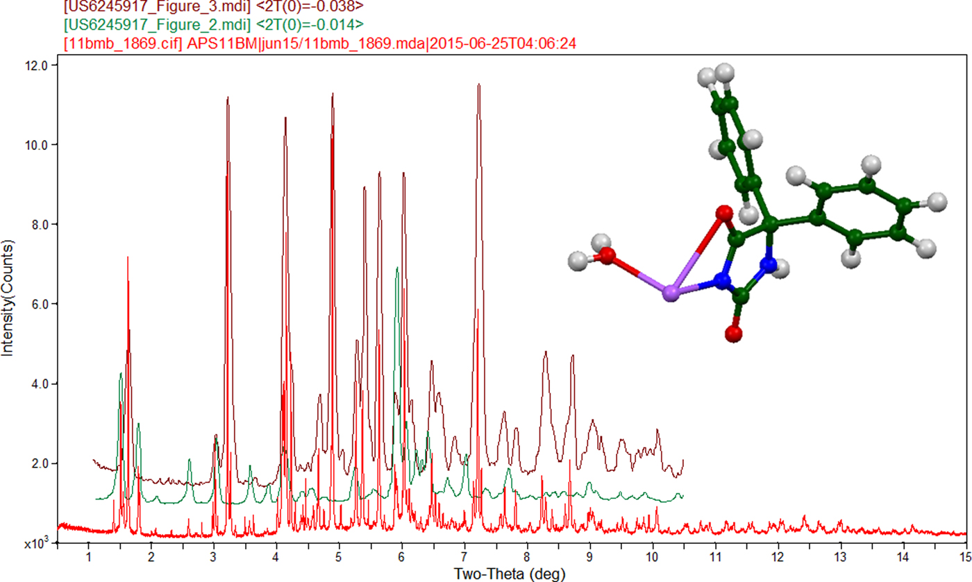
Phenytoin is an anticonvulsant, and is used to prevent and control seizures. A common brand name containing the sodium salt is Dilantin®. A commercial reagent sample (USP lot I0L257) was analyzed in this study and found to be comprised primarily of phenytoin sodium monohydrate (Bosch et al., Reference Bosch, Johnson and Stahl2001), as well as minor amounts of phenytoin sodium anhydrate and at least one other crystalline compound (see Figure 1). The Bosch patent contained the atom coordinates for phenytoin sodium monohydrate from a single crystal structure determined at 173 K. This structure and the data shown in Figure 1(c) (Figure 3 of the Bosch patent) were used to index the synchrotron pattern collected as part of this study at 295 K, and a Rietveld refinement was carried out to determine the room temperature unit cell. The structure was optimized using density functional theory techniques (Dovesi et al., Reference Dovesi, Orlando, Erba, Zicovich-Wilson, Civalleri, Casassa, Maschio, Ferrabone, De La Pierre, D-Arco, Noël, Causà and Kirtman2014). At 295 K, phenytoin sodium monohydrate crystallizes in the monoclinic space group P21/c, with a = 15.6729(4), b = 6.2193(1), c = 15.9063(3) Å, β = 111.856(2)̊, V = 1439.02(3) Å3, and Z = 4. The diffraction data were collected on beamline 11-BM (λ = 0.414163 Å) at the Advanced Photon Source, Argonne National Laboratory. The thermal expansion between 173 and 295 K is anisotropic; the expansion is 0.94, 0.20, and 0.67% along a, b, and c, respectively. The cell volume is 4.73% larger at room temperature. The Na is 5-coordinate (NaNO4), and distorted square pyramidal. The Na coordination polyhedra share corners to form double chains along the b-axis. The amino group N6-H22 forms a strong hydrogen bond to the ketone oxygen atom O2. The water molecule is coordinated to the Na, and participates in weak O-H⋯O and O-H⋯C hydrogen bonds. Also contributing to the crystal energy are C-H⋯O hydrogen bonds. Phenyl-phenyl interactions are prominent in the crystal packing. The DFT-optimized structure has been submitted to ICDD for calculation of a powder pattern for the Powder Diffraction File.
(Colour online) (a) Synchrotron powder diffraction pattern of commercial phenytoin sodium, (b) Figure 2 of US Patent 6,245,917, a laboratory X-ray powder diffraction pattern of phenytoin sodium anhydrate and (c) Figure 3 of US Patent 6,245,917, a laboratory X-ray powder diffraction pattern of phenytoin sodium monohydrate. The patent patterns were digitized and rescaled in 2θ to match the synchrotron wavelength of 0.414163 Å.

SUPPLEMENTARY MATERIAL
The supplementary material for this article can be found at https://doi.org/10.1017/S0885715618000246
Acknowledgements
Use of the Advanced Photon Source at Argonne National Laboratory was supported by the US Department of Energy, Office of Science, Office of Basic Energy Sciences, under Contract No. DE-AC02-06CH11357. This work was partially supported by the International Centre for Diffraction Data. We thank Lynn Ribaud and Saul Lapidus for their assistance in data collection.