1. Introduction
Lower Jurassic ichthyosaurs are known from thousands of specimens, although not all of them are sufficiently complete to assign to a species. Most Lower Jurassic genera can, however, be distinguished from one another by a few key postcranial elements, such as the forefin, humerus, coracoid or pelvic girdle, in the absence of a skull. In fact, the genus Ichthyosaurus has an unusual forefin that easily distinguishes it from all other genera. The anterior digital bifurcation (shared only with Protoichthyosaurus, Lomax, Massare & Mistry, Reference Lomax, Massare and Mistry2017) and a prominent digit V gives the forefin a unique morphology (Motani, Reference Motani1999). Forefins of Ichthyosaurus are extremely variable in the digit that bifurcates (digit II or III), the location of the bifurcation on the digit, the number of bifurcations in the fin and the relative size of the branches of the bifurcation, even within a species. Although an isolated forefin can be identified as Ichthyosaurus, species assignment within the genus usually requires a well-preserved skull. Thus, identification of postcranial features that are unique to a particular species of Ichthyosaurus can provide additional criteria for species identification that can be applied to partial skeletons or ones with poorly preserved skulls.
Hindfins have been largely ignored in ichthyosaur taxonomy, probably in part because they are less frequently preserved than skulls and anterior portions of the postcranium. Hindfins are much smaller than forefins in some Jurassic ichthyosaurs, and so are more vulnerable to disarticulation and transport by currents before fossilization. When compared with the forefin, even comprehensive phylogenetic analyses incorporate relatively few hindfin characters (e.g. Maxwell, Zammit & Druckenmiller, Reference Maxwell, Zammit and Druckenmiller2012; Fischer et al. Reference Fischer, Appleby, Naish, Liston, Riding, Brindley and Godefroit2013; Ji et al. Reference Ji, Jiang, Motani, Rieppel, Hao and Sun2016). This research explores the potential for using hindfin morphology to distinguish species of the Lower Jurassic genus Ichthyosaurus.
The genus presently has six species: Ichthyosaurus communis, I. breviceps, I. conybeari, I. anningae, I. larkini and I. somersetensis, distinguished largely by features of the skull (McGowan, Reference McGowan1974; Lomax & Massare, Reference Lomax and Massare2015, Reference Lomax and Massare2017; Massare & Lomax, Reference Massare and Lomax2017). Ichthyosaurus is thus one of the most speciose genera of parvipelvian ichthyosaurs. In fact, many of the genera recognized recently are monotypic, making Ichthyosaurus seem even more unusual for its large number of species. Moreover, several new genera of parvipelvian ichthyosaurs are presently known from just one specimen (e.g. Malawania, Fischer et al. Reference Fischer, Appleby, Naish, Liston, Riding, Brindley and Godefroit2013; Muiscasaurus, Maxwell et al. Reference Maxwell, Dick, Padilla and Parra2016; Keilhauia, Delsett et al. Reference Delsett, Roberts, Druckenmiller and Hurum2017). In contrast, hundreds of well-preserved specimens of Ichthyosaurus are in museum collections worldwide, especially in the UK.
Most of the fairly complete skeletons of Ichthyosaurus were collected in the nineteenth century, mainly from the west Dorset coast (Lyme Regis–Charmouth area) and quarries in Somerset (near the village of Street), although the genus is known from elsewhere (Massare & Lomax, Reference Massare and Lomax2017). This study examined nearly 100 specimens of Ichthyosaurus with hindfins that are sufficiently well preserved to provide morphological information. The hindfins display two distinct morphologies of the mesopodium (tarsus), as a result of a bifurcation of digit II. The large number of specimens has resulted in a more complete picture of hindfin variation, which would not have been possible with a small sample size. What could have been interpreted as unique morphologies are actually end-members of a continuum of individual variation. This has implications for identifying unique diagnostic characters (autapomorphies) based on limited fossil material.
Institutional abbreviations: ANSP – Academy of Natural Science of Drexel University, Philadelphia, Pennsylvania, USA; BGS – British Geological Survey, Keyworth, Nottingham, UK; BELUM – Ulster Museum, National Museum of Northern Ireland, Belfast, UK; BRLSI – Bath Royal Literary and Scientific Institution, UK; BRSMG – Bristol City Museum and Art Gallery, UK; BRSUG – University of Bristol, UK; BU – Lapworth Museum of Geology, University of Birmingham, UK; CAMSM – Sedgwick Museum, Cambridge University, UK; CC – Cliffe Castle Museum, Bradford, UK; CCHC – Charmouth Heritage Coast Centre, Dorset, UK; CLC – Cheltenham Ladies College, UK; CHMUS – Charterhouse School Museum, Surrey, UK; CMNH – Cleveland Museum of Natural History, Ohio, USA; DBYMU – Derby Museum and Art Gallery, UK; DORCM – Dorset County Museum, UK; FMNH – Field Museum, Chicago, Illinois, USA; GLAHM – Hunterian Museum, University of Glasgow, UK; GLRCM – Gloucester City Museum, UK; HEM – Haslemere Educational Museum, Surrey, UK; LEICT – Leicester Arts and Museums Service (New Walk Museum and Art Gallery), UK; MANCH – Manchester Museum, The University of Manchester, UK; LMG – Leeds Museums and Galleries, UK; NHMUK (formerly BMNH) – The Natural History Museum, London, UK; NLMH – Niedersächsisches Landesmuseum (Lower Saxony State Museum), Hannover, Germany; NMING – National Museum of Ireland, Dublin, Ireland; NMW – National Museum of Wales, Cardiff, UK; NOTNH – Nottingham Natural History Museum (Wollaton Hall), UK; OUMNH – Oxford University Museum of Natural History, UK; PETMG – Peterborough Museum and Art Gallery, UK; RAMM – Royal Albert Memorial Museum, Exeter, UK; ROM – Royal Ontario Museum, Toronto, Canada; SHEFFM – Weston Park Museum, Sheffield, UK; SMNS – Staatliches Museum für Naturkunde (Stuttgart State Museum of Natural History), Germany; SOMAG (formerly AGC) – Alfred Gillett Collection, cared for by the Alfred Gillett Trust (C & J Clark Ltd), Street, UK; TM – Teylers Museum, Haarlem, Netherlands; TTNCM – Somerset County Museum, Taunton, UK; USNM – National Museum of Natural History, Smithsonian Institution, Washington DC, USA; WARMS – Warwickshire Museum, Warwick, UK; WFIS – Wagner Free Institute of Science, Philadelphia, Pennsylvania, USA; WOSMG – Worcester City Art Gallery and Museum, UK; YPM – Peabody Museum, Yale University, New Haven, Connecticut, USA.
2. Materials
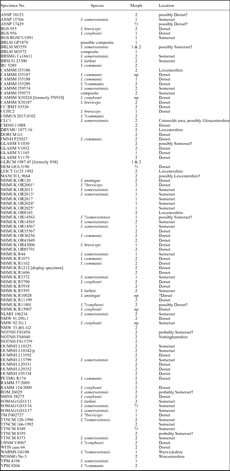
We examined 99 specimens of Ichthyosaurus with at least one hindfin that provided morphological information (Table 1). Twenty-four specimens had two informative hindfins. Most hindfins were associated with a skeleton or significant portion of a skeleton, but some specimens were isolated or associated with pelvic bones and/or a few centra (ANSP 10123, BRLSI GP1870, GLAHM V1165, GLRCM 1987.45 (formerly GLRCM 938), MANCH L9664, NHMUK OR35567, NHMUK R5790, NHMUK R5918, NOTNH FS3450, OUMNH J.29351, OUMNH J.95338 and RAMM 124–2009). Other specimens with partial hindfins exist, but they are too incomplete and/or too disarticulated to provide morphological information. Many of the specimens examined, however, were not sufficiently complete to assign to species (Table 1).
Specimens that were used in this study with at least one informative hindfin

Note: Two specimens display a different morphotype on each fin. Location is based on museum records or specimen labels, except as noted. Species identifications based on evaluations by authors.
Abbreviations: Morph – morphotype; np – mesopodium not preserved. ? species or morphotype identification is uncertain. † assessment by the authors, based on preservation.
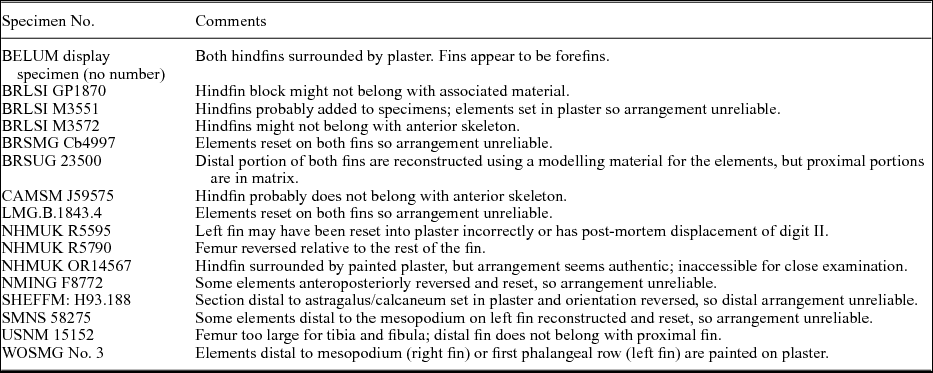
Six specimens were not included in the analysis because the authenticity of the arrangement of bones in the hindfins is questionable (Table 2). BRSMG Cb4997 is a fairly complete skeleton of I. somersetensis with both the left and right hindfin preserved. All of the fin elements, however, are set in a filler material. We cannot assess whether the current position of the individual bones accurately reflects their original arrangement, so this specimen was not included in this analysis. Likewise, the hindfins of LMG.B.1843.4 have been rebuilt, and so the specimen was not included. A similar situation occurs in BRLSI M3551, a composite specimen in which both hindfins were probably added to the specimen (Massare & Lomax, Reference Massare and Lomax2016a). The hindfins themselves are on opposite sides of the vertebral column and surrounded by plaster. It is unclear whether the individual bones are in plaster or matrix. A proximal bone of one fin (?left) is too large to belong with the other elements and it is lying on the ‘matrix’ rather than within it. Furthermore, the femur is cracked across the shaft and the proximal portion is rotated relative to the distal portion. Because of these issues, both fins of BRSLI M3551 were excluded from this analysis. A fourth questionable hindfin is NMING F8772, in which the posterior branch of digit II has some elements that are notched and oriented with the notches facing posteriorly rather than anteriorly. Considering that only elements of the leading edge can be notched, it is clear that these elements have been altered from their original orientation. This calls into question whether other elements have been reset into a different arrangement as well. Thus, this specimen was also excluded from the analysis. Another specimen excluded is USNM 15152, a composite of probably three individuals. The entire fin is surrounded by plaster and it is unclear whether individual bones are in matrix. The femur is too large for the size of the tibia and fibula, and the elements of the distal half of the fin are probably from a forefin, or at least from a larger individual than the proximal fin. The last specimen excluded is an unnumbered skeleton on display at BELUM. Both hindfins are surrounded by plaster, and the portions distal to the femora appear to be forefins that were added to the specimen.
Composite, unreliable or reconstructed specimens

Some composite specimens were included in this study because hindfins were unaltered and preserved in matrix. See text for additional information.
Three other specimens included in this research might be parts of composite specimens (Table 2). BRLSI GP1870 comprises three blocks of matrix: one with a hindfin, another with a portion of forefin and the third with some articulated caudal vertebrae, all of which are set in plaster within a wooden frame. The lack of direct association suggests that they might not belong to the same individual, but this is impossible to determine solely by visual examination. BRLSI M3572 is another suspicious specimen in which the skeleton appears to be pieced together from more than one individual (Massare & Lomax, Reference Massare and Lomax2016a). In particular, the caudal portion of the vertebral column and the hindfins might not belong to the same individual as the skull and pectoral region or the dorsal vertebral column. One hindfin is adjacent to articulated caudal centra whereas the other is on an isolated block of matrix, surrounded by plaster. The two hindfins have the same morphology and the matrix appears similar, so they probably belong to the same individual, although not necessarily the same individual as the anterior skeleton. In both BRLSI GP1870 and BRLSI M3572, the hindfins themselves are in blocks of matrix and appear to maintain the original arrangement of elements, so the specimens are included in the analysis. CAMSM J59575 is almost certainly a composite specimen in which the anterior and posterior portions of the skeleton do not belong to the same individual (Massare & Lomax, Reference Massare and Lomax2016a). The bone colour is different on the presacral skeleton compared to the hindfin and caudal centra, and the blocks containing these bones are set in plaster and separated from the presacral portion of the skeleton. Although the hindfin of CAMSM J59575 is surrounded entirely by plaster, the hindfin might be at least partially in matrix. The arrangement of bones is consistent with what occurs in other specimens, and so this specimen was included in the analysis. Because of the issue of composites, these three specimens could not be assigned to a species.
Six other specimens were included in the study, but have been partially reconstructed, and require some explanation (Table 2). SHEFFM: H93.188 has both hindfins preserved, but the left (upper) is disarticulated and does not provide any useful information, whereas the right (lower) has been incorrectly rebuilt, in part. The mesopodium is almost complete, with space for metatarsal two, which is missing. Distal to this, the fin is articulated, but set in plaster, with only portions of each digit preserved. The reset piece is, however, reversed relative to the rest of the fin, with the anterior edge facing posteriorly, identified by notching. Furthermore, the bone colour is different from the main specimen, so its authenticity can be questioned. We considered only the mesopodium morphology in this study. BRSUG 25300, the holotype of I. larkini, also has some reconstruction (Lomax & Massare, Reference Lomax and Massare2017). The distal portions of both fins have been reconstructed from plaster and have a much darker colour than the bone, but the proximal portion of each fin is authentic and provides useful morphological data. Similarly, some of the phalanges distal to the mesopodium of the left hindfin of SMNS 58275 have been restored and partially reset. NHMUK OR14567 is a practically complete skeleton that has the right hindfin preserved. The hindfin is surrounded by paint, which makes it suspicious. It is also on display, high on the wall and behind glass, so a close inspection is not possible. Nevertheless, the arrangement of bones appears to be authentic and so we include it here. The distal portion of each fin of WOSMG No. 3 has been painted to illustrate a complete hindfin. The mesopodium, however, is original bone imbedded in matrix, and so we have included the specimen in this study. Finally, in NHMUK R5790, an isolated hindfin, the femur is reversed relative to the rest of the hindfin, with the anterior end pointing posteriorly. The femur has probably been reset incorrectly.
3. Hindfin development and homology
The primitive reptilian hindfin has been greatly modified in parvipelvian ichthyosaurs. As in all other Merriamosauria, parvipelvians have lost digit I in the hindfin (Ji et al. Reference Ji, Jiang, Motani, Rieppel, Hao and Sun2016). The tibia and fibula are polygonal bones that lack a shaft or even an anteroposterior constriction. Polygonal tarsal and metatarsals have broad contacts with adjacent bones, sometimes interlocking, and thus make the determination of homologies challenging. The problem is complicated by the presence of accessory digits (e.g. Ophthalmosaurus, Platypterygius) or digital bifurcations (e.g. Ichthyosaurus, Protoichthyosaurus). Additionally, Ichthyosaurus has lost digit V in the hindfin, as have other Lower Jurassic ichthyosaurs (e.g. Leptonectes, Eurhinosaurus), although Lomax & Massare (Reference Lomax and Massare2017) incorrectly identified a digit V in I. larkini and I. somersetensis. In reptiles, digit I is the first to be lost when digit reduction occurs, and, if reduction continues, digit V is the next one to be lost (Bakker et al. Reference Bakker, Fowler, Oude, Dondorp, Garrido Navas, Horbanczuk, Sire, Szczerbinska and Richardson2013). Because of digital bifurcations, however, Ichthyosaurus has more than just these three digits in the hindfin (see Section 6b).
Because the limbs of ichthyosaurs are so derived compared to those of terrestrial reptiles, three terminologies have been used to identify elements of the mesopodium in parvipelvian hindfins. The three elements in the second row (immediately distal to the tibia and fibula) have been referred to as the centrale, astragalus and calcaneum (Caldwell, Reference Caldwell1997); tibiale, intermedium or astragalus, and fibulare (Zammit, Norris & Kear, Reference Zammit, Norris and Kear2010; Massare & Lomax, Reference Massare and Lomax2016b; Lomax & Massare, Reference Lomax and Massare2017); or distal tarsal two, astragalus and calcaneum (Fernández, Reference Fernández2007; Maxwell, Reference Maxwell2012). McGowan & Motani (Reference McGowan and Motani2003, p. 57) noted the lack of consensus and used two different terminologies in diagrams of hindfins (McGowan & Motani, Reference McGowan and Motani2003, figs 67, 70).
The important difference, however, is not merely in assigning names of bones. The identification of the anterior bone in the second row as the tibiale or distal tarsal two has implications for development of the fin, because the process that forms each bone is quite different. In embryonic development, the tibiale forms from segmentation of the tibia, whereas tarsal two would form in the digital arch from distal tarsal three (Shubin & Alberch, Reference Shubin, Alberch, Hecht, Wallace and Prance1986). In extant reptiles (crocodiles, turtles, lizards), a condensation that will eventually become the femur segments distally and bifurcates to form condensations that will become the tibia and fibula. The fibula condensation similarly bifurcates into condensations for an intermedium and fibulare (Shubin & Alberch, Reference Shubin, Alberch, Hecht, Wallace and Prance1986). Although Fabrezi, Abdala & Martínez Oliver (Reference Fabrezi, Abdala and Martínez Oliver2007) recognized a condensation for the radiale in the forefin, a similar condensation does not occur in the hindfin (Shubin & Alberch, Reference Shubin, Alberch, Hecht, Wallace and Prance1986; Fabrezi, Abdala & Martínez Oliver, Reference Fabrezi, Abdala and Martínez Oliver2007). This contrasts with development in salamanders and birds, where a condensation that will eventually form the tibiale is present distal to the tibia (Shubin & Alberch, Reference Shubin, Alberch, Hecht, Wallace and Prance1986). The interpretation of homology, and thus terminology, depends on the pattern of development that ichthyosaurs followed.
Phylogenetic analyses have placed the Ichthyopterygia with basal diapsids (Motani, Minoura & Ando, Reference Motani, Minoura and Ando1998) or among the parareptiles (Maisch, Reference Maisch2010). The latter hypothesized an origin from a more primitive ancestral form than the former. Both studies hypothesized the origin of Ichthyopterygia before the divergence of archosauromorphs (which includes crocodiles) and lepidosauromorphs (which includes lizards). More recently, Scheyer et al. (Reference Scheyer, Neenan, Bodogan, Furrer, Obrist and Palmondon2017, fig. 12) proposed a phylogeny in which archosauromorphs, lepidosauromorphs and Ichthyopterygia diverged at the same time. All three phylogenetic hypotheses, however, place the group within the Reptilia. Key characteristics of the reptilian tarsus are recognized in many early reptiles (e.g. Rieppel, Reference Rieppel1993; O'Keefe et al. Reference O'Keefe, Sidor, Larsson, Maga and Ide2006), suggesting that the reptilian developmental pattern was well established by the time the ichthyosaurs diverged. The hindfin of the Middle Triassic ichthyosaur Mixosaurus retains all five digits, and both distal tarsals one and two contact the tibia (Caldwell, Reference Caldwell1997; Maxwell, Reference Maxwell2012). Only two proximal tarsals (astragalus, calcaneum) occur in this primitive ichthyosaur, the typical reptilian pattern. Interestingly, Caldwell (Reference Caldwell1997) suggested that the most anterior, proximal tarsal in Stenopterygius could be a centrale that did not fuse with the intermedium to form the astragalus, although he acknowledged that it could also be distal tarsal two. Whether hindfin development in parvipelvians differed from the primitive reptilian pattern has yet to be established (but see Zverkov, Reference Zverkov2017).
This work uses a conservative interpretation, which assumes that the ichthyosaurian tarsus developed in the primitive reptilian pattern and that the pattern is retained even in derived taxa. Thus, in parvipelvian ichthyosaurs, the second row of the hindfin includes distal tarsal two, the astragalus and the calcaneum, and the third row typically includes metatarsal two, distal tarsal three and distal tarsal four. In Ichthyosaurus, however, a bifurcation in digit II commonly occurs, resulting in four elements in the third row (see Section 5). An issue with this interpretation is that distal tarsals two and three do not contact one another in many specimens of Ichthyosaurus, as well as other parvipelvian taxa (McGowan & Motani, Reference McGowan and Motani2003, fig. 70; Maxwell, Reference Maxwell2012, fig. 1). However, in Mixosaurus, the distal tarsals contact those of adjacent digits along a broad contact. Such a contact is expected considering how the distal tarsals develop from the digital arch (Shubin & Alberch, Reference Shubin, Alberch, Hecht, Wallace and Prance1986). In Ichthyosaurus, and many parvipelvians, however, metatarsal two is often between distal tarsals two and three. Such changes in positions of distal tarsals could be related to the difference in shape of the tibia and fibula between Mixosaurus (elongated, with a distinct shaft) and Ichthyosaurus (polygonal). Alternately, a faster growth rate of metatarsal two relative to distal tarsal two could also result in separation of the two distal tarsals.
A clarification of the term ‘contact’ is also required. Ichthyosaurian fins often have gaps between elements either because of post-mortem disarticulation or because of the presence of uncalcified tissue in life. Juveniles, in particular, often lack an interlocking mosaic of fin elements (Johnson, Reference Johnson1977; but see Lomax et al. Reference Lomax, Larkin, Boomer, Dey and Copestake2017). In spite of gaps between bones, the presence of facets on adjacent elements often indicates that bones were in contact or have the potential to be in contact if the gaps were eliminated by additional growth or re-articulation. This work uses the term ‘contact’ in an imprecise sense to mean both physically touching and having the potential to physically touch an adjacent bone if gaps between elements are eliminated.
4. Femur morphology in Ichthyosaurus
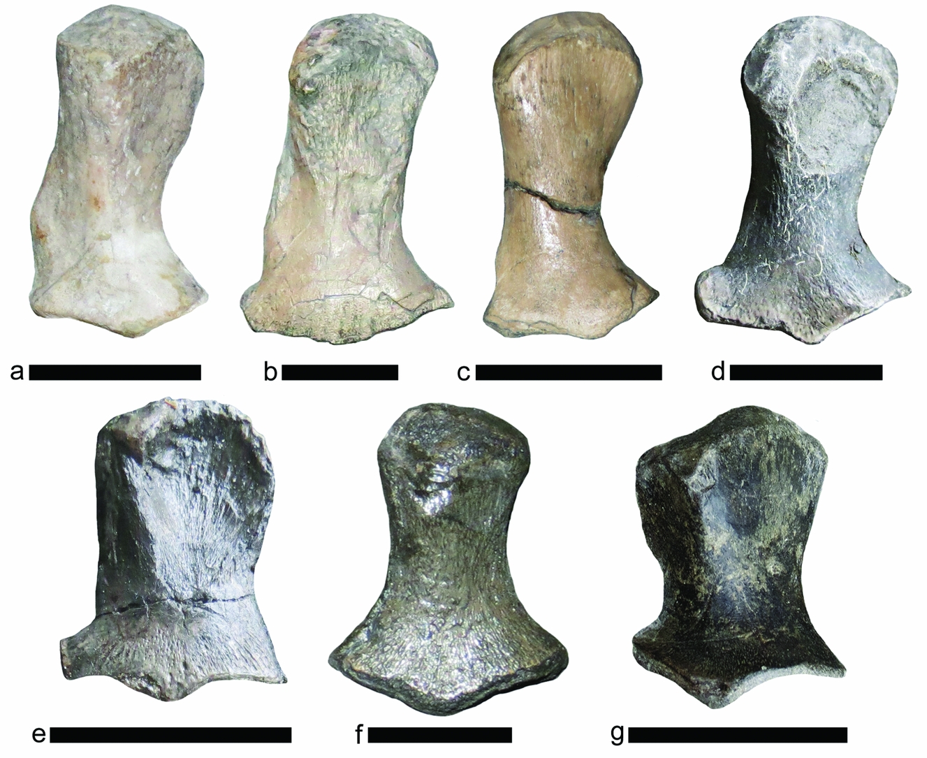
In general, the femur of Ichthyosaurus is more elongated than the humerus, with a narrow shaft and a distal end that is flattened dorsoventrally and expanded anteroposteriorly (Fig. 1). The amount of anteroposterior expansion varies depending on the size and orientation of the facets for the tibia and fibula. Thus, in dorsal or ventral view, the distal end is wider than the proximal end, but the difference in widths varies within a species. Minor differences in the orientation of the femur also influence the assessment of relative widths. Ichthyosaurus anningae differs from the other species in having a much less elongated femur in which the distal and proximal ends are equally wide, making the femur shape similar to that of a humerus (Fig. 1g). This species can thus be distinguished from the others by femur morphology, as well as the relative size of the femur compared to the humerus (Lomax & Massare, Reference Lomax and Massare2015).
Femora of the six species of Ichthyosaurus, anterior to the left. (a) I. larkini (BRSUG 25300) right femur in ventral view. (b) I. somersetensis (NLMH 106234, reversed) right femur in dorsal view. (c) I. somersetensis (TTNCM 8373) left femur in dorsal view. (d) I. communis neotype (NHMUK R1162) left femur in dorsal view. (e) I. breviceps holotype (NHMUK OR43006) left femur in dorsal view. (f) I. conybeari (NMW 93.5G.2, reversed), ?right femur in dorsal view. (g) I. anningae holotype (DONMG:1983.98) ?left femur in dorsal view. Scale bars in (a–e) equal 3 cm. Scale bar in (f) equals 1 cm. Scale bar in (g) equals 2 cm. Scale is estimated for (d) and (e) because specimens are on display behind glass.

In Ichthyosaurus larkini and I. somersetensis, the anterior edge of the shaft is nearly straight, although some specimens have an expansion, or bulge, in at least the distal half of the shaft that gives it a convex curvature (e.g. BRSUG 25300, I. larkini; BRSMG Cb4997, I. somersetensis; Fig. 1a–c). The posterior edge of the shaft is concavely curved, giving the femur a distinct, asymmetric shape in dorsal or ventral view. This asymmetry can distinguish I. larkini and I. somersetensis from the other species, although not from each other. In contrast, I. communis, I. breviceps and I. conybeari have femora with more symmetric shapes in dorsal or ventral views, owing, in part, to a similar curvature of the anterior and posterior edges (Fig. 1d–f). I. conybeari usually has a distinctly narrow head and shaft, and a relatively broad expansion of the distal end in dorsal or ventral view (Fig. 1f). The distal end is much wider than the proximal end, more so than in other species, and this feature can sometimes distinguish I. conybeari from the other species.
In all species of Ichthyosaurus, the ventral process of the femur is centrally located and is more prominent than the dorsal process (Maxwell, Zammit & Druckenmiller, Reference Maxwell, Zammit and Druckenmiller2012, fig. 1). The dorsal process, however, appears to be offset anteriorly in all species, although it can sometimes be slightly more centrally located, and its prominence also varies. For example, one specimen of Ichthyosaurus somersetensis (NHMUK OR2013*) has a dorsal process that is offset anteriorly; whereas another specimen (SOMAG/GEO/17) has a dorsal process that is slightly more centrally located, although still anteriorly offset. Such a minor difference could be the result of the femur orientation. Thus, the processes are not particularly useful in referring a specimen to species.
The relative size of the tibia and fibula are not useful in distinguishing species except for I. conybeari, which has a much larger fibula than tibia, both proximodistally and anteroposteriorly. I. somersetensis often has a fibula that is somewhat larger, at least anteroposteriorly, than the tibia (e.g. NHMUK OR2013*, NLMH 106234), but the difference is not of the magnitude seen in I. conybeari. In general, the relative sizes of the tibia and fibula vary within a species, although in I. anningae, no sufficiently well-preserved specimens are known. The facets on the femur for the tibia and fibula are approximately the same length, even in I. conybeari. Variations in the size of facets arise mainly because of differences in the orientation of the femur. The angle between the facets also varies within species. The most striking example is in I. somersetensis, where in some specimens, a proximal deflection of the fibula facet occurs, with the angle between the facets being nearly 90° in some specimens (e.g. BRSMG Cb4997, TTNCM 8373; Fig. 1c). This feature is not ontogenetic, because neither the smallest (NHMUK R44) nor the largest (NLMH 106234 (Fig. 1b), NHMUK 2013*) specimens of the species display this feature. Notably, in the holotype of I. larkini, one femur has facets at nearly a right angle (Fig. 1a), but the other femur does not, so the angle is likely related to differences in orientation.
5. Mesopodium morphology in Ichthyosaurus
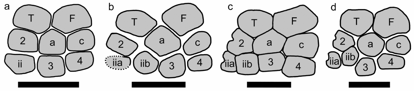
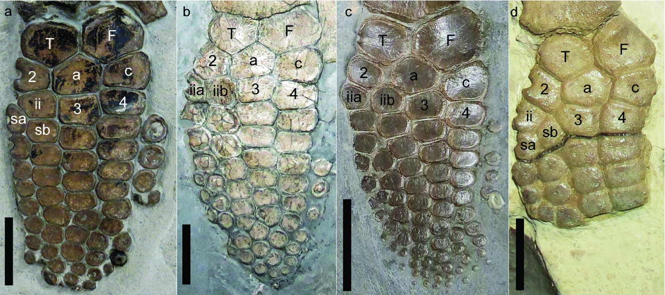
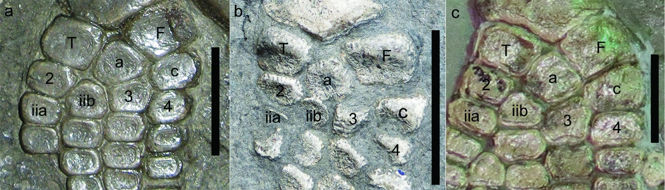
The mesopodium of Ichthyosaurus has one of two morphotypes, distinguished by the number of elements in the third row. Morphotype 1 has three elements: metatarsal two, distal tarsal three and distal tarsal four, which are immediately distal to distal tarsal two, the astragalus and the calcaneum, respectively (Fig. 2a). Nearly all specimens showing Morphotype 1 have a bifurcation in the first phalangeal row of digit II. The exception is WARMS G6188, in which the right hindfin has the usual bifurcation in the first phalangeal row, but the left hindfin has the proximal bifurcation in the third phalangeal row. A bifurcation of phalanges that results in additional lines of elements is polyphalangy (Cooper & Dawson, Reference Cooper and Dawson2009). Morphotype 2 has a bifurcation in the metatarsal row of digit II, resulting in four elements in the third row (Fig. 2b–d). Thus the third row has two metatarsal elements associated with digit II, distal tarsal three and distal tarsal four. A bifurcation in the metatarsal row resulting in an additional metatarsal is polydactyly (Cooper & Dawson, Reference Cooper and Dawson2009). Our contention is that in Ichthyosaurus, the bifurcation process is essentially the same, but the position at which it occurs on digit II is the result of individual variation. Thus, the distinction between polydactyly and polyphalangy is not useful for the unusual hindfin (and forefin) morphology found in Ichthyosaurus.
Ichthyosaurus hindfin morphotypes. (a) Morphotype 1 (NHMUK OR43006), with three elements in the third row. (b) Morphotype 2 (NHMUK R1162), with four elements in third row. Metatarsal two-b and distal tarsal three have equal contact with the astragalus. Dashed line indicates inferred outline of metatarsal two-a that is partially overlain. (c) Morphotype 2 variation (NLMH 106234, right hindfin, reversed), with distal tarsal three having broad contact with astragalus and metatarsal two-b having minor to no contact. (d) Morphotype 2 variation (NHMUK R5595 right hindfin, reversed), with three elements in contact with the astragalus. Anterior is to the left in all specimens. Scale bars in (a), (b) and (d) equal 2 cm. Scale bar in (c) equals 3 cm. Scales in (a), (b) and (d) are estimates because the specimens are on display behind glass. Abbreviations: 2 – distal tarsal two; 3 – distal tarsal three; 4 – distal tarsal four; a – astragalus; c – calcaneum; F – fibula; iia – anterior metatarsal of digit II; iib – posterior metatarsal of digit II; T – tibia.

Should Ichthyosaurus be separated into two genera, one displaying polydactyly and the other not? This might have been the conclusion were fewer specimens available for examination, but in reality, the situation is more complex. Morphotype 1 is the common morphology in I. breviceps, I. conybeari and I. somersetensis. However, all six species of Ichthyosaurus show Morphotype 2 in at least some specimens. Ichthyosaurus communis and I. larkini seem to show this morphotype exclusively, but the latter species is known from relatively few specimens. The only specimen of I. anningae with the proximal hindfin exposed also shows this morphology. Because three species show both morphotypes, we argue that the difference is the result of individual variation. Without the exceptionally large sample size, this might not have been recognized. Moreover, two of the 24 specimens that preserve both hindfins (BRLSI M3559, GLRCM 1987.45) show a different morphotype on each fin. Neither of these is a composite specimen, although composites occur in historic collections (Massare & Lomax, Reference Massare and Lomax2016a). This further suggests that the two morphotypes, although seemingly distinct, reflect individual variation. Thus, the difference between Morphotype 1 and 2 is largely whether the bifurcation of digit II occurs in the first phalangeal row of digit II (Morphotype 1) or in the metatarsal row (Morphotype 2). Similarly, in the forefin of Ichthyosaurus, the proximal bifurcation can be in the metacarpal row or one of the phalangeal rows (not always the first) of either digit II or digit III (Motani, Reference Motani1999, fig. 6). Moreover, the position of the bifurcation can vary between the left and right fin of an individual, although rarely (e.g. SOMAG/GEO11, ANSP 15766). So having a similar kind of variation in the hindfin is not unexpected.
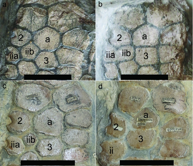
In limb development, a bifurcation results in two equivalent branches, so in the hindfin, both branches of the bifurcation represent digit II (Oster et al. Reference Oster, Shubin, Murray and Alberch1988; Motani, Reference Motani1999). In both the hindfin and forefin, however, one branch of the bifurcated digit can be oriented such that it appears to be a continuation of the original digit (Fig. 3a, d; Motani, Reference Motani1999, fig. 6c, d; Lomax, Massare & Mistry, Reference Lomax, Massare and Mistry2017, fig. 11a, b), but this is not always the case (Fig. 3b, c; Lomax, Massare & Mistry, Reference Lomax, Massare and Mistry2017, fig. 11c, d). Furthermore, one branch can have smaller elements than the other (Fig. 3a; Motani, Reference Motani1999, fig. 6e, f), although this is more common on distal bifurcations (Fig. 3b). Irrespective of size or orientation, this work considers the two branches to be equivalent subdigits of digit II. In Morphotype 2, owing to the bifurcation, two metatarsals are associated with digit II, referred to herein as metatarsal two-a and two-b. This results in four metatarsals in the hindfin. Neither metatarsal one nor five is present, as might have been inferred from four metatarsal elements if only a portion of the variation in the hindfins was examined. An alternate explanation, however, is that the posterior branch of the bifurcation is a continuation of digit II, whereas the anterior branch is a supernumerary digit. This interpretation is consistent with the posterior to anterior sequence of branching of the digital arch during limb development (Shubin & Alberch, Reference Shubin, Alberch, Hecht, Wallace and Prance1986), so it also has some basis in development. However, there are a few cases where the anterior branch seems to be the continuation of digit II (e.g. Fig. 3d), which is why we favour interpreting the two subdigits as equal.
Variation in the orientation and size of subdigits resulting from a bifurcation of digit II. Anterior to the left. (a) WARMS G6188 (Morphotype 1), right hindfin (reversed) in which subdigit-b is more in line with proximal elements of digit II than is subdigit-a. Note that subdigit-a has somewhat smaller elements than subdigit-b. (b) NLMH 106234 (Morphotype 2), right hindfin (reversed) showing the two subdigits of digit II symmetrically arranged with respect to distal tarsal two. Note the presence of a third subdigit in the fourth phalangeal row. (c) SMNS 58275 (Morphotype 2), right hindfin, in which neither subdigit is in line with proximal elements of digit II. (d) CLC1 (Morphotype 1), left hindfin in which subdigit-a is more in line with the proximal elements of digit II than is subdigit-b. Scale bars in (a), (c) and (d) equal 2 cm. Scale bar in (b) equals 3 cm. Scale for (d) is estimated because the specimen is on display behind glass. Abbreviations: 2 – distal tarsal two; 3 – distal tarsal three; 4 – distal tarsal four; a – astragalus; c – calcaneum; F – fibula; iia – anterior metatarsal of digit II; iib – posterior metatarsal of digit II; sa – anterior subdigit of digit II; sb – posterior subdigit of digit II; T – tibia.

Three variations occur in Morphotype 2, mainly differing in the number and extent of contact of elements in the third row with the astragalus (Fig. 2b–d). Some specimens have two small metatarsals in digit II that are directly distal to distal tarsal two, with metatarsal two-b having little or no contact with the astragalus (Figs 2c, 4a, b). Distal tarsal three has a broad contact with the astragalus similar to that which occurs in Morphotype 1. In some specimens of I. communis, on the other hand, both metatarsal two-b and distal tarsal three have fairly broad contacts with the astragalus. The contacts are approximately the same length (Fig. 2b). These two variations seem to be distinct morphologies, and if only a specimen or two were known, one could conclude that each species had a diagnostic hindfin morphology. However, although many specimens of I. somersetensis have little or no contact between metatarsal two-b and the astragalus, others have contacts with the astragalus of varying amounts (Fig. 4a–c). Similarly, although some specimens of I. communis have two equally large elements and broad, symmetric contacts of metatarsal two-b and distal tarsal three with the astragalus, other specimens of I. communis have a distal tarsal three contact that is somewhat longer than the metatarsal contact (e.g. Fig. 4d). The size of distal tarsal three relative to the adjacent metatarsal seems to be the controlling factor in the extent of contact with the astragalus, and could reflect individual variation in relative growth rates. Thus, the seemingly distinct morphologies of I. somersetensis and I. communis are end-members of a continuum of variation in the relative size of the contacts between metatarsal two-b, distal tarsal three and the astragalus. In fact, two specimens of Ichthyosaurus (OUMNH J.29351, NHMUK OR41849) preserve two hindfins, each with a different variation of Morphotype 2. Neither is a composite specimen.
The continuum of variation in Morphotype 2, all with four elements in the third row. (a) CAMSM J59574, distal tarsal three with broad contact with astragalus and metatarsal two-b with no contact. (b) BRSMG Ce16611, distal tarsal three with broad contact with astragalus and metatarsal two-b with minor contact. (c) OUMNH J.13799 (reversed), distal tarsal three with broad contact with astragalus and metatarsal two-b with much shorter contact with astragalus. (d) NHMUK R1073, distal tarsal three with slightly longer contact with astragalus than that of metatarsal two-b. Scale bars in (a) and (c) equal 3 cm. Scale bar in (b) equals 4 cm, based on a cast of the specimen at LEICT. Scale bar in (d) equals 2 cm but is estimated because the specimen is on display behind glass. Abbreviations: 3 – distal tarsal three; a – astragalus; iib – metatarsal of posterior subdigit of digit II.

When Morphotype 2 occurs in I. somersetensis, typically distal tarsal three has a broad contact with the astragalus, with metatarsal two-b having only a minor contact, if any, with the astragalus (Figs 2c, 4a, b). A similar morphology also occurs in I. conybeari (SMNS 58275, NHMUK R5790) and I. breviceps (CAMSM X50187, NHMUK OR2001*), although less commonly than Morphotype 1. Without several specimens of each species, the less common morphology would not have been evident, and mesopodium morphology might have been considered a species-specific character rather than a matter of individual variation.
In a third variation of Morphotype 2, metatarsal two-b, distal tarsal three and distal tarsal four have fairly broad and nearly equal contacts with the astragalus, at least on well-articulated specimens (e.g. NHMUK R5595, NHMUK OR8165 left fin). Metatarsal two-b is more proximally located such that it nearly separates distal tarsal two from the astragalus (Fig. 2d). It appears to be the common morphology of I. larkini, but only four specimens of the species are known, three of which preserve a hindfin. Lomax & Massare (Reference Lomax and Massare2017, p. 12) misidentified the digit count in I. larkini and identified the element that almost separates tarsal two (their tibiale) from the astragalus as the proximal element of digit III, whereas it is actually metatarsal two-b. Two additional hindfins have this morphology (NOTNH: FS3450 and NOTNH: FS4940). They could be I. larkini, but additional diagnostic material is lacking, so they cannot be assigned unequivocally to the species. As in the other variations of Morphotype 2, the relative size of the contacts with the astragalus varies considerably. The contact between the astragalus and distal tarsal three is longer than the contact with metatarsal two-b and with distal tarsal four in NOTNH FS4940 and BRSUG 25300, compared to Figure 2d. In fact, two variations of this morphology are preserved in BRSUG 25300 (the holotype of I. larkini): in one hindfin distal tarsal four has a significant contact with the astragalus, whereas in the other fin, tarsal four has only a slight, if any, contact with the astragalus, even accounting for taphonomic displacement. A slight contact between distal tarsal four and the astragalus occurs in Morphotype 1 and other variations of Morphotype 2, so it is not unique to the I. larkini variation of Morphotype 2. One specimen of Ichthyosaurus that preserves two hindfins (NHMUK OR8165; Fig. 5a, b) has the I. larkini variation in one hindfin and another variation of Morphotype 2 in the other fin; specifically, metatarsal two-b does not have a broad contact with the astragalus nor does it nearly separate it from distal tarsal two. This suggests that the morphology of I. larkini is another variation of Morphotype 2, but we have not yet identified specimens of other species with this variation. Although this variation seems to be unique to I. larkini, too few specimens are known to adequately evaluate it. To complicate the picture, another specimen with two hindfins (GLRCM 1987.45; Fig. 5c, d) has one fin with the ‘I. larkini’ morphology but the other fin displays Morphotype 1, with three elements in the third row. Unfortunately, GLRCM 1987.45 is not sufficiently complete to assign to a species. Thus, Morphotype 1 might occur in I. larkini, or equally likely, the I. larkini variation of Morphotype 2 might occur in another species. This suggests that this variation of Morphotype 2 cannot be used on its own to unequivocally refer a specimen to I. larkini.
Specimens with two different morphologies in the hindfins. (a) NHMUK OR8165, left hindfin (reversed) showing one variation of Morphotype 2, with four elements in the third row and three elements in contact with the astragalus. (b) NHMUK OR8165, right hindfin showing another variation of Morphotype 2, with four elements in the third row and one element (distal tarsal three) in broad contact with the astragalus. (c) GLRCM 1987.45, ?left hindfin showing one variation of Morphotype 2, with four elements in the third row and three elements in contact with the astragalus. Specimen had previously been identified as a forefin, but the propodial shape and size indicate that it as a hindfin. (d) GLRCM 1987.45, ?right hindfin (reversed) showing Morphotype 1, with three elements in the third row and distal tarsal three in broad contact with the astragalus. Scale bars equal 3 cm, but the scales for (a) and (b) are estimates because the specimens are on display behind glass. Abbreviations: 2 – distal tarsal two; 3 – distal tarsal three; a – astragalus; ii – metatarsal two; iia – metatarsal of the anterior subdigit of digit II; iib – metatarsal of posterior subdigit of digit II.

6. Other features of the hindfin
6.a. Notching of anterior elements
Notching on one or more elements in the anterior digit of the hindfin occurs in many Lower Jurassic ichthyosaurian genera including Ichthyosaurus. Notching has been interpreted as a retained primitive feature homologous to the shaft in long bones of the tetrapod limb (Motani, Reference Motani1999) or as a structure to protect nerves or blood vessels (Johnson, Reference Johnson1979; Caldwell, Reference Caldwell1997). Maxwell, Scheyer & Fowler (Reference Maxwell, Scheyer and Fowler2014) found that periosteal bone is present at the anterior end of notched elements, whereas it is lacking at the posterior end. They suggested that this related to increased stresses on the leading edge of the forefin. Thus, notching might be related to the way the fin was used during locomotion. The slight correlation between the number of notched elements and the size of the individual suggests an ontogenetic component of notching (Johnson, Reference Johnson1977; Maxwell, Scheyer & Fowler, Reference Maxwell, Scheyer and Fowler2014) and supports the hypothesis that notching is related to fin use throughout life. In Ichthyosaurus, notching is more common in the hindfin than in the forefin (Massare & Lomax, Reference Massare and Lomax2016b). In Stenopterygius, notching is more variable in the hindfin than in the forefin (Maxwell, Scheyer & Fowler, Reference Maxwell, Scheyer and Fowler2014).
Massare & Lomax (Reference Massare and Lomax2016b) argued that notching in the fins of Ichthyosaurus probably had no taxonomic significance in elements other than the tibia, but this study has found that some species of Ichthyosaurus have notched elements whereas others do not. Ichthyosaurus somersetensis almost always has notching in the hindfin. Of 21 specimens of I. somersetensis recognized in this research (Table 1), only one (ROM 26029) might lack notching in the hindfin. Distal tarsal two lacks a notch, but the anterior element in the metatarsal row is probably missing and the first phalanx is damaged. Most specimens of I. somersetensis have a notch in the distal tarsal or metatarsal or both, and sometimes in one or two phalanges as well. All known specimens of I. larkini with a hindfin show notching in at least one element, but with so few specimens, it is impossible to say whether this always occurs. On the other hand, no known specimens of I. communis, I. breviceps or I. anningae have notched elements in the hindfin (or the forefin). Only two specimens of I. anningae, however, preserve incomplete hindfins (NHMUK OR120 and NHMUK R10028), and although neither shows notching, with so few specimens, the variation in this feature cannot be assessed. Ichthyosaurus somersetensis is the common species from Somerset, whereas I. communis is the common species from Dorset (i.e. the Lyme Regis–Charmouth area). Ichthyosaurus larkini is known only from Somerset, whereas I. breviceps and I. anningae are exclusively from Dorset. As a result of the difference in species abundances, notching of anterior elements of the hindfins is rare on specimens from Dorset, but occurs commonly on specimens from Somerset, as noted by McGowan (Reference McGowan1974) and Massare & Lomax (Reference Massare and Lomax2016b).
Ichthyosaurus conybeari, which is an uncommon species from both Somerset and Dorset (McGowan, Reference McGowan1974; Massare & Lomax, Reference Massare and Lomax2016b), almost always has a notched tibia, the only species of Ichthyosaurus to show notching in the zeugopodium. A notched tibia is present in very small specimens (CAMSM X50224, femur length = 1.3 cm), even in the absence of notching in the forefin. A larger specimen (BGS 956, femur length 1.9 cm) has a notched tibia and distal tarsal two, as well as forefin notching. A much larger specimen (SMNS 58275, femur length = 3.5 cm) also has a notched tibia and two notched elements in the right forefin. A notched tibia appears to be a unique character for the species, at least within Ichthyosaurus. If this is the case, then OUMNH J.10301 could also be referred to I. conybeari. This would be the largest example of the species known, with an estimated femur length greater than 6 cm. OUMNH J.10301 is a historic specimen in which the matrix block with the hindfin is isolated from at least two other blocks, all within a wooden frame. The entire specimen might be a composite (Massare & Lomax, Reference Massare and Lomax2016a). The picture is more complicated, however. Skull and humerus morphology suggest that NHMUK R11801 and USNM V4697 are probably I. conybeari, but both lack a notched tibia in the articulated hindfin that is preserved. The latter, however, has an odd morphology in the hindfin (see Section 7). So although the presence of a notched tibia is indicative of I. conybeari, it would appear that the lack of a notched tibia does not completely exclude that possibility. Although both I. somersetensis and I. larkini have notched elements in the hindfin, the tibia is never notched in those species.
6.b. Number of digits
An issue arises in how to characterize the number of digits in the hindfin given that digit II can have a bifurcation in the metatarsal row. Applying the definition of digits as elements arising from a metatarsal/metacarpal (McGowan, Reference McGowan1972) creates complications in species diagnoses. For example, specimens of I. conybeari with Morphotype 1 would have three digits corresponding to metatarsals two, three and four; whereas specimens with Morphotype 2 would have four digits, corresponding to metatarsals two-a, two-b, three and four. Irrespective of the morphotype, all specimens of I. conybeari have similar hindfins, with five columns of elements (including a distal bifurcation of digit II). An additional complication is that the fourth digit arising from the metatarsal bifurcation is neither digit I nor digit V, making Ichthyosaurus distinctly different from a taxon with digit I or V in the hindfin (e.g. Mixosaurus). A strict application of the definition creates potential errors in phylogenetic analysis as well because the four digit hindfin in some species of Ichthyosaurus is not plesiomorphic. Cooper et al. (Reference Cooper, Berta, Dawson and Reidenberg2007, fig. 1) distinguished between digital rays (metatarsal plus phalanges) and digits (phalanges only), but those definitions create a similar issue in characterizing hindfins of Ichthyosaurus.
Thus, the standard terminology does not work well. All Ichthyosaurus species have three primary digits in the hindfin, digits II, III and IV, with digit II having two subdigits resulting from a proximal bifurcation in either the first phalangeal row (Morphotype I) or the metatarsal row (Morphotype 2). Most specimens have a third subdigit of digit II, resulting from a distal phalangeal bifurcation. Subdigits arise from bifurcations. They are distinct from pre- and post-axial accessory digits, which do not arise from the tarsus/carpus (McGowan, Reference McGowan1972) nor from a digital bifurcation. Previous workers have largely not distinguished between primary digits and subdigits, counting them together to describe the number of digits in the hindfin (e.g. Lomax & Massare, Reference Lomax and Massare2017) and forefin (e.g. McGowan & Motani, Reference McGowan and Motani2003; Ji et al. Reference Ji, Jiang, Motani, Rieppel, Hao and Sun2016; Massare & Lomax, Reference Massare and Lomax2017). Similarly, following Cooper et al. (Reference Cooper, Berta, Dawson and Reidenberg2007) and Cooper & Dawson (Reference Cooper and Dawson2009), we use the term ‘digits’ here to include both primary digits and subdigits. We exclude accessory digits from the counts that follow.
Thus, the common morphology of the hindfin of all species of Ichthyosaurus, for both morphotypes, has a total of five digits, the result of bifurcations in digit II. The two morphotypes are consistent, although different, in the position of the proximal bifurcation, either in the first phalangeal row (Morphotype 1) or in the metatarsal row (Morphotype 2). Usually a second bifurcation occurs distally, which results in five digits. However, the fin must be complete to the fifth phalangeal row in order to confirm the digit count because the distal bifurcation often occurs in the fourth or fifth phalangeal row of digit II (e.g. NHMUK R3372, NLMH 106234, OUMNH J.29351), especially in I. somersetensis.
Exceptions to this pattern occur. Two specimens of Ichthyosaurus have six digits. PETMG R174 (I. communis) and YPM 9204 (possibly I. communis) have bifurcations in the metatarsal, first or second phalanx, and fifth or sixth phalanx of digit II. Less unusual occurrences are specimens with fairly complete hindfins that have only four digits, although this could be a preservation issue. Collection or preparation could have missed a small distal bifurcation. In particular, the lack of a distal bifurcation on historic specimens needs to be evaluated carefully. The anterior edge of some hindfins is at the edge of the matrix, presenting the possibility that some fin elements, especially small ones, could have been accidentally overlooked or intentionally removed to show a better defined fin. Thus, a distal bifurcation, especially one with small elements, cannot be entirely ruled out on historic specimens with only four digits, even in what appears to be a fairly complete hindfin. That being said, some specimens that are well preserved and sufficiently complete have hindfins that lack the distal bifurcation (e.g. BRLSI GP1870, CAMSM J59574, NHMUK R11199, NHMUK OR43006, TTNCM 8359 and WARMS G6188; possibly also CAMSM J35188 and NHMUK OR2013 (not OR2013*)). Those that can be assigned to a species are either I. breviceps or I. somersetensis, but several specimens cannot be identified to species.
Although some forefins of Ichthyosaurus have a pre-axial (anterior) accessory digit (e.g. NHMUK OR43006), hindfins seem to lack them. Sometimes one subdigit of digit II has larger elements than the other, possibly as a result of unequal division of embryonic tissue or different growth rates. This does not change the developmental equivalence of the two subdigits. When this occurs, however, it is most often the anterior subdigit that is smaller (Fig. 3a). Poor preservation or disarticulation could make it difficult to distinguish a subdigit from a pre-axial accessory. This is especially problematic for distal bifurcations if one subdigit has small, rounded elements (Fig. 3b), if the proximally adjacent element lacks a distinct facet or if only a few elements are present. It is often a judgement call as to whether small anterior elements belong to a subdigit or a pre-axial accessory digit. We have not been able to unequivocally identify a pre-axial accessory digit on any hindfin (contrary to Massare & Lomax, Reference Massare and Lomax2014), so we conclude that pre-axial accessory digits are absent or rare on hindfins of Ichthyosaurus.
In contrast, post-axial (posterior) accessory digits are easier to recognize. The elements are round or oval, whereas digit IV always has larger, rectangular phalanges, at least in the proximal half of the fin. The presence of one post-axial accessory digit seems to be the typical condition in hindfins of Ichthyosaurus, although they are present on less than half of the specimens examined. A post-axial accessory digit occurs in all species, possibly even I. anningae (NHMUK R10028). Post-axial accessory digits are often poorly preserved, incomplete or absent because of decisions made during preparation, especially on historic specimens. So, the lack of a post-axial accessory needs to be viewed initially with some scepticism.
7. Unusual hindfin morphologies in Ichthyosaurus
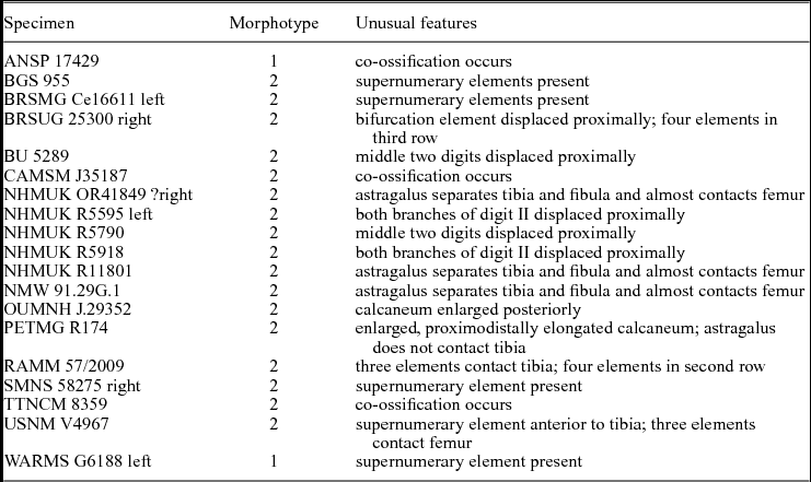
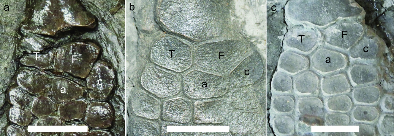
Several hindfins show unusual morphologies, some of which might be pathology (Table 3). Had there been fewer or less complete specimens to examine, some of these morphologies could have been interpreted as sufficiently unique to be a new taxon. PETMG R174, a specimen of Ichthyosaurus communis, lacks a contact between the astragalus and the tibia (Fig. 6a). In addition, the calcaneum is proximodistally elongated and anteroposteriorly short. It is posterior to the fibula for much of its length. The hindfin otherwise has the Morphotype 2 arrangement of elements. A less aberrant, but similar, arrangement occurs in OUMNH J.29352. The astragalus has a minor contact with the tibia and the calcaneum is proximodistally elongated only in the posterior portion and it is not anteroposteriorly short (Fig. 6b). The calcaneum is, however, similarly posterior to most of the fibula. Another specimen, CHMUS 2015.0102, has a posteriorly expanded, but small, calcaneum and a posteriorly reduced fibula (Fig. 6c), but not to the extent displayed in OUMNH J.29352. The astragalus has a broader contact with the tibia and fibula. Thus, the three specimens display a sequence of morphologies that grade from a highly unusual arrangement to one that is more similar to the typical morphology (compare Fig. 6c to Fig. 4b, c). It suggests that even the morphology of PETMG R174 is just an odd variation.
Hindfins with unusual morphologies

Note: ‘left’ and ‘right’ indicate the left or right hindfin on specimens that preserve both.
An example of gradation in morphologies in proximal fin elements. Anterior to the left. (a) The hindfin of PETMG R174 has an oval, proximodistally elongated calcaneum and the tibia and astragalus lack contact. (b) The hindfin of OUMNH J.29352 has a similarly shaped astragalus, a short contact between the tibia and astragalus and a calcaneum that is anteroposteriorly elongated only in the posterior portion. (c) The hindfin of CHMUS 2015.0102 (reversed) has a calcaneum, astragalus and fibula shaped similarly to (b) but the relative sizes and arrangement of elements are more like that of a typical Ichthyosaurus hindfin. Image courtesy of Charterhouse School Archive. Scale bar in (a) equals 3 cm. Scale bars in (b) and (c) equal 1 cm. Abbreviations: a – astragalus; c – calcaneum; F – fibula; T – tibia.

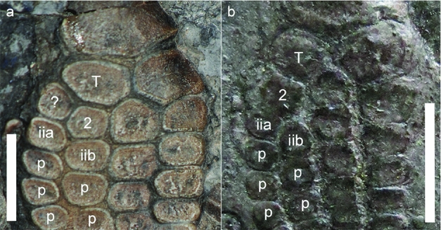
Another unusual hindfin morphology is that of RAMM 57/2009, which is clearly Ichthyosaurus based on the rest of the skeleton. The hindfin has four elements in the second row, with three elements in contact with the tibia (Fig. 7a). In addition to the facet for the astragalus, the tibia has two other facets, one more anteriorly oriented than the other. This arrangement could be interpreted as a bifurcation in the distal tarsal row. The matrix is darker around the fin than elsewhere on the block, so it is possible that it was reset, but the facets on adjacent elements suggest that the arrangement is authentic. Taphonomic displacement is possible, but the facets and the lack of disarticulation suggest that this is not displacement. No other hindfin of Ichthyosaurus shows this morphology in which the bifurcation of digit II seems to have occurred more proximally than usual. The anterior branch of digit II curves ‘outwards’ as if displaced anteriorly by the posterior branch. Also, for a given position on the proximal half of the fin, there are more elements in the anterior branch than in the posterior branch of digit II (Fig. 7a). Both of these features also occur in DORCM G1, BGS 955 and NHMUK R11199 (Fig. 7b), although these specimens have the usual Morphotype 2 bifurcation in the metatarsal row. In all three specimens, the relative position of the proximal elements is the same: metatarsal two-a is slightly more proximally located than metatarsal two-b (Fig. 7a, b). The major difference is that the bifurcation apparently occurs in a more proximal row in RAMM 57/2009 than in the others. As with the previous example, the unusual morphology of RAMM 57/2009 shares similarities with fins having a more typical morphology.
Another unusual morphology of the hindfin. Anterior to the left. (a) RAMM 57/2009, left hindfin showing two elements immediately distal to the tibia, possibly the result of a bifurcation in digit II. The anterior subdigit curves ‘outwards’ (anteriorly) and metatarsal two-a is more proximally located than metatarsal two-b. (b) NHMUK R11199, left hindfin, showing a bifurcation of digit II in the metatarsal row. The anterior subdigit curves ‘outwards’ (anteriorly), and metatarsal two-a is more proximally located than metatarsal two-b. Scale bar in (a) equals 2 cm. Scale bar in (b) equals 1 cm but is estimated because the specimen is on display behind glass. Abbreviations: 2 – distal tarsal two; iia – anterior metatarsal of digit II; iib – posterior metatarsal of digit II; p – phalanx; T – tibia; ? – homology unknown.

In NHMUK R11801, a small articulated skeleton, the astragalus is positioned so far proximally that it separates the tibia and fibula and almost contacts the femur. Distal tarsal two and the calcaneum seem to be pushed anteriorly and posteriorly, respectively, by metatarsal two-b and distal tarsal three (Fig. 8a). Disarticulation and displacement can produce this arrangement (e.g. BU 5289; Fig. 8b), but NHMUK R11801 is not disarticulated. In isolation, the arrangement of elements could be interpreted as having three elements in the first row and four elements in the second and third rows, thus an entirely new morphology. In fact, a contact between the astragalus and femur occurs in Maiaspondylus (Druckenmiller & Maxwell, Reference Druckenmiller and Maxwell2010, table b1) and Platypterygius hercynicus (Kolb & Sander, Reference Kolb and Sander2009; Fischer, Reference Fischer2012). Other derived ichthyosaurs (Aegirosaurus, Brachypterygius, Maiaspondylus) have a comparable contact between the humerus and the intermedium in the forefin (Bardet & Fernández, Reference Bardet and Fernández2000; McGowan & Motani, Reference McGowan and Motani2003; Maxwell & Caldwell, Reference Maxwell and Caldwell2006). However, the morphology of NHMUK R11801 is yet another ‘extreme’ variation of a common arrangement of Morphotype 2. NMW 91.29G.1 also has an astragalus that nearly contacts the femur, but metatarsal two-b is somewhat smaller than in NHMUK R11801, and so the calcaneum and tarsal two are closer to the astragalus (Fig. 8c). The second row of three elements is better defined, and distal tarsal two and the calcaneum are not displaced outwards as much. This is also the case in one fin of NHMUK OR41849, where the astragalus practically contacts the femur, separating the tibia from the fibula. The other fin also shows the separation but the astragalus is not as proximally located. In NHMUK R1162 the astragalus is positioned slightly more distally (Fig. 2b), and the tibia and fibula are closer together, a more typical arrangement of elements.
Hindfins in which the astragalus separates the tibia and fibula and almost contacts the femur. (a) NHMUK R11801, left hindfin, showing digit III and the posterior subdigit of digit II more proximally positioned than usual. The fin appears to have three elements in the first row and four elements in the second row because tarsal two and the calcaneum are shifted anteriorly and posteriorly, respectively. (b) BU5289, right hindfin (reversed), with a similar morphology as (a). However, it is probably due to post-mortem displacement in this specimen. Note, that some elements are dorsoventrally rotated and partially buried. (c) NMW 91.29G.1, ?left hindfin (reversed), showing the astragalus almost in contact with the femur, but digit III and the posterior subdigit of digit II do not displace distal tarsal two and the calcaneum as in (a). Scale bars in (a) and (b) equal 1 cm. Scale bar in (c) equals 2 cm. Abbreviations: 2 – distal tarsal two; 3 – distal tarsal three; 4 – distal tarsal four; a – astragalus; c – calcaneum; F – fibula; iia – anterior metatarsal of digit II; iib – posterior metatarsal of digit II; T – tibia.

USNM V4967 is another unusual morphology, where a fairly large, supernumerary element is anterior to the tibia and in contact with the femur (Fig. 9a). The tibia is smaller than usual relative to the fibula, so it is possible that the supernumerary bone restricted its growth. Maxwell (Reference Maxwell2012, p. 550) reported a somewhat similar, but more common, anomaly in Stenopterygius quadriscissus, in which a single element contacts the femur anterior to the tibia. USNM V4967 is otherwise a typical example of Morphotype 2, with a proximal bifurcation in the metatarsal row and a more distal bifurcation in the second phalangeal row. Other features of the skeleton identify this specimen as Ichthyosaurus, but as with PETMG R174, RAMM 57/2009, NMW 91.29G.1 and NHMUK R11801, the hindfin might have been considered a new taxon had it occurred in isolation. This makes it clear that fragmentary or isolated material does not usually provide sufficient information to justify a new taxon (e.g. as in Brusatte et al. Reference Brusatte, Young, Challands, Clark, Fischer, Fraser, Liston, Macfadyen, Ross, Walsh and Wilkinson2015).
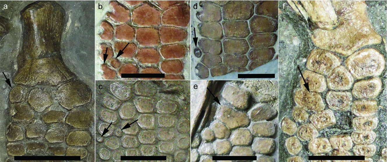
Examples of supernumerary elements and co-ossifications. Anterior to the left. (a) USNM V4967, showing a fairly large, supernumerary bone (arrow) that is anterior to the tibia and in contact with the distal end of the femur. (b) BRSMG Ce16611, arrows point to two supernumerary elements in digit II. (c) BGS 955, arrows point to two small supernumerary elements. (d) WARMS G6188, arrow points to a supernumerary element anterior to digit II. (e) ANSP 17429 (reversed), arrow points to a co-ossification of elements from digits II and III, possibly metatarsal three and metatarsal two-b. (f) TTNCM 8359 (reversed), arrow points to a co-ossification of the proximal element of the two metatarsals of digit II. The other hindfin (not figured) shows Morphotype 2. Scale bars in (a) and (b) equal 3 cm. Scale bars in (c) and (f) equal 1 cm. Scale bars in (d) and (e) equal 2 cm.

Several specimens have smaller, more distally located supernumerary elements in the fin, in the form of a small round bone between two of the digits (Fig. 9b–d). These elements are usually associated with digit II. BRSMG Ce16611 has a supernumerary element in digit II, between the first and second phalanges, and a second, smaller isolated element anterior to it (Fig. 9b). BGS 955 has a small, round supernumerary element in digit II between the first and second phalangeal rows and another anterior to the second phalanx of digit II (Fig. 9c). The right hindfin of SMNS 58275 has a supernumerary element between the fourth and fifth phalangeal rows of digit II. The left fin of WARMS G6188 has a supernumerary element anterior to and between phalanges one and two of digit II, proximal to where the distal bifurcation occurs (Fig. 9d). Similar supernumerary elements occur occasionally in the forefins of Ichthyosaurus (pers. obs.) and have been reported in forefins and hindfins of Stenopterygius (Maxwell, Reference Maxwell2012), and in whales (Cooper & Dawson, Reference Cooper and Dawson2009).
Another irregularity in a few hindfins is co-ossification of adjacent elements, which has also been reported in forefins of Ichthyosaurus (Motani, Reference Motani1999; Lomax, Massare & Mistry, Reference Lomax, Massare and Mistry2017), Mixosaurus (Maxwell, Reference Maxwell2012), Stenopterygius (Johnson, Reference Johnson1979; Maxwell, Reference Maxwell2012), Undorosaurus (Arkhangelsky & Zverkov, Reference Arkhangelsky and Zverkov2014, fig. 1c) and in the hindfin of Suevoleviathan (Maisch, Reference Maisch1998; pers. obs. SMNS 15390), as well as whales (Cooper & Dawson, Reference Cooper and Dawson2009). In the hindfin of Ichthyosaurus, a co-ossification can be recognized as a large, anteroposteriorly elongated, irregular element. Thus, in TTNCM 8359 (Fig. 9f), the elongated bone in digit II is probably a co-ossification of metatarsals two-a and two-b. So what was originally a bifurcation in the metatarsal row of Morphotype 2 mimics Morphotype 1 because of the co-ossification. ANSP 17429 (Fig. 9e) also shows two co-ossified elements between digits II and III, possibly metatarsal three and metatarsal two-b, but the fin is somewhat disarticulated and identification of elements is difficult. The proximal portion of the fin is missing in CAMSM J35187, but a co-ossification of phalanges of the two branches of digit II occurs in the third or fourth phalangeal row.
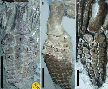
Post-mortem displacement of digits can also result in odd morphologies that could, in isolation, be mistaken for new taxa. Four specimens have a portion of the hindfin shifted proximally. In NHMUK R5918, an isolated hindfin, distal tarsal two has been rotated and moved anterior to the tibia and the two branches of digit II have also been shifted proximally (Fig. 10a), probably a taphonomic effect. Note that the space between the posterior branch of digit II and digit III is wider than between digit III and IV, suggesting post-mortem disarticulation. The left hindfin of NHMUK R5595 (Fig. 10c), a nearly complete skeleton of I. larkini (Lomax & Massare, Reference Lomax and Massare2017), shows a similar shift, but distal tarsal two has not been rotated and digit II is not shifted proximally as much as in NHMUK R5918. This, however, might not be taphonomic, but instead, the result of decisions made during preparation, because most of the fin appears to be set in a filler material. Unfortunately, the specimen is not accessible for close examination. BRSUG 25300, the holotype of I. larkini, also shows what might be post-mortem displacement on the right hindfin (posterior fin on specimen). Metatarsal two-b has been displaced proximally so that it is between distal tarsal two and the astragalus, resulting in what could be interpreted as four elements in the second row (Fig. 10b). Although metatarsal two-b in the left fin of the specimen partly separates the astragalus and distal tarsal two, it is more distally positioned and is clearly an element of the third row. Post-mortem displacement resulted in an odd morphology in one of the two fins. A similar displacement occurs in NHMUK R5790, a small, isolated hindfin. Metatarsal two-b is between the astragalus and distal tarsal two, allowing a similar interpretation of four elements in the second row. The wide separation between elements in NHMUK R5790 makes it clear that this odd arrangement is the result of post-mortem disarticulation and displacement.
Examples of post-mortem displacement that produces unusual morphologies. (a) NHMUK R5918, an isolated hindfin, in which distal tarsal two has been rotated and moved proximally so that it is anterior to the tibia. This also shifts both branches of digit II proximally relative to digit III. (b) BRSUG 25300, left hindfin, in which metatarsal two-b has been shifted so that it is almost in contact with the tibia. Darker elements of the distal portion of the fin are entirely reconstructed. (c) NHMUK R5595, left hindfin, in which distal tarsal two and both branches of digit II have been moved proximally, similar to (a). All scale bars equal 3 cm, but scale for (c) is an estimate because the specimen is on display behind glass. Abbreviations: 2 – distal tarsal two; 3 – distal tarsal three; 4 – distal tarsal four; a – astragalus; c – calcaneum; F – fibula; iia – anterior metatarsal of digit II; iib – posterior metatarsal of digit II; T – tibia.

8. Discussion and conclusion
Ichthyosaurus has three primary digits (II, III, IV) in the hindfin, a feature it shares with Temnodontosaurus, Suevoleviathan, Leptonectes, Eurhinosaurus, Stenopterygius and Hauffiopteryx (Maisch, Reference Maisch1998; McGowan & Motani, Reference McGowan and Motani2003; Maxwell, Reference Maxwell2012, fig. 2). The hindfins of the other Lower Jurassic genera, Excalibosaurus, Wahlisaurus and Protoichthyosaurus, are too incomplete to determine the total digit count. Only Ichthyosaurus is known to have an anterior digital bifurcation in the hindfin (but see below), which increases the digit count to as high as six, although five is more typical. The digital bifurcation can distinguish hindfins of Ichthyosaurus from most genera. However, incomplete hindfins present a problem in recognizing Ichthyosaurus. Morphotype 1 is seen in other genera, so if only the first three rows of elements are preserved, Ichthyosaurus is not easily distinguished from other Lower Jurassic genera, although the femora might be distinctive. Thus, the taxonomic usefulness of hindfin morphology has some limitations.
Another limitation is that the hindfin of Protoichthyosaurus shares similarities with that of Ichthyosaurus, a sister taxon (Lomax, Massare & Mistry, Reference Lomax, Massare and Mistry2017). Furthermore, both genera have an anterior digital bifurcation in the forefin, which suggests the possibility that Protoichthyosaurus also has a bifurcation in the hindfin. Only three specimens of Protoichthyosaurus with a hindfin or femur are known (Lomax, Massare & Mistry, Reference Lomax, Massare and Mistry2017). BRLSI M3555 is the only specimen to preserve a complete femur, which has a long shaft with a narrow proximal end and a very wide distal end. It is similar to that of I. conybeari but the shaft is more elongated and the distal expansion is broader. The femur on SOMAG/GEO/12 is not as elongated and the shaft is wider, although the proximal end is damaged and poorly preserved. With only two specimens the variation in the elongation cannot be assessed. Only SOMAG/GEO/12 preserves the mesopodium, which displays Morphotype 1. The more distal portion of the fin is partially disarticulated, and anterior elements are too scattered to ascertain the presence or absence of a bifurcation. A third specimen of Protoichthyosaurus (BRLSI M3563) preserves a hindfin, but the specimen is a composite. Only the right forefin and associated scapula and ribs definitely belong together and can be assigned to Protoichthyosaurus. The rest of the skeleton might not belong to the same individual or the same species (Massare & Lomax, Reference Massare and Lomax2016a; Lomax, Massare & Mistry, Reference Lomax, Massare and Mistry2017). Thus, based on currently recognized specimens, the hindfin of Protoichthyosaurus cannot be distinguished from that of Ichthyosaurus.
Within Ichthyosaurus, species cannot be identified unequivocally from a well-preserved hindfin, although certain morphologies are more common in some species than others (Table 4). Morphotype 1 definitely occurs in I. breviceps, I. conybeari and I. somersetensis, although I. anningae and I. larkini are known from few specimens, and so this morphotype could possibly occur in those species. Combined with the presence/absence of notching, I. breviceps, I. conybeari and I. somersetensis can be distinguished with some degree of confidence if a specimen displays Morphotype 1. A hindfin without notching is more likely in I. breviceps than the others. A notched tibia almost certainly indicates I. conybeari. Any Ichthyosaurus hindfin with a notched tibia can be assigned to this species, at least based on currently known specimens. A notched distal tarsal two, metatarsal and/or phalanges is most likely I. somersetensis. Other features of the skull and postcranium, however, are needed to verify these preliminary identifications.
Morphologic characteristics of hindfins of species of Ichthyosaurus

Bifur = total number of bifurcations in digit II. Fins must be complete to at least the fifth phalangeal row to assess the presence or absence of a distal bifurcation.
The occurrence of Morphotype 2, in which the bifurcation is in the metatarsal row, presents more uncertainty in identifications based only on a hindfin. If metatarsal two-b nearly separates distal tarsal two from the astragalus, and three elements contact the astragalus (Fig. 2d), then the specimen could be I. larkini, the only species that has been unequivocally identified with this morphology. However, this variation might also occur in I. somersetensis, because some specimens with this variation cannot be assigned to a species. I. larkini and I. somersetensis have similarly shaped femora as well. If distal tarsal three has a broad contact with the astragalus and metatarsal two-b has only a slight, if any, contact with the astragalus (Fig. 2c), then the specimen could be I. somersetensis, I. conybeari or I. breviceps. The presence of a notched distal tarsal two or more distal notching would most likely be I. somersetensis; a notched tibia would indicate I. conybeari; and a lack of notching would most likely be I. breviceps. A hindfin in which metatarsal two-b and distal tarsal three have broad, nearly equal contacts with the astragalus (Fig. 2b) indicates that the specimen is most likely I. communis or I. anningae, in the absence of notched elements. As with Morphotype 1, additional features of the skull or humerus shape are needed to confirm these preliminary identifications. Thus, although morphotypes are not unique to a species, combinations of hindfin characters can suggest the most likely species assignment for a particular hindfin morphology. However, intermediate morphologies exist in the extent of contact of the astragalus and metatarsal two-b. In these cases, the hindfin morphology has no taxonomic utility; other features are required even for a preliminary species assignment.
This research has also shown the effect of large sample sizes on the identification of taxonomically distinct morphological characters. Without several fairly complete specimens of each species, the occurrence of both morphotypes in I. somersetensis, I. breviceps and I. conybeari could not have been recognized. It would have appeared that the two morphotypes were diagnostic of two different sets of species within Ichthyosaurus. Hindfin morphology would have appeared much less variable and much more distinct with fewer specimens. The presence or absence of a bifurcation in the metatarsal row could be seen as a major difference among species or as individual variation, depending on how many specimens are known/examined. This raises doubts about the validity of new taxa based on limited material.
Gaps between morphologies are much larger when fewer specimens are available. With only one or two specimens, intermediate morphologies that grade from one form to another are unknown. More specimens result in more variation, and apparent gaps between morphologies are filled in, as is the case here with hindfin morphology. Distinct characters can be more easily defined if only one or two specimens are known because intermediate morphologies are missing. This raises the question of whether a particular morphology is distinct or only seems to be because of a small sample size. Character matrices and cladistic analyses cannot answer that question.
Cladistics can sort out many characters simultaneously, but variation is not always assessed with the same level of detail as defining the character states themselves. One individual from a new location or an unusual horizon can be examined in such detail that often numerous ‘unique’ features can be identified. In fact, it is probably easier to identify such features on a single individual than on a dozen individuals, especially considering that multiple specimens will vary in orientation, preservation and completeness. To put it in a different context, a new specimen could look quite different from the type specimens of known species, but if one examines several specimens to get a better picture of variation, the new specimen might be within the variation of a known species. This is especially true of isolated elements, e.g. isolated hindfins that appear to have unique morphologies. Character matrices that are based solely on type specimens will inevitably justify the erection of new taxa from isolated material. Variation must be adequately assessed by comparing many specimens, rather than just examining type specimens or citing exclusively from the literature. Otherwise, the decision to erect a new taxon is not substantially different from what was done in the nineteenth century, when new species were erected on the basis of a single individual (often from a new location or horizon) if it showed a small difference in morphology from previously known specimens.
Acknowledgements
We thank the following individuals and their respective institutions for access to and help with specimens: E. Daeschler and E. Gilmore (ANSP); M. Simms (BELUM); P. Shepherd, L. Neep and S. Harris (BGS); M. Williams (BRLSI); D. Hutchinson, I. Gladstone and the late R. Vaughan (BRSMG); C. Hildebrandt, J. Hanson and M. Benton (BRSUG); J. Clatworthy (BU); M. Riley and S. Finney (CAMSM); G. McGowan (CC); P. Davidson (CCHC); R. Roberts (CLC); S. Bailey (DBYMU); P. Robinson (DONMG); J. Cripps (DORCM); W. Simpson (FMNH); N. Clark and R. Smith (GLAHM); A. Smith and D. Rice (GLRCM); R. Neller (HEM); M. Evans (LEICT); D. Gelsthorpe and R. Petts (MANCH); N. Owen (LMG); S. Chapman, P. Barrett, L. Steel and R. Hansen (NHMUK); A. Richter (NLMH); N. Monaghan (NMING); C. Howells (NMW); A. Smith (NOTNH); E. Howlett and H. Ketchum (OUMNH); G. Wass and J. Liston (PETMG); K. Seymour (ROM); A. McLean (SHEFFM); E. Maxwell (SMNS); D. Parsons (TTNCM); H.-D. Sues, D. Bohaska and A. Millhouse (USNM); J. Radley and L. McCoy (WARMS); S. Glassman, (WFIS); G. Phillips and R. Roden (WOSMG); D. Brinkman (YPM). We also thank R. Clark, C. Berry and the Alfred Gillett Trust, Street, for access to the Alfred Gillett Collection (SOMAG). We are grateful to C. Smith and S. Jackson (CHMUS), A. McGee (CMNH), H. Morgenroth (RAMM) and H. Voogd (TM) for providing images of specimens in their respective collections that we have not been able to examine. Finally, we greatly appreciate the reviews provided by N. Zverkov, E. Maxwell and J. Lawrence Wujek, which significantly improved the paper. DRL's research was covered in part by a PGR, Dean's Doctoral Scholarship Award from the University of Manchester.