Introduction
Hyperthyroidism is associated with a number of psychiatric disorders including mood disorders, schizophrenia-like psychoses and delirium (Reference Lu, Lee, Tsai, Hu and Sim1). Symptoms such as anxiety and irritability are common in Graves' disease, which is the most common cause of hyperthyroidism. Subjects with Graves' disease also reported significantly worse memory, attention, planning and productivity while hyperthyroid than prior to onset of a hyperthyroid state (Reference Stern, Robinson, Thorner, Arruda, Prohaska and Prange2). There is one reported case of a patient with long standing depressive disorder who subsequently developed catatonia related to Graves' disease (Reference Farah and McCall3).
Encephalopathy associated with autoimmune thyroid disease (EAATD) has been an increasingly recognised neuropsychiatric manifestation of autoimmune thyroid disease. It was initially described in Hashimoto's disease (Reference Brain, Jellinek and Ball4) and more recently in Graves' disease as well (Reference Saito, Fujita, Miyakoshi, Arai and Nagai5,Reference Canton, de Fabregas, Tintore, Mesa, Codina and Simo6). The predominant clinical features of EAATD described in a series of 145 patients included seizures, multifocal myoclonus, altered sensorium or impairment of consciousness, cognitive deficits including memory loss, psychotic symptoms such as hallucinations and occasionally headaches or focal neurological deficits like aphasia (Reference Tamagno, Federspil and Murialdo7). There is a report of an adolescent girl with Hashimoto's Encephalopathy presenting with treatment resistant psychotic symptoms (Reference Mahmud, Lteif, Renaud, Reed and Brands8). Liu et al. (Reference Liu, Tseng and Lin9) have reported a case of Hashimoto's Encephalopathy presenting with features suggestive of depression. Here, we present the case of a lady with Graves' disease who was brought to medical attention in a catatonic state. She had clinical and laboratory features suggestive of EAATD.
Case history
A 48-year-old lady with a premorbidly well adjusted personality, with no past or family history of psychiatric illness presented to us in August 2011. She had a history of two episodes of generalized tonic-clonic seizures about 15 years ago for which she was treated with anti-epileptic medication for a period of about 5 years and had no recurrence of seizures subsequently.
She presented to the emergency services of our hospital with a history of an acute illness of 3 weeks' duration, characterised by agitated behaviour, talking irrelevantly and inability to recognise her family members. In the 4 days prior to presentation, she also had a history of poor oral intake, maintaining the same posture for several hours a day, staring in the same direction and episodes of fever. A detailed history after admission revealed that the patient had been having significant weight loss and infrequent periods over the past 2 years. The patient could have been pre-menopausal given her age. There was a history of frequent bowel movements (4–6/day) in the last 3 weeks.
Examination
At admission, the patient had a pulse rate of 160/min, blood pressure of 110/60 mm Hg along with catatonic signs of mutism, staring, posturing, mitgehen, rigidity and withdrawal. She also had thyromegaly. Her pulse rate was persistently high at 120–140/min over the next few days. She also had evidence of mild ophthalmopathy. Her tendon jerks were symmetrically exaggerated. Her Bush-Francis catatonia rating score was 13 at admission.
Investigations
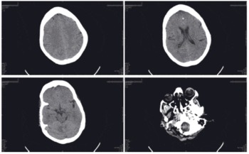
The patient underwent blood tests which showed normal renal and hepatic function and serum electrolytes. The hemogram and complete blood count were normal. Fasting blood glucose was 77 mg%. Cerebrospinal fluid analysis was also normal. Computed tomography (CT) scan of the brain (Fig. 1) showed non-specific mild diffuse oedema. There was a calcified lesion in medial frontal lobe that could explain the past history of seizures. These findings were considered non-specific and hence the patient was referred to psychiatry for management of the catatonic state in the absence of evidence of central nervous system infections, structural pathology or metabolic derangements in the tests done in the emergency department.
CT of the brain shows non-specific changes of diffuse oedema, manifested by sulcal effacement (top left) and a loss of subarachnoid space around the mid-brain (bottom left). There is a calcified lesion in the left medial frontal lobe suggestive of an old cysticercal lesion – a probable explanation of the seizures in the past (top right). Proptosis is also evident in the scan (bottom right).

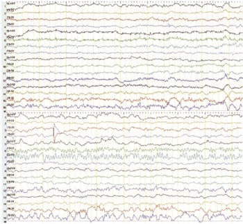
Further investigation reports were available over the next 1 week. The patient tested negative for human immunodeficiency virus (HIV) infection. Sepsis was ruled out by negative urine microscopic examination, blood and urine cultures. The electroencephalogram (EEG) (Fig. 2) showed a brief period of delta rhythm in the right temporal leads on an otherwise normal background rhythm. Her thyroid profile was suggestive of hyperthyroidism with thyroid stimulating hormone (TSH) = 0.08 uIU/ml (0.5–8.9), free T3 (fT3) = 14.84 pg/ml (2.3–4.2) and free T4 (fT4) = 6.64 ng/dl (0.89–1.76). An ultrasound of the neck showed a diffuse thyroid enlargement. A 99mTc pertechnate thyroid scan was done which revealed homogeneously increased tracer uptake in an asymmetrically enlarged thyroid, suggestive of Grave's disease. Anti-thyroid (anti-TG) antibody profile showed elevated anti-thyroglobulin antibodies 1430 IU/ml (positive being >325 IU/ml) and anti-thyroid peroxidase (anti-TPO) antibodies 77.4 U/ml (positive being >50 U/ml). Anti-TSH receptor antibodies could not be tested due to non-availability of the test locally and strong suggestion of Graves' disease on basis of clinical feature and scintigraphy studies.
EEG of the same patient was normal for most part of the 25 min record (top). However, there was a brief period of right temporal delta rhythm (bottom-Channels 15 & 16). The patient was on lorazepam 1 mg i.v. q 6 h on the day this EEG was taken, hence the background activity is fast.

Differential diagnosis
The working diagnosis at admission was one of organic catatonia. The history of fever, disorientation, diarrhoea and presence of fleeting visual hallucinations and delusions were strongly suggestive of organic aetiology and not of functional acute psychosis. Metabolic causes, systemic infections and neuroinfection were ruled out by means of tests done in the emergency department. The presence of tachycardia, mild ophthalmopathy and brisk deep tendon reflexes made us suspect hyperthyroidism as a possible cause. However, as hyperthyroidism seldom presents with delirium or catatonia, we considered a possibility of EAATD which was confirmed on the basis of anti-thyroid antibodies.
Treatment and in-hospital course
Pending the results of thyroid hormone assays and given the catatonic syndrome, a lorazepam trial was initiated at a dose of 1 mg i.v. q 6 h on day 1 which resulted in complete resolution of the catatonic syndrome within 24 h. She continued to remain in delirium with occasional visual illusions and hallucinations, disorientation and sundowning for the next 1 week till 1 mg risperidone was added at bedtime. Lorazepam was subsequently tapered to a dose of 2 mg bedtime for sleep. After consultation with the endocrinologist, carbimazole 15 mg thrice daily and propranolol at a dose of 40 mg/day were added to maintain a pulse rate of about 80/min.
Catatonia was treated with lorazepam as it is a standard practice. Subsequently, the delirium was treated symptomatically with risperidone pending reports of investigations. Since the patient showed complete improvement and resolution of delirium within 1 week (before the results of thyroid profile and anti-thyroid antibodies were available) we did not consider steroid treatment. The patient was well oriented at discharge, with a mini-mental status examination (MMSE) score of 22 out of 28 points administered, which was appropriate given her illiterate status. Brief lobar functions testing revealed mild inattention as the only cognitive deficit.
Post-discharge follow-up
The patient reported for follow-up with restoration of weight. There were no features of delirium subsequently. There was a brief period of elevated mood, increased grooming and increased activity suggestive of hypomania. Risperidone dosage was increased to 2 mg/day with which these symptoms resolved within 1 week. Thyroid hormone estimation is not available at follow-up.
Discussion
This case describes a patient with undiagnosed Graves' disease with systemic manifestations of 2 years' duration presenting with acute onset of neuropsychiatric symptoms in form of altered behaviour and catatonia. There were clinical pointers to an organic cause of catatonia during the acute phase of her illness, namely, disorientation, fever and diarrhoea. The initial work up had ruled out central nervous system infections and hepatic or renal failure or electrolyte disturbances as a cause of her catatonia. CT findings were non-specific and did not reveal major infarcts. Detailed medical history, presence of a thyromegaly, ophthalmopathy, exaggerated reflexes and persistent tachycardia alerted us to a possibility of thyroid disease as a possible cause of her catatonia.
Tamagno et al. (Reference Tamagno, Federspil and Murialdo7) have laid down criteria to diagnose EAATD and described the various commonly encountered clinical features in this entity. The major features include an acute or sub-acute onset of neuropsychiatric symptoms in the presence of autoimmune thyroid disease. The thyroid hormone status is insufficient to explain the degree of neuropsychiatric symptoms. Other causes of encephalopathy must be ruled out and there may be clinical improvement with steroid treatment.
Our patient had manifestations of hyperthyroidism for 2 years, however, the neuropsychiatric manifestations of delirium and her presentation in a catatonic state are of 3 weeks duration and acute in nature.
Her thyroid hormone levels are in the hyperthyroid range. Such a degree of hyperthyroidism commonly presents with anxiety, irritability, mood changes, inattention, memory disturbances and difficulty in planning. The occurrence of delirium and catatonia is indicative of a more severe neuropsychiatric disturbance that is not explained by the degree of her hyperthyroidism.
Her pre-menopausal state is a period of risk for acute psychosis. However, the occurrence of delirium with visual hallucinations, fluctuating symptoms and sundowning favour an organic aetiology for the behavioural disturbance, namely EAATD. Other organic aetiologies such as hepatic or uraemic encephalopathy, diabetic ketoacidosis, systemic or neurological infections were ruled out by normal laboratory tests. Creutzfeldt-Jakob disease was considered unlikely in view of absence of classical EEG findings of periodically occurring triphasic waves and improvement in the patient's condition.
Elevated anti-thyroid antibody titres are the hallmark of EAATD as they are markers for autoimmunity in these patients. Anti-TPO antibodies were found to be positive in all 14 cases of Graves' disease (also positive in our patient), with anti-TG positivity in only 7 out of 11 cases (strongly positive on our patient) and anti-TSH receptor antibodies were positive in 9 out of 10 cases (could not be done in our patient) in a review by Tamagno et al. (Reference Tamagno, Celik and Simo10). Though we could not get an anti-TSH receptor antibody level, the clinical picture and laboratory and radiological findings are classical of Graves' disease.
EAATD has been clinically characterised in recent literature (Reference Canton, de Fabregas, Tintore, Mesa, Codina and Simo6). EAATD has been described to have predominantly neurological symptoms like seizures and loss of consciousness (51%), cognitive deterioration and memory loss (48%), myoclonus (32%) and psychotic symptoms (26%) among other symptoms (Reference Tamagno, Federspil and Murialdo7). There have been case reports of psychiatric symptoms in these patients (Reference Mahmud, Lteif, Renaud, Reed and Brands8,Reference Liu, Tseng and Lin9). Although our patient had seizures in the past, it was almost a decade before onset of symptoms of thyroid disease and is unlikely to be related to the current autoimmune thyroid disease. CT scan showed a calcified lesion in the left medial frontal cortex, making a now dormant cysticercal infection the most likely cause of the past seizures of which the patient has had no recurrence.
EEG abnormalities, usually a generalised background slowing, have been shown to occur in EAATD in 12 out of 13 patients with Graves' disease (Reference Tamagno, Celik and Simo10). However, one patient as reported by Canton et al. (Reference Canton, de Fabregas, Tintore, Mesa, Codina and Simo6) had frontal slow waves on EEG. In our patient, the EEG was taken after the initiation of lorazepam treatment and showed a fast beta background activity. Benzodiazepines such as lorazepam are known to cause fast background activity on EEG. There was one brief period of temporal delta rhythm lasting about 4 s. This was a non-specific EEG change suggestive of encephalopathy. The adolescent girl with treatment resistant psychosis described by Mahmud et al. (Reference Mahmud, Lteif, Renaud, Reed and Brands8) had a normal EEG with markedly raised anti-TPO antibody titres. A recently reported case of depression occurring in a patient with Hashimoto's encephalopathy (Reference Liu, Tseng and Lin9) also showed EEG with disorganised alpha activity with brief runs of temporal delta activity. Hence, the absence of the generalised background slowing described in most cases of EAATD with neurological manifestations may not be seen in cases presenting with psychiatric symptoms.
Except for the lack of proof for steroid responsiveness, our patient seemed to satisfy criteria for probable EAATD as proposed by Tamagno et al. (Reference Tamagno, Federspil and Murialdo7). However, this phenomenon of improvement of the encephalopathy without a trial of steroids has been reported earlier (Reference Tateishi, Iguchi, Kimura, Aoki, Uemura and Shibazaki11) and does not necessarily exclude the diagnosis of EAATD. There is a case of failure of immunosuppressant therapy in EAATD which later remitted after thyroidectomy (Reference Yuceyar, Karadeniz and Erdogan12). Another case of Graves' disease associated encephalopathy has responded to thyroidectomy (Reference Gelosa, DiFrancesco and Tremolizzo13).
Conclusions
Our patient is a case of probable EAATD presenting in catatonia. She had clinical features of thyroid disease of 2 years' duration with opthalmopathy and laboratory and radiological evidence in favour of Graves' disease with anti-thyroid antibodies positive in the serum. However, her current presentation was an acute onset of delirium without any features suggestive of a mood disorder or acute psychotic episode. There was no past history or family history of psychiatric illness. The neuropsychiatric manifestations of catatonia or delirium are severe in nature and out of proportion to the degree of hyperthyroidism in our patient. The CT scan showed non-specific abnormalities which have been described by Tamagno et al. (Reference Tamagno, Federspil and Murialdo7) and the EEG showed abnormalities consistent with previous descriptions of EAATD with psychiatric manifestations (Reference Mahmud, Lteif, Renaud, Reed and Brands8,Reference Liu, Tseng and Lin9).
The diagnostic criteria for this entity of EAATD still needs to be finalised in view of the evidence available in literature and steroid responsiveness need not be an essential criteria for its diagnosis (Reference Tamagno14). There is also a need for consensus among experts in matters of the protean clinical manifestations, diagnostic criteria, hints about aetiology and pathogenesis as well as treatment of this condition (Reference Berger15).
A high index of suspicion must be maintained for organic aetiology in cases presenting with catatonia. In case of severe neuropsychiatric manifestations occurring in a patient with thyroid disease, autoimmune thyroid disease and EAATD must be strongly considered.