Cardiac tumours can be either primary, or secondary to a metastasis from a distant tumour such as a hypernephroma, ovarian mature cystic teratoma, hepatocellular carcinoma,Reference Lin, Chiu, Chen Chien, Wang and Chu1 melanoma, and many others. Such tumours are rare in children, and about one-third of those thus afflicted are asymptomatic.Reference Ohashi, Asakura, Sakamoto, Shimizu and Yoshida2, Reference Dulac, Plat and Taktak3 When present, symptoms depend on the size and location of the tumour, which can create a variety of clinical findings, such as heart murmurs, chest pain, tachyarrythmias, and congestive cardiac failure. The arrhythmias are thought to be caused by the infiltration of the tumour cells into the myocardium, leading to its distortion and formation of new conducting accessory pathways.Reference Paraskevaidis, Triantafilou, Karatzas and Kremastinos4, Reference Parmley, Salley, Williams and Head5 The tumours tend to increase in size slowly, albeit that they may cause cavitary obstruction leading to haemodynamic compromise.
The most common benign cardiac tumours are myxomas, rhabdomyomas, and fibromas, with the latter variants being most common in children. When found, fibromas are usually present on the left ventricular free wall, with fewer than one-tenth involving the atriums, even then with a low risk of embolization. Nowadays, the tumours are usually diagnosed with echocardiography, magnetic resonance imaging, and cardiac catheterization.Reference De Cobelli, Esposito and Mellone6, Reference Natarajan, Vijayanagar, Ecstein, Bognolo and Toole7 When found, surgical excision is the treatment of choice, being indicated when the tumour causes either arrhythmias or intracardiac obstruction.Reference Dulac, Plat and Taktak3, Reference Williams, Danielson, McGoon, Feldt and Edwards8 We describe here the finding of a giant ventricular fibroma co-existing with an atrial septal defect in an adolescent female.
Case report
Both the atrial septal defect and the right ventricular tumour were diagnosed shortly after birth of our patient, which occurred in Montenegro. During her childhood years, the girl was asymptomatic, and Holter monitoring proved negative for any arrhythmia. She was not taking any medications, and was followed-up by regular echocardiographic examinations. Her cardiologist decided to refer her to our centre in Monaco when the size of her mass was shown to be increasing by echocardiography, and when she started reporting dyspnoea on exertion. She denied any palpitations or loss of consciousness.
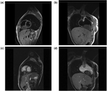
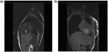
Physical examination revealed a child of normal development, with a murmur audible on auscultation. Her electrocardiogram was normal. Echocardiography revealed a defect in the oval fossa permitting left to right interatrial shunting, and an inhomogeneous giant right ventricular mass. The left ventricle, ejection fraction, and cardiac valves were all normal. There was a trivial tricuspid regurgitation, and systolic right ventricular pressure was estimated at 33 millimetres of mercury. There was moderate volume overload of the right ventricle, albeit without any acceleration of flow in the right ventricular outflow tract. The inferior caval vein was not dilated. Cardiac magnetic resonance imaging using Gadolinium contrast with late enhancement confirmed the presence of the defect in the oval fossa, which measured 15 millimetres in diameter, along with the other normal findings. The right ventricle, however, although exhibiting normal systolic function, was dilated, and contained a mass measuring 59 by 41 by 41 millimetres attached to its inferior segment. The mass was sparing the tricuspid valve and the apex (Fig. 1), was isosignal on T1 sequences, weak hyposignal on T2 sequences, and showed an intense signal in the late phase of enhancement (Fig. 2).
Cross-sectional and longitudinal magnetic resonance imaging view of the heart before (a, b) and after (c, d) injection of contrast, showing a mass in the right ventricle.

Magnetic resonance imaging views of the heart during the late phase of enhancement. The cross-sectional (a) and longitudinal (b) cuts show a mass in the right ventricle, with its characteristic features of intense late enhancement with central hypointensity compared with the normal myocardium consistent with the diagnosis of a fibroma. This was confirmed by the surgical biopsy.

After evaluation of the clinical state of the patient, and the results the investigations, the surgical team decided that a surgical excision of the tumour should not be attempted, mainly because it was causing neither any cavitary obstruction nor cardiac arrhythmias. Instead, it was decided surgically to close the atrial septal defect, and simultaneously to biopsy the tumour. This revealed the tumour to be a fibroma. The atrial septal defect was closed successfully, without any complications. Subsequent echocardiography revealed obliteration of the interatrial shunt, the right ventricular systolic pressure being measured at 26 millimetres of mercury, with no right ventricular inflow or outflow obstruction. The dyspnoea and cardiac murmur disappeared, and the patient was discharged home receiving aspirin at 75 mg daily for 6 months, with a recommendation for regular follow-up, including echocardiography, by her referring cardiologist.
Comment
Magnetic resonance imaging is a valuable tool in the diagnosis of a cardiac fibroma.
Surgical excision of the tumour should be considered for selected patients, but unless indicated on the basis of symptoms, is not mandatory. Should surgical excision be deferred, however, careful follow-up is essential.
Acknowledgments
We thank Sami Dimassi for the excellent technical support.