INTRODUCTION
Entamoeba histolytica is an important protozoan parasite of humans, that is transmitted via the faecal-oral route. This parasite is responsible for approximately 50 million cases of invasive amoebiasis each year (WHO, 1997). E. histolytica usually resides in the large intestine, and can persist there for months or years and cause an asymptomatic intestinal infection. However, this amoeba is often invasive and can also penetrate the intestinal epithelium and cause ulcerative colitis or disseminate into other organs, most commonly the liver, leading to abscess formation.
The genome sequence of E. histolytica has been available since 2005 (Loftus et al. 2005; http://www.genedb.org/genedb/ehistolytica/; http://www.tigr.org/tdb/e2k1/eha1/). This genome contains approximately 10000 genes and, interestingly, about 30% of them do not currently have any homologues in public gene databases. Analysis of the genomic data provides evidence for lateral gene transfer of bacterial genes into the E. histolytica genome (Loftus et al. 2005). Also, most of the genes linked to the mitochondrial amino acid biosynthesis pathways are lacking, and the oxidative stress proteins discovered in E. histolytica are usually associated with those described for various anaerobic prokaryotes (Loftus et al. 2005). Furthermore, the analysis of the E. histolytica genome reveals an intriguing redundancy, exemplified by the presence of 30 homologues of the intermediate subunit of the GalGalNAc lectin, multiple new genes encoding pore-forming peptides and approximately 40 cysteine proteinases (Loftus et al. 2005).
The availability of the E. histolytica genome sequence opens up a range of unique scientific opportunities. For instance, it is now possible to study the transcription and expression profiles of E. histolytica genes by microarray and proteomic analyses, respectively. However, currently there are very limited data for most genes expressed under different culture or environmental conditions and in different strains.
To study the transcriptome of E. histolytica, MacFarlane et al. (2005a,b) developed a genomic DNA microarray containing approximately 2000 genes. These authors showed that 31–47% of the genes are expressed under ‘standard’ culture conditions. Cultivation under ‘heat shock’ conditions induced an upregulation of 17% of the genes, whereas cultivation in the presence of Caco-2 cells had no significant influence on the transcriptional profile (MacFarlane et al. 2005a,b; Shah et al. 2005). Recently, this microarray was used for a comparison of the expression levels between different virulent and non-virulent Entamoeba species and strains (E. histolytica HM-1:IMSS, E. histolytica Rahman, and E. dispar). Some genes with known roles in pathogenicity and stress response had a decreased expression in non-pathogenic E. histolytica (see MacFarlane and Singh, 2006). In another microarray analysis, employing a ‘shotgun’ genomic DNA microarray, collagen activated and non-activated trophozoites were compared (Debnath et al. 2004). Fourteen differentially regulated genes were analysed further by sequencing. Gene products identified in this way were predicted to be involved in cytoskeleton rearrangements, vesicle formation, phagocytosis, protein degradation, stress response and/or ribosomal assembly, and included cysteine proteinases, ribosomal proteins, HSP70 and myosin 1B (Debnath et al. 2004).
Differential expression can also be studied at the protein level, an approach that is likely to be of great relevance for understanding regulatory processes or mechanisms. Thus far, 4 proteomic studies of E. histolytica have been conducted (Marion et al. 2005; Okada et al. 2005, 2006; Leitsch et al. 2005), of which 3 have explored the protein composition of purified phagosomes. Different proteins involved in uptake-processes were identified, including those involved in signalling, endocytosis, cytoskeleton rearrangement, vesicular trafficking and surface recognition (Marion et al. 2005; Okada et al. 2005, 2006). A two-dimensional gel electrophoresis (2-DE) protocol was developed for E. histolytica, which allows the reproducible display of protein expression profiles for this organism (Leitsch et al. 2005). Approximately 1500 protein spots were detected by silver-staining, and 10 landmark spots of the respective Coomassie-stained gels were identified by matrix-assisted laser desorption/ionization time of flight (MALDI-TOF) mass spectrometry or protein sequencing (Leitsch et al. 2005).
In the present study, a practical and reproducible procedure for the solubilization of E. histolytica proteins and their 2-DE analysis is reported. Of the proteins resolved, approximately 100 proteins were subjected to analysis by MALDI-TOF mass spectrometry. The resultant peptide mass fingerprints were analysed by comparison with data available in the E. histolytica genome and NCBI databases. The present investigation is considered to pave the way for the construction of 2-DE reference maps for E. histolytica.
MATERIALS AND METHODS
Cultivation of cells and extract preparation
Trophozoites of the E. histolytica isolate HM-1:IMSS were cultured axenically in TYI-S-33 medium in plastic tissue culture flasks (Diamond et al. 1978). For the experiments, 2×10⁶ trophozoites were cultivated for 48 h in 250 ml flasks. The trophozoites were harvested after being chilled on ice for 5 min and sedimented at 430 g at 4 °C for 5 min. The resultant pellet was washed twice in phosphate-buffered saline (PBS, 6·7 mM NaHPO4, 3·3 mM NaH2PO4, 140 mM NaCl, pH 7·2). Immediately prior to cell lysis, the cysteine proteinase inhibitor E-64 was added (final concentration 50 μM) to the cell pellet. Subsequently, the cells were lysed via 4 freeze-thaw cycles in CO2/ethanol. Immediately after the last cycle, E-64 was added (same concentration) to the extract, which was centrifuged for 1 h at 100000 g (4 °C). The supernatant (PBS-soluble extract) was stored at −70 °C. The PBS-insoluble fraction was resuspended directly in 600 μl of rehydration buffer prior to storage at −70 °C.
Two-dimensional gel electrophoresis
First dimension IEF was performed at 20 °C using 18 cm immobilized pH gradient (IPG) strips (Amersham Biosciences), applying a linear pH range of 4 to 7. The maximum current setting was 50 μA/strip using the IPGphor Isoelectric Focusing Unit (Amersham Biosciences). The strips were rehydrated for a minimum of 12 h in ceramic strip holders using 100 μl of sample diluted with 250 μl of rehydration buffer (10 M thiourea, 2·5% CHAPS, 0·01% Coomassie Brilliant blue, 0·5% IPG-buffer of the respective pH gradient, and 1 mM dithiothreitol. Approximately 0·5 mg of protein was used. A voltage of 30 V was applied during rehydration (10 h). After rehydration, IEF was carried out under the following conditions: 1000 V for 1 h, 3000 V for 1 h and 8000 V for 12 h. The focusing was terminated between 90000 and 91000 Vh. Subsequently, the strips were equilibrated in 50 mM Tris pH 8·8, 6 M urea, 30% glycerol, 2% SDS, 0·03% bromphenol blue supplemented with 65 mM dithiothreitol (15 min) and then in 135 mM iodoacetamide (15 min). After transferring the strips to 12·5% sodium dodecyl-sulphate (SDS) polyacrylamide gels, they were sealed in place with 0·5% agarose in 25 mM Tris-base, 0·19 M glycine, 0·1% SDS, 0·01% bromphenol blue.
In the second dimension, electrophoresis was performed using 25 mM Tris-base, 0·19 M glycine, 0·1% SDS as the running buffer for 18 h at 10 mA and 4 °C. A pre-stained molecular mass marker (low range, Bio-Rad) was used as a standard. The gels were stained with colloidal Coomassie Brilliant Blue (CBB) staining of proteins (Neuhoff et al. 1988). Briefly, gels were incubated in 1·4% phosphoric acid, 8% ammonium sulfate, 20% methanol, 2% Coomassie Brilliant Blue G-250 for 12 h at 22 °C. Destaining was performed in 25% methanol at room temperature for 8 h. Gels were stored at 4 °C in 25% ammonium sulfate.
In-gel digestion of proteins
Protein spots were excised from gels, washed with 50% acetonitrile in 25 mM ammonium bicarbonate, dehydrated in acetonitrile and dried in a vacuum centrifuge. The gel pieces were rehydrated in 10 μl of 5 mM ammonium bicarbonate, containing 300 ng of trypsin (proteomic grade, Roche Diagnostics, Mannheim, Germany). After 15 min, 10 μl of 5 mM ammonium bicarbonate were added to ensure that the gel pieces were wet during tryptic cleavage (37 °C, overnight). To extract the peptides, 20 μl of 0·3% trifluoroacetic acid (TFA) in acetonitrile were added, and the samples were sonicated for 10 min. The supernatant was removed and dried under vacuum. The peptides were redissolved in 8 μl of 0·1% TFA, 5% acetonitrile in water. Prior to mass spectrometry analysis, the peptides were purified using a reversed-phase minicolumn filled in a micropipette tip (ZipTip C18, Millipore, Bedford, MA, USA), according to the manufacturer's protocol. The peptides were eluted from ZipTips using 2·5 μl of matrix solution (7 mg/ml α-cyano-4-hydroxycinnamic acid (Sigma), dissolved in 60% acetonitrile, 0·12% TFA in water and spotted directly on a target plate and air-dried.
Mass spectrometry
MALDI-mass spectrophotometric measurements were performed using a Voyager-DE STR BioSpectrometry Workstation (Perseptive Biosystems, Inc., Framingham, USA) in the reflection mode at an acceleration voltage of 20 kV, 70% grid voltage and a delay of 200 ns. Each spectrum obtained was the mean of 256 laser shots. Mass spectra were calibrated using known autolytic fragments of trypsin as internal standards. The peptide masses were measured as monoisotopic masses. The resultant spectra were processed utilizing the Data Explorer Software (version 3.5.00). Alternatively, an Axima CFR MALDI-TOF spectrometer (Shimadzu Biotech GmbH, Duisburg, Germany) was used. Depending on spectra quality, 200–400 laser shots were accumulated. Control of the spectrometer and spectra processing was carried out employing the Axima CFR software version 2.2.1.
Database analysis
Proteins were identified using the program MS-Fit (http://prospector.ucsf.edu) by comparison with data publicly available in the non-redundant, annotated E. histolytica (TIGR; http//www.tigr.org/tdb/e2K1/eha1) and NCBI databases. The parameters for the search were as follows: (i) the modifications on cysteine residues by carboxyamidomethylation were set as obligate, (ii) methionine oxidation was considered as a partial modification and (iii) one was used as the maximum missed tryptic cleavage sites. The monoisotopic masses were considered and the mass tolerance was set at 0·07 Da. To determine the certainty of the analysis, the MOWSE (Molecular Weight Search) score analysis of MS-Fit was used, based on the scoring system described by Pappin et al. (1993). For all proteins, the concordance between the theoretical and the experimental isoelectric points and molecular masses as well as a coverage of the protein of more than 20% were essential criteria.
RESULTS AND DISCUSSION
Resolution, sensitivity and reproducibility of 2-DE separations
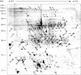
Entamoeba histolytica contains large amounts of cysteine proteinases, which have to be inhibited rapidly to obtain reproducible and reliable results. Therefore, the presence of the specific cysteine proteinase inhibitor E-64 before and directly after the freeze-thaw cycles of the E. histolytica extract was highly beneficial for both resolution and reproducibility in the 2-DE analysis (data not shown). Also, the addition of 10 M thiourea to the rehydration buffer in the solubilization step of both fractions (PBS-soluble proteins as well as the PBS-insoluble proteins) resulted in a greater number of detectable protein species in the 2-DE analysis compared with the solubilization in urea (data not shown). The PBS-soluble as well as the PBS-insoluble protein fractions were subjected to 2-DE analysis. An amount of 500 μg protein per 18 cm IPG strip was shown to be optimal for CBB-stained gels. Representative 2-DE gels of different batches of soluble protein fractions from E. histolytica trophozoites are shown in Fig. 1. The extracts were separated over a pH range of 4 to 7, stained with CBB and analysed. The reproducibility of the sample preparation, gel electrophoresis and staining was verified by running sequential replicate gels (at least 5 times) using protein extracts prepared from different cultures of E. histolytica trophozoites. Gels run under identical conditions gave almost identical results with respect to spot pattern and relative intensities (Fig. 1). Most of the amoebic proteins separated within a pH range of 4 to 7 have a pI of 5 to 6. A total of approximately 400 CBB-stained proteins could be detected when the PBS-soluble protein extract was separated in a 2-DE using 18 cm IPG strips and a linear pH gradient. In the PBS-insoluble fraction, about 500 protein species could be detected using a Coomassie Blue stain (data not shown).

Fig. 1. (A) Representative gels displaying the 2-DE analysis of 2 independent extract preparations from PBS-soluble proteins from Entamoeba histolytica. The proteins (500 μg) were separated on linear IPG strips (pH 4–7), followed by 12·5% SDS-PAGE. The gels were stained with Coomassie Blue. Molecular mass standards are indicated on the left. (B) Comparison of 4 enlarged partial 2-DE gels (compared area (B) is marked in (A) showing PBS-soluble proteins derived from 4 independent extract preparations.
Protein identification
Using the Coomassie-stained gels of PBS–soluble extracts, 95 protein spots were randomly isolated, in-gel digested, and analysed by MALDI-TOF mass spectrometry. For 90 of these 95 spots, quality peptide mass fingerprint spectra were obtained by MALDI-TOF MS analyses. For 10 spots (# 3, 9, 40, 44, 62, 60, 66, 71, 81, 88; Fig. 2), no protein sequence matches were found in the E. histolytica or the NCBI databases. While 63 protein spots related unequivocally to unique proteins (Table 1), for 17 proteins, 2 or more entries were found (Table 2). Multiple entries may result from the presence of 2 or more proteins within 1 protein spot or, alternatively, different proteins may have similar molecular mass fingerprints.

Fig. 2. The 2-DE reference map of proteins from the PBS-soluble extract from Entamoeba histolytica. The proteins (500 μg) were separated on linear IPG strips (pH 4–7), followed by 12·5% SDS-PAGE. The gels were stained with Coomassie Blue. Numbers indicate protein spots that were subjected to MALDI-TOF mass spectrometry. The resultant peptide mass fingerprints were analysed and compared with the E. histolytica genome and the NCBI databases to predict the identity of the proteins. Spot numbers refer to proteins listed in Tables 1 and 2. Isoelectric point (top) and molecular mass (left) are indicated.
Table 1. List of Entamoeba histolytica proteins analysed by MALDI-TOF mass spectrometry (The identified proteins can be grouped into 7 functional categories as listed in the table. Accession numbers are from the NCBI protein database (http://www.ncbi.nlm.nih.gov/entrez/query.fcgi?CMD=search&DB=protein). Apparent pI and molecular weight (MW) were calculated using MacVector 7.2.2 software. T, theoretical; E, experimental.)

Table 2. List of Entamoeba histolytica proteins analysed by MALDI-TOF mass spectrometry with two or more entries in the database (Accession numbers are from the NCBI protein database (http://www.ncbi.nlm.nih.gov/entrez/query.fcgi?CMD=search&DB=protein). Apparent pI and molecular weight (MW) were calculated using MacVector 7.2.2 software. T, theoretical; E, experimental.)

The 63 proteins identified represented 41 different molecules of known function, and 6 proteins were recorded previously as hypothetical proteins in the databases interrogated (Table 1). These proteins could be grouped into 7 categories of functionally related molecules (cytoskeletal proteins, glycolysis, surface-associated proteins, RNA/DNA metabolism, ubiquitin-proteasome pathway, vesicular trafficking and signal transduction) (Table 1).
Proteins associated with the cytoskeleton
The cytoskeleton associated proteins actin, actophorin, myosin calcium-binding light chain, coactosin, actin depolymerization factor and a kinesin-like protein have been reported previously in a phagosome study (Marion et al. 2005). Actin depolymerization factors and actophorin are members of the ADF/cofilin protein family (Moon and Dubrin, 1995). These proteins are involved in the turnover of actin filaments in dynamic regions of the cell (Maciver et al. 1998; Bamburg, 1999). Coactosin seems to bind the barbed ends of actin filaments, thus sterically hindering the binding of capping proteins (Rohrig et al. 1995). Kinesin is a member of the microtubule-based ATPase motor proteins which perform force-generating processes, such as organelle transport and chromosome segregation (Kull et al. 1996).
Enzymes involved in glycolysis and glycogen metabolism
Some enzymes of the glycolytic pathway, including aldehyde-alcohol dehydrogenase, NADP-dependent alcohol dehydrogenase, enolase, fructose-1·6-bisphosphate aldolase, pyruvate kinase, pyruvate phosphate dikinase and glycogen phosphorylase were identified. The aldehyde-alcohol dehydrogenase is a bifunctional 95 kDa polypeptide, having both acetaldehyde dehydrogenase and alcohol dehydrogenase activities. Each enzymatic property is located to a different part of the molecule (Bruchhaus and Tannich, 1994; Yang et al. 1994; Chen et al. 2004). In the present study, a protein with a molecular mass of 38 kDa was detected. All peptide mass fingerprints determined for this protein matched only the aldehyde dehydrogenase domain of the bifunctional enzyme. Thus, the question arises as to whether this molecule is the product of protein degradation during the extraction process or whether the bifunctional protein is proteolytically processed in E. histolytica.
Surface-associated proteins
Three putative surface-associated proteins, the 170 kDa subunit of the galactose-inhibitable lectin, a BsA-like leucine rich repeat protein and a cysteine proteinase were identified in the PBS-soluble protein extract. Lectin is one of the most important surface molecules of E. histolytica. It is a 260 kDa heterodimer, consisting of the 170 kDa transmembrane (heavy) and a GPI-anchored 35 kDa (light) subunits. These subunits are linked by disulphide bonds (Cheng et al. 2001). The functional activity of lectin has been shown to be involved in host cell binding, cytotoxicity, complement resistance and virulence (Mann, 2002; Frederick and Petri, 2005).
Recently, proteins with homology to BspA leucine rich repeat (LRR) proteins were identified (Davis et al. 2006). For organisms other than E. histolytica, it has been shown that such proteins are surface-associated and that the LRR motif is linked to fibronectin binding (Sharma et al. 1998; Kobe and Kajava, 2001). In the genome of E. histolytica, more than 80 genes coding for BspA-like proteins have been identified (http://www.tigr.org/tdb/e2k1/eha1/). Further characterization of one family member confirmed the surface localization for E. histolytica, although no transmembrane domain has been predicted (Davis et al. 2006). The BspA-like protein identified herein has a molecular mass of approximately 20 kDa, which is in marked contrast to a previous study (Davis et al. 2006) describing a protein of about 60 kDa. These proteins have a sequence identity of around 50%.
In addition, a cysteine proteinase was identified which contains an N-terminal, putative transmembrane domain. As for the acetaldehyde/alcohol dehydrogenase identified, only a portion of 11 kDa was detected, possibly as a result of protein degradation.
Proteins of the ubiquitin-proteasome system
Entamoeba contains a ubiquitin-proteasome system. Some proteins involved in this system, including 2 proteasome alpha subunits, HSP70, ubiquitin, an ubiquitin-related protein, an ubiquitin-conjugating enzyme and an ubiquitin-protein ligase E3 component, were discovered using the present proteomic approach.
Signalling proteins and intracellular trafficking
Small GTP-binding proteins are common to all eukaryotes. They are involved in important processes, such as intracellular trafficking, cell proliferation and cytoskeleton rearrangement. The superfamily of GTP-binding proteins is classified into 5 families (i.e. Ras, Rho/Rac, Rab, Sar/Arf and Ran). Ninety-one putative Rab proteins have been identified in the E. histolytica genome database (Saito-Nakano et al. 2005). In the present investigation, molecules RabK1, RabF4, and RabX28 were identified. For all 3 of these molecules, no obvious homologues were detected in other organisms for which data are available. In addition to the 3 Ras family members, 1 Rho family member was discovered. It is known that about 30 Rho proteins are represented in the E. histolytica genome database (http://www.tigr.org/tdb/e2k1/eha1/). The activity of Ras and Rho proteins is modulated by 3 classes of proteins, namely GTPase activating protein (GAP), GDP-dissociation stimulator (GDS) and GDP-dissociation inhibitor (GDI) (Takai et al. 1994). This proteomic approach revealed 1 Rab and 1 Rho GDP dissociation/exchange inhibitor. Based on the E. histolytica genome (http://www.tigr.org/tdb/e2k1/eha1/), 3 genes of the Rab dissociation inhibitor but only 1 gene of the Rho GDP exchange inhibitor are known. In a recent proteomic study of the phagosome of E. histolytica, several different small GTPases (e.g. Rab1, 5, 7A, 7b, 7C, 7E, 8, 11B, 11C, X17) and other proteins involved in vesicular trafficking were identified (Okada et al. 2006). Surprisingly, none of these proteins were recovered in the present investigation.
Proteins involved in signal transduction
Herein 1 kinase and 1 phosphatase, proposed to be involved in signal transduction, were identified. The kinase has homology to serine/threonine kinases and belongs to a large family of more than 300 members represented in the E. histolytica genome. For the serine/threonine phosphatases, genes of 66 members exist within the E. histolytica genome (http://www.genedb.org/genedb/ehistolytica/).
Proteins involved in RNA/DNA metabolism
Three proteins predicted to be involved in RNA or DNA metabolism, including a helicase, DNA polymerase and a putative signal recognition particle, were identified.
In conclusion, the combined use of the present proteomic approach together with comparative analyses in publicly available genome sequence data for E. histolytica and other organisms has identified a range of E. histolytica proteins which had not been reported previously to be expressed in this parasite. Clearly, the present proteomic analysis provides a foundation for the identification of novel proteins from E. histolytica which are crucial for the development, survival and pathogenicity of this parasite, and should also lead to a better understanding of the molecular biology of E. histolytica and its relationship with its host.
This work was supported by the Deutsche Forschungsgemeinschaft (DFG), BR 1744/7-1. We would like to thank T. W. Gilberger and T. Roeder for critical reading of the manuscript.