


Rubinstein–Taybi syndrome (official abbreviation RSTS, but RTS is more frequently used) is a rare syndrome with a frequency of 1 in 100 000 to 1 in 125 000 affected newborns (OMIM database 180849). The syndrome is characterised by mental retardation, growth retardation and a particular dysmorphology. The syndrome is autosomal dominant but reports of transmission are exceedingly rare; the overwhelming majority of cases are caused by de novo mutations (Refs Reference Hennekam1, Reference Marion, Garcia and Karasik2, Reference Petrij3). The first gene implicated in causing the syndrome is CREBBP (also called CBP; OMIM 600140), which encodes a cAMP-response-element binding protein (CREB) binding protein (Ref. Reference Petrij4).
Mutations in CREBBP causing RSTS range from very large deletions, removing hundreds of kilobases, to deletions that remove only a single exon (Ref. Reference Petrij3). Translocations and pericentric inversions have also been found (Refs Reference Imaizumi and Kuroki5, Reference Tommerup, van der Hagen and Heiberg6, Reference Lacombe7, Reference Breuning8). However, the majority of mutations are point mutations, small deletions or duplications ranging from one to several base pairs that may cause premature translation stops, amino acid substitutions or splicing defects (Table 1). The occurrence of deletions that remove the entire gene indicate that the syndrome is caused by haploinsufficiency.
Point mutations and small deletions and insertions in CREBBP and EP300 published to datea
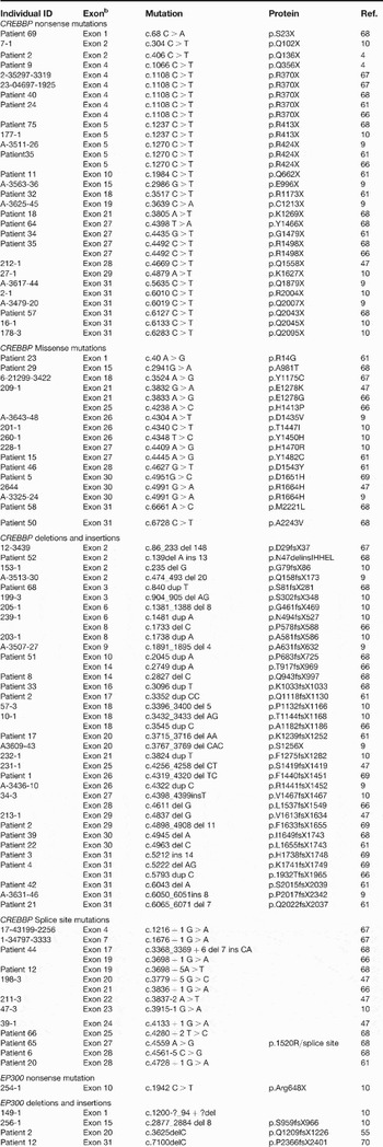
aIn CREBBP there are 31 nonsense mutations of which four are recurrent mutations, giving a total of 23 different nonsense mutations. Of the 17 missense mutations, one is recurrent and 16 are therefore different. There are 37 deletions or insertions, making this the largest group of mutations. Nearly all insertions are actually duplications. Fourteen mutations lead to a putative splicing defect. Interestingly, 13 of these are situated at a splice site but one is a substitution that leads to a change of amino acid but is also predicted to cause a splicing defect. In EP300, one nonsense mutation and four deletions have been found. The extent of the single exon deletion of exon 1 is not known.
bNote that CREBBP and EP300 exon numbers are in line with the literature but differ by –1 from actual exon numbers.
Mutations in CREBBP can account for 50% of all cases at best (Ref. Reference Bartsch9). This led to the conclusion that at least one other gene is involved. Indeed, mutations causing RSTS have also been found in EP300 (OMIM 602700), which encodes a product that is homologous to CREBBP (Ref. Reference Roelfsema10). Mutations in other genes may yet be found.
Clinical features
RSTS presents at birth, with certain characteristics becoming more apparent as the patient ages. Dysmorphic features can be manifest in the face, hands and feet (Refs Reference Rubinstein and Taybi11, Reference Michail, Matsoukas and Theodorou12). Prenatal growth is normal but growth retardation is apparent within a few months of birth and requires attention to avoid overfeeding. Standard growth curves have been drawn based on 95 RSTS patients and these act as a guide to the clinician (Ref. Reference Stevens and Bhakta13).
The facial appearance is striking and includes microcephaly, down-slanting palpebral (eyelid) fissures, broad nasal bridge, beaked nose with the nasal septum extending below the nostrils, highly arched palate and mild micrognathia (Fig. 1). Other abnormalities of the head include eye anomalies such as nasolacrimal duct obstruction, ptosis of the eyelids, congenital or juvenile glaucoma and refractive errors. Furthermore, oro-dental features, such as talon cusps, screwdriver incisors, cross bites and crowding, are frequent (Refs Reference Hennekam14, Reference Wiley15). The eye anomalies lead to visual problems in the overwhelming majority of RSTS patients and require extra attention from the physician (Refs Reference Hennekam14, Reference Rubinstein16, Reference Hennekam, Boogaard and Doorne17).
Physical features of Rubinstein–Taybi syndrome. Facial characteristics, broad thumbs and distal phalanges, and partial duplication of the big toes are clearly visible. Courtesy of Dr. R.C.M. Hennekam (UCL, London, UK). Pictures were obtained and are reproduced with informed consent.
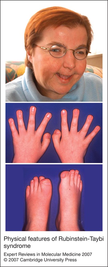
The other striking dysmorphic features of RSTS concern the hands and feet; more specifically, the thumbs and big toes can be broader than normal (Fig. 1). Because of this, the syndrome is sometimes referred to as broad thumb and halux syndrome. However, the broadness is quite variable – the circumference of the thumbs can be almost twice the normal size but they can also be normal. Broad thumbs and big toes are seen in most, but not all, RSTS patients (Refs Reference Hennekam14, Reference Hennekam18). Strikingly, most of the patients that were recently identified as carriers of EP300 mutations have normal big toes (Ref. Reference Roelfsema10). All fingers may be broad, not just the thumbs, and persistent fetal pads may also be found. Broadening of digital bones is not restricted to the distal ones and a small number of patients have short, very broad metatarsal bones (Ref. Reference Petrij3). Additionally, angulation of the thumbs is also regularly seen (Ref. Reference Hennekam19). Severe angulation may require surgical treatment.
Perhaps less striking, but nevertheless serious, are the abnormalities of internal organs found in some patients. Approximately a third of all patients have a heart malformation, such as a patent ductus arteriosus, atrial or ventricular septal defect, coarctation or pulmonic stenosis (Ref. Reference Stevens and Bhakta13). Kidney abnormalities may also be found, and hypospadia is seen in some male patients (Ref. Reference Hennekam14). Secondary risk seizures (28% seizures, 57-66% abnormal EEG) can occur (Refs Reference Hennekam14, Reference Wiley15) and additional signs, such as behavioural problems and autistic traits, as well as medical complications, have also been observed. Guidelines based on the available clinical information, with recommendations for specific surveillance and interventions, have been produced to help in the care of individuals with RSTS (Ref. Reference Wiley15).
In general, mental retardation in RSTS is typically moderate to severe, with IQ ranging from <25 to 79. There may be some decline in IQ with increasing age (Ref. Reference Hennekam20). Patients are born with mental retardation and it is unclear whether or not this progresses throughout life.
One intriguing phenomenon is the fact that RSTS patients are prone to develop tumours. These tumours show a pattern of neural and developmental origin (Ref. Reference Miller and Rubinstein21). Tumours reported in RSTS patients include neuroblastoma, medulloblastoma, oligodendroglioma, meningioma, pheochromocytoma, rhabdomyosarcoma, leiomyosarcoma, seminoma, odontoma, choristoma and pilomatrixomas (Refs Reference Hennekam14, Reference Hennekam, Stevens and van de Kamp22, Reference Sobel and Woerner23, Reference Bonioli and Bellini24, Reference Bilir, Bilir and Wilson25, Reference Cambiaghi26, Reference Urlinger27, Reference Russell, Hoffman and Bain28, Reference Jonas, Heilbron and Ablin29).
Keloid scar tissue, which can continue to grow beyond original wound boundaries, is also frequently seen in RSTS patients (Refs Reference Goodfellow, Emmerson and Calvert30, Reference Rohlfing, Lewis and Singleton31). The itching that results from these keloids can be quite severe. Other dermatological findings are capillary angioma, hirsutism and hypertrichosis.
As almost all cases of RSTS are caused by de novo mutations, the recurrence risk is negligible. However, one of the parents may be an asymptomatic mosaic carrier of a mutation. This phenomenon, which can obviously also play a role in other congenital genetic disorders, should be well known to genetic counsellors. Counsellors providing information to the parents of the patient will inform them of the progression of the disorder and especially point out the increased risk of cancer.
CREBBP and p300
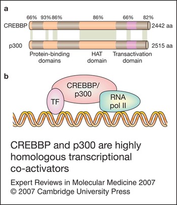
CREBBP and p300 are ubiquitously expressed homologous proteins that act as transcriptional co-activators (Fig. 2) (Refs Reference Kwok32, Reference Lundblad33, Reference Chrivia34). CREBBP comprises 2442 amino acids and p300 is 2415 amino acids long. The genes are highly conserved but invertebrates have only one orthologous gene (Ref. Reference Shi and Mello35). Homologous proteins have also been found in the plant Arabidopsis thaliana (Ref. Reference Bordoli36). CREBBP and p300 have a transactivation domain but they do not specifically bind to DNA. Both proteins can interact with a large number of transcription factors, and several domains involved in protein interaction can be distinguished: zinc finger domains, a CREB-binding domain (KIX domain), a bromodomain and a transactivation domain (Refs Reference Giles, Peters and Breuning37, Reference Goodman and Smolik38). Transcription factors downstream of various pathways need to compete for the limited amount of CREBBP or p300 available in the nucleus. It is thought that in this way CREBBP and p300 act as integrators of signals from these different pathways. The protein forms a physical bridge between the DNA-binding transcription factors and the RNA polymerase II complex (Fig. 2). Apart from this bridging or scaffolding function, CREBBP and p300 also serve as histone acetyltransferases (HATs) (Refs Reference Bannister and Kouzarides39, Reference Ogryzko40), targeting histone H3 and H4. Acetylation of histones opens up the chromatin structure – a process essential for gene expression. CREBBP and p300 also acetylate a range of other proteins, including p53 (Ref. Reference Gu and Roeder41).
CREBBP and p300 are highly homologous transcriptional co-activators. (a) Homology between cAMP-response-element binding protein (CREB) binding protein (CREBBP) and p300. Percentages indicate identity of amino acids between CREBBP and p300, for different domains. The overall identity of amino acids is 61%. Important domains, such as protein-binding domains, the histone acetyl transferase (HAT) domain and the transactivation domain are indicated. (b) CREBBP and p300 interact with a transcription factor (TF) and with the RNA polymerase II complex. The transcription factor binds to DNA, whereas CREBBP and p300 will not. Other proteins, such as the p300/CREBBP-associated factor (P/CAF), can form part of this complex. Various transcription factors compete with each other for the limited amount of CREBBP or p300 in the nucleus. In this way, the transcriptional co-activators act as integrators of signalling pathways.
CREBBP and p300 do not belong to a superfamily of similar proteins that show HAT activity (Ref. Reference Roth, Denu and Allis42). There are many other proteins with HAT activity, such as the MYST family (MYST1-MYST4 also known as MOF, HBO1, MOZ and MORF) and basal transcription factors such as TAFII250. However, these proteins do not show any significant homology and they generally have diverse functions in addition to the HAT activity.
Various mouse models have been generated that have a complete or partial knockout of Crebbp or Ep300. Crebbp-knockout models display some similarities to the features described in RSTS (Refs Reference Kung43, Reference Oike44). However, some differences are also obvious – no heart problems have been described in these mice. Homozygous knockout mice for both genes are not viable and the embryos show extensive haemorrhaging because of defective blood vessel formation (Refs Reference Tanaka45, Reference Yao46). Interestingly, double heterozygous mice are also not viable, showing the same type of malformation as the homozygous knockout mice (Ref. Reference Yao46).
Although CREBBP and p300 are probably largely redundant, there are subtle but clear differences between the two proteins (Refs Reference Yao46, Reference Kalkhoven47, Reference Kasper48). The proteins show a differential subcellular localisation during oocyte and early embryo development and have similar, but not completely overlapping, expression patterns during embryogenesis (Refs. Reference Kwok, Liu and Smith49, Reference Partanen, Motoyama and Hui50). It is difficult to untangle the differences in their roles in signalling pathways. However, experiments with F9 teratocarcinoma cell lines showed that retinoic acid signalling is p300 dependent and does not require CREBBP, whereas cAMP signalling depends on CREBBP and not p300 (Refs Reference Kawasaki51, Reference Ugai, Uchida, Kawasaki and Yokoyama52). Experiments with transgenic mice revealed the importance of the specific acetyl transferase function of p300 in myogenesis, whereas the acetyl transferase function of Crebbp does not appear to be necessary for muscle formation (Ref. Reference Roth53). Conditional knockout mice for Ep300 or Crebbp both showed a decrease of CD4+ CD8+ double-positive cells but an increase in CD8+ cells was reported in the case of the Crebbp-knockout mice (Ref. Reference Kasper48). The fact that skeletal abnormalities found in heterozygous Crebbp-knockout mice have not been reported for heterozygous Ep300-knockout mice is also quite intriguing (Refs Reference Yao46, Reference Tanaka54). Interestingly, in humans with RSTS caused by EP300 mutations, the skeletal features seem to be milder and patients often have normal feet and hands (Ref. Reference Bartholdi55). Despite all these differences, there is no indication that CREBBP and p300 differ in their roles as HATs and there is a large overlap in the range of other proteins that they can acetylate.
Chromosomal abnormalities
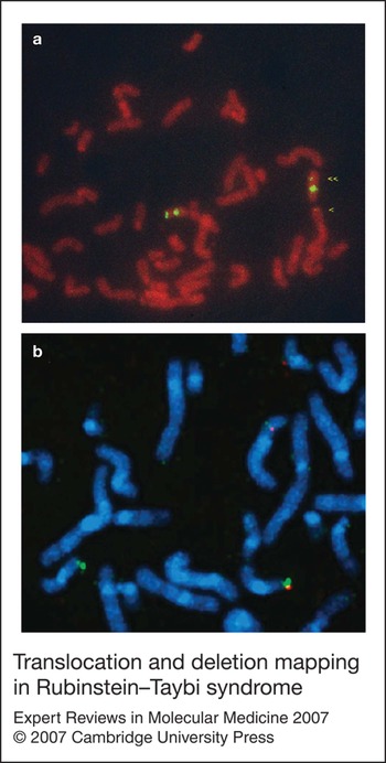
Chromosomal translocations and inversions are generally of great help in identifying genes involved in genetic disorders, and RSTS is no exception. When CREBBP was reported as a causative gene for RSTS, four translocations and two inversions within the gene were already known (Ref. Reference Petrij3). All these rearrangements have a breakpoint in the CREBBP gene. Since then, only one other chromosomal translocation has been found, despite exhaustive fluorescent in situ hybridisation (FISH) experiments using five overlapping cosmids spanning the entire gene (Fig. 3) (Refs Reference Petrij3, Reference Petrij56). New translocations or inversions should have been detected with this approach, but none was found. The breakpoints of six out of seven translocations and inversions reported to date, reside in a region of approximately 10 kb surrounding exon 2. A smaller section of about 5 kb within this region proved to be very difficult to clone (Fig. 4) (Ref. Reference Giles57). Interestingly, in contrast to the germline mutations reported in RSTS patients, CREBBP and EP300 are also involved in somatic translocations associated with leukaemia as fusion partners of MOZ, MYST or MLL (Ref. Reference Troke, Kindle, Collins and Heery58).
Translocation and deletion mapping in Rubinstein–Taybi syndrome. (a) Fluorescent in situ hybridisation (FISH) on metaphase spreads of blood cells from a Rubinstein–Taybi syndrome (RSTS) patient with a t(1;16) breakpoint (Refs 3, 85). Cosmid RT166 spans the breakpoint. The single arrowhead indicates the derivative chromosome 1 and the double arrowhead indicates the derivative chromosome 16 with both the chromosome 16 α-satellite (probe pHUR195) and RT166 signals, present (Ref. 3). (b) Metaphase spread of an RSTS patient with a microdeletion in the CREBBP gene. On the deletion chromosome, cosmid RT102 (red) is deleted whereas cosmid RT166 (green) is still present. Signals for both cosmids can be seen on the normal chromosome.
Location of mutations in CREBBP associated with Rubinstein–Taybi syndrome. (a) Model of the CREBBP gene with its exons represented as vertical bars. The entire gene spans a region of approximately 154 kb. Indicated are the translocation and inversion breakpoints as published by Petrij et al. (Ref. 3). The breakpoints are indicated as blue bars. The first one in intron 1 is a t(1;16). The breakpoints in intron 2 are inv(16), t(16;20), t(7;16), t(2;16) and another inv(16). All these breakpoints reside in an area that is considered to be unstable (highlighted in red). In intron 16 there is a t(2;16) breakpoint. (b) Overview of splice mutations, point mutations and small deletions or insertions found in CREBBP mRNA. The coding part is green and the blue region represents the histone acetyltransferase (HAT) domain. Splice sites are indicated on the upper mRNA. The mutation indicated in red is an amino acid change but this probably leads to aberrant splicing rather than to a missense mutation (Ref. 68). The exact nature of all the mutations is described in Table 1. UTR, untranslated region; CREBBP, cAMP-response-element binding protein (CREB) binding protein; MLPA, multiplex ligation-dependent probe amplification. Note that CREBBP and EP300 exon numbers are in line with the literature but differ by −1 from the actual exon numbers because of a 5′ non-coding exon.
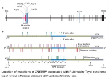
One might expect that the majority of deletions also originate from this apparently unstable region in CREBBP, but this is not the case. A relatively small rearrangement, which deletes only the second exon, seems not to be related to instability (the small size sets it apart within the group of deletions detected so far). In addition, it might be that only a few deletions begin or end in this region. Deletions in general tend to be large and quite a few deletions remove the entire gene. Three very large deletions, involving up to 3 Mb, have been described in severe RSTS, which results in death in infancy. All deletions include CREBBP and the neighbouring TRAP1 and DNASE1 genes. In particular, DNASE1, a gene associated with systemic lupus erythematosus (SLE), seems a good candidate gene for some of the additional features in these patients. Bartsch and co-workers identified this particular phenotype and named it 16p13.3 deletion syndrome (Ref. Reference Bartsch59).
Recently, a new technique has been developed for detection of deletions – multiplex ligation-dependent probe amplification (MLPA) – which can detect copy number changes, such as deletions and duplications, in DNA rather than on metaphase chromosome spreads (Refs Reference Roelfsema10, Reference Schouten60). The technique itself is a multiplex PCR performed using probe pairs hybridised to the DNA of the patient. Not only can large deletions be detected, but small deletions, previously requiring Southern blotting, can be also be detected.
There are also some clear advantages for FISH, which is the method of choice for detecting translocations and inversions and may be superior to MLPA in the detection of mosaic carriers. Three deletions that occur in a mosaic fashion have recently been described (Ref. Reference Bentivegna61).
When the CREBBP gene was first identified in RSTS, chromosomes from patients were analysed with FISH and deletions were found. This led to estimates of the proportion of patients that can be attributed to microdeletions ranging from 25% to 10% (Refs Reference Petrij3, Reference Bartsch62). Remarkably, microdeletions have not been found in high numbers since. It is possible that there was some bias in patient ascertainment leading to this initial high number of microdeletions. The percentage of RSTS patients that can be ascribed to microdeletions should therefore be treated with caution.
Other methods that have been used for detection of microdeletions in CREBBP include: quantitative real-time PCR; analysis of polymorphic microsatellite markers in intron 1 (MS1), intron 2 (MS2 and MS4) and intron 13 (D16S3065) (Ref. Reference Coupry63); and multiplex PCR/liquid chromatography assay (MP/LC) (Ref Reference Udaka64). These methods share the same advantage with MLPA in that they can be used on relatively small amounts of genomic DNA.
The mutation spectrum
A variety of techniques has been applied successfully to detect small mutations: denaturing gradient gel electrophoresis (DGGE), single-stranded conformation polymorphism analysis (SSCP), denaturing high-performance liquid chromatography (DHPLC) and direct sequencing (Refs Reference Bartsch9, Reference Roelfsema10, Reference Bentivegna61, Reference Kalkhoven65, Reference Udaka66, Reference Bartsch67, Reference Coupry68, Reference Murata69). The majority of mutations in CREBBP and EP300 found by these methods are small deletions, duplications/insertions or point mutations predicting premature stop codons. In addition, several missense variants have been isolated that are probably disease-causing mutations because they could be confirmed by functional tests or identified as de novo mutations.
The largest fractions of mutations found in CREBBP are small deletions or duplications/insertions – usually of one or two base pairs – and point mutations. These mutations can be found in coding sequences within the gene but are often found in splice sites at the intron–exon boundary. Almost all mutations lead to a premature translation stop of the mRNA. The resulting truncated protein is likely to be stable – in some cases this is confirmed by western blotting – but the truncated protein does not function (Refs Reference Petrij56, Reference Kalkhoven65). The mutations that lead to a putative truncated protein can be found scattered throughout the gene (Fig. 4): there is no clear clustering of mutations. However, there are noteworthy spots within the CREBBP gene where recurrent mutations have been found (Refs Reference Udaka64, Reference Kalkhoven65, Reference Udaka66, Reference Bartsch67, Reference Coupry68). They are always transitions, and one of these, c.1108 C> T in exon 4 has been reported five times. The 11 bp deletion c.4898_4908 del 11 reported by Murata et al. (Ref. Reference Murata69) is the same patient as reported by Petrij et al. (Ref. Reference Petrij56).
In missense mutations, a substitution leads to a completely different amino acid in the protein, which can have devastating effects. Those found in RSTS patients obviously lead to a partial or complete loss of protein function, which is enough to cause the syndrome. The subtle nature of amino acid substitutions makes it difficult to establish whether the substitution of a single amino acid is actually causing the disease. RSTS, in this respect, has as an advantage, because nearly all mutations occur de novo. Screening the genes of the parents will therefore indicate whether the sequence variant is an acquired mutation or a polymorphism.
An example of one mutation that is probably a neutral polymorphism rather than disease-causing is p.N1978S. This was reported as a recurrent mutation but has been unveiled as a polymorphism by Bentivegna and co-workers (Ref. Reference Bentivegna61), who found this amino acid change in a patient who also had a mutation leading to a premature stop codon and, moreover, found the same change in one of the 50 normal individuals they used as a test panel.
In contrast to other mutations, most missense mutations found thus far are clustered in CREBBP in the region that encodes the HAT domain. This indicates that loss of HAT activity may already be sufficient to cause RSTS. Although there is clear clustering of missense mutations in the HAT domain, not all missense mutations that have been reported are situated there (Fig. 4). Obviously, there is more than one way to disrupt the function of CREBBP in such a manner that it results in RSTS. These other missense mutations could help to identify additional important domains. For example, the missense mutation p.R14G in a putative nuclear localisation signal has been reported (Ref. Reference Bentivegna61).
Thorough screening of CREBBP by several groups has resulted in a best detection rate of approximately 50% of RSTS cases, meaning that a large number of patients have mutations in another gene. An obvious candidate is the homologous EP300 gene. No mutations were found at the EP300 locus using FISH, upon analysis of a small number of patients (J.H. Roelfsema and D.J.M. Peters, unpublished). The gene has been thoroughly scanned using MLPA and point mutation detection methods, such as DGGE, SSCP analysis and direct sequencing, as for the CREBBP gene. Five such mutations have been found (Table 1). Four lead to a putative truncated protein and one – a deletion of the first exon and a large part of the promoter region, if not the entire promoter – presumably leads to the complete loss of the gene (Refs. Reference Roelfsema10, Reference Bartholdi55, Reference Zimmermann70).
An important question in genetic heterogeneity is whether there are phenotypical differences. RSTS, like any other syndrome, has characteristics that differ between patients. RSTS patients with missense mutations have not been reported to have a less-pronounced phenotype compared with the average RSTS phenotype, with the exception of one patient (Ref. Reference Bartsch9). However, there are reports that patients with large deletions are diagnosed at an earlier age than are patients with other types of mutation. The relatively low number of reports of patients with deletions may therefore be a result of ascertainment bias. Small mutations may result in an aberrant protein that could still function to suppress some of the RSTS characteristics. Alternatively, larger deletions may cause the loss of important sequences other than the coding sequences of CREBBP. The latter hypothesis may be the more plausible one, owing to evidence that truncated Crebbp in the mouse actually has a dominant-negative effect, which compounds the effects of haploinsufficiency (Ref. Reference Oike44). However, one should bear in mind that the earlier presentation of RSTS in patients with deletion mutations has not been subjected to a rigorous statistical analysis.
Although the number of patients with a mutation in EP300 is low – only five to date – there is a picture emerging of a less-prominent phenotype in these patients. Three out of the five have normal feet, and the typical hand dysmorphology is not very prominent in these patients (Refs. Reference Roelfsema10, Reference Bartholdi55, Reference Zimmermann70).
Reduced HAT activity causes RSTS
Although not all reported missense mutations reside in the HAT domain of CREBBP, a domain that ranges at least from amino acids 1173 to 1849, clearly there is some clustering in this area. This domain is therefore likely to be essential, and any loss of its function can lead to RSTS. Murata and co-workers identified the de novo mutation R1378P, which suggested that reduced HAT activity is indeed sufficient to cause RSTS (Ref. Reference Murata69). To perform functional analysis of the missense mutation they constructed fusion proteins of GST with wild-type mouse Crebbp and a mutated Crebbp similar to the mutation found in the patient. They subsequently looked at the transcriptional co-activation potency of the wild-type and mutated Crebbp using a Cre–LacZ reporter gene. Upon stimulation with forskolin, the wild-type Crebbp fusion protein induced LacZ expression but the mutated fusion protein failed to do so. This has been confirmed using a variety of techniques (Ref. Reference Kalkhoven65), which suggest that some RSTS patients have approximately 50% of the CREBBP-HAT activity levels found in healthy individuals. Functional analysis of two RSTS-associated mutations in the plant homeo domain (PHD) finger of the HAT domain showed that they lacked in vitro acetyltransferase activity towards histones and CREBBP itself, and displayed reduced co-activator function for the transcription factor CREB. Importantly, Epstein–Barr virus (EBV)-transformed lymphoblastoid cells from a patient with a deletion in the PHD finger of exon 22 showed approximately 50% less endogenous CBP HAT activity compared with control cells (Ref. Reference Kalkhoven65) .
Mental retardation and reduced HAT activity
Given the dysmorphology of the face and head of RSTS patients one might expect that the mental retardation seen in these patients is a result of an unknown architectural defect in the brain. There is, however, no evidence for this hypothesis. In fact, very little is known about the brains of RSTS patients. In addition, no dysmorphology in the brain of heterozygous Crebbp-knockout mice has been found (Refs Reference Oike44, Reference Tanaka45).
The process of learning and memory involves gene activity in neuronal cells and it is well established that these genes are, at least partially, under the regulation of CREB-mediated kinase pathways involving protein kinase A (PKA), mitogen-activated protein kinase (MAPK) and Ca2+/calmodulin-dependent protein kinase IV (CaMKIV) (Refs Reference Bito, Deisseroth and Tsien71, Reference Abel72, Reference Atkins73). This means that CREBBP is directly involved in these pathways. Bourtchouladze and co-workers have performed interesting experiments with Crebbp-knockout mice concerning their cognitive abilities (Ref. Reference Bourtchouladze74). They confirmed the poor performance of heterozygous Crebbp-knockout mice in long-term memory tests. By enhancing the CREB pathway, using phosphodiesterase 4 inhibitors, they could ameliorate the defective long-term memory in a dose-dependent manner. The importance of the CREB pathway is also indicated by the work of Wood et al. (Ref. Reference Wood75), who analysed mice carrying inactivating mutations in the CREB-binding (KIX) domain of CREBBP – CBPKIX/KIX mice – and showed that the KIX domain is required for long-term memory storage. They also observed that that the expression of specific CREB target genes is reduced in the hippocampi of these mice during memory consolidation.
Another important aspect in the process of gene expression related to learning and memory is acetylation. For functional analysis, Crebbp-knockout mice always have to be heterozygous, just as in RSTS, because homozygous Crebbp-knockout mice die during development (Refs Reference Kung43, Reference Tanaka54, Reference Oike76). Alarcon and co-workers therefore used a standard knockout mouse with one Crebbp gene rendered inactive (Ref. Reference Alarcon77). They subjected the mice to various experiments using the fear-conditioning test and the Morris water maze to test the cognitive abilities of the mice. Hippocampal long-term potentiation is impaired in heterozygous Crebbp-knockout mice (Ref. Reference Alarcon77), as described by Oike and co-workers (Ref. Reference Oike44), as well as in transgenic mice expressing a truncated form of Crebbp lacking the HAT domain and C-terminus (Ref. Reference Korzus, Rosenfeld and Mayford78). The mice performed poorly in long-term memory compared with wild-type mice in the fear conditioning test but spatial memory, tested using the Morris water maze, was not significantly different. Long-term memory, in contrast to short-term memory, relies, in part, on gene expression. Changes in gene expression accompanied by the altered chromatin state needed for long-term memory probably rely on the HAT activity of Crebbp. Acetylation levels of histone 2B were approximately 30% lower in hippocampal tissue extracts of heterozygous Crebbp-knockout mice compared with those from wild-type mice (Ref. Reference Alarcon77). The long-term-potentiation defect in heterozygous Crebbp-knockout mice could be partially reversed by crossing in a constitutionally active Crebbp-expressing mouse line. The authors suggest that the incompleteness of the rescue can be explained by the reduced HAT activity that is still present in the resultant mouse line (Ref. Reference Alarcon77). Treatment of the Crebbp-knockout mice with histone deacetylase inhibitors ameliorated the long-term potentiation defect and increased the acetylation levels of hippocampal extracts.
Similar results were presented in a paper by Korzus and co-workers (Ref. Reference Wood79). This group, however, did not make use of a knockout mouse but they generated a transgenic mouse. The transgene conditionally expresses Crebbp that has been mutated specifically at the HAT domain by two missense mutations (Y1540A and F1541A). Administering doxycyclin to the adult mouse leads to Tet expression under the control of a Ca2+/calmodulin-dependent protein kinase II (CaMKII) promoter and this leads to expression of the aberrant Crebbp that will compete with the normal protein. The advantage of this intricate system is that reduced HAT activity of Crebbp only takes place at a specific time at a specific place. This, for instance, rules out any defect that may have arisen in the brain during development. Decreased Fos expression indicated that the system worked in the mice. In the visual-paired comparison test, mice that expressed the transgene performed poorly compared with the mice with no expression of the transgene and wild-type mice. This was, however, only the case after 24 h and not after 30 min, indicating that only the long-term memory was impaired, not the short-term memory. In contrast to the results from Alarcon and co-workers, these mice also performed less well in the Morris water maze. Switching the transgene off again led to a complete reversal of cognitive abilities. The same was true during administration of histone deacetylase inhibitors. There is a remarkable difference between the two papers concerning the spatial memory of the mice. In all likelihood this can be explained by the different models that the groups used. They both, nonetheless, indicate that reduced HAT activity leads to impaired cognitive function that can be ameliorated by histone deacetylase inhibitors. Studies on the different mouse models have been reviewed by Josselyn (Ref. Reference Josselyn80).
Tumour development in RSTS
CREBBP and EP300 have been dubbed tumour suppressor genes (Refs Reference Giles, Peters and Breuning37, Reference Goodman and Smolik38). However, the tumour suppressor activity of these genes is not certain. Evidence gathered from sporadic tumor samples indicates that CREBBP and EP300 are not mutated as often as the gene that encodes p53. Mutations in both CREBBP and EP300 have been found in tumour tissue (Refs. Reference Gayther81, Reference Ozdag82, Reference Kishimoto83). Crebbp- and Ep300-knockout mice show some phenotypic differences. No tumours have been reported for the heterozygous Ep300-knockout mice but some heterozygous Crebbp-knockout mice produce lymphomas (Refs Reference Kung43, Reference Yao46). These lymphomas have been investigated and some show an inactivation of the remaining Crebbp gene.
Can the tumours that arise in RSTS patients be explained by the lack of the tumour suppressor activity of CREBBP or p300? The tumours reported in RSTS patients are rare tumours, which are typically found in children. They belong to a group that originates from developmental and neural tumours (Ref. Reference Miller and Rubinstein21). It is difficult to obtain tumour material from RSTS patients and a thorough investigation that may reveal a second, somatic inactivation of the CREBBP gene or EP300 is therefore hard to establish. Somatic inactivation in tumours of RSTS patients, which would be the classical hallmark of tumour suppressor activity, has yet to be reported. Perhaps the tumours in RSTS patient arise as a result of the lack of HAT activity, just like the other phenomena found in RSTS patients.
Concluding remarks and clinical implications
RSTS is a disorder that is initially diagnosed by a clinical geneticist. Additional steps in diagnosis may include autoradiograms of hand and feet and molecular diagnostics. A more detailed molecular strategy to aid in diagnosis has been proposed by Hennekam (Ref. Reference Hennekam84). Mutation analysis will result in the detection of an identified mutation in only around 50% of cases. FISH is usually the first choice of many geneticists searching for mutations – this will identify large gene rearrangements. Our first method of choice would be MLPA. This can be performed on genomic DNA isolated from peripheral blood and does not require metaphase spreads. If the MLPA is carried out with enough probes, it will also be able to detect smaller deletions – eliminating the need for Southern blotting. FISH retains some advantages over MLPA, such as the possibility of detecting rare translocations and inversions as well as deletions in patients who are mosaics.
The second step in molecular diagnosis should be screening CREBBP for point mutations and small deletions or insertions. One might consider screening the remaining patients for mutations in the EP300 gene; however, very few mutations have been found. Because MLPA is so easy, one may consider screening EP300 by this method before even commencing to point mutation detection in the CREBBP gene. The last step should be screening EP300 for point mutations and small deletions or insertions.
If our finding that the mutations in EP300 are ten times less frequent than mutations in CREBBP is true for all RSTS patient DNA sample collections, this leaves a large number of patients that need further investigation. It seems unlikely that the low number of mutations is a result of technical shortcomings. Several independent research groups have been active over the past years searching for mutations using different methods with similar results. One explanation could be that the mutations reside in parts of the genes that are not screened, such as non-coding regulatory sequences. A second possibility is that the mutations are difficult to detect because patients are mosaics. The recent finding of mosaic patients indicates that this possibility is not far-fetched. Nevertheless, a lot of missing mutations would need to be explained by a phenomenon that is generally considered rare. The third option is that the mutations reside in other genes. Given the fact that loss of HAT activity of the aberrant CREBBP alone is sufficient to cause RSTS it seems only logical to start looking for mutations in those genes that encode other proteins with HAT activity.
Acknowledgements and funding
We thank R. Hennekam for providing the pictures of a patient with RSTS, J.G. Dauwerse for FISH pictures, the anonymous reviewers for helpful comments and the Dutch Cancer Society for financial support (KWF RUL2000-2218).



