Absence of a hypothetical hepatic factor passing through the pulmonary circulation is supposed to be causative for arterio-venous fistula development with subsequent cyanosis after a classical unidirectional Glenn operation (partial cavo-pulmonary anastomosis between the superior caval vein and right pulmonary artery).Reference Hoffman1 Redirection of hepatic blood flow into the pulmonary arteries usually may cause fistulas to close,Reference Hoffman1 resulting in an increase of the arterial oxygen saturation in these patients. We report on a successful endovascular recanalisation of the ligated right pulmonary artery origin in a patient with Ebstein’s anomaly 9 years after unidirectional Glenn procedure with the use of radiofrequency energy perforation and consecutive covered stent implantation. After redirection of hepatic venous blood into pulmonary circulation, spontaneous closure of the arterio-venous fistulas was observed.
Case report
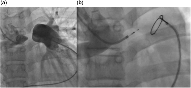
A 12-year-old girl (150 cm, 38.0 kg) with Ebstein’s anomaly, who at the age of 3 years was managed with an additional unidirectional partial cavo-pulmonary anastomosis and surgical ligation of the right pulmonary artery origin 1 week after an unsuccessful tricuspid valvuloplasty. After 9 years, the child was scheduled for cardiac catheterisation due to significant central cyanosis (SaO2 80–85%, Hb 15.0 g/dl). At diagnostic cardiac catheterisation, the arterial blood saturation was measured with 82%. The blood pressure in the right pulmonary artery was in the normal range (mean: 13 mmHg). Angiography showed a wide partial cavo-pulmonary anastomosis without any sign of stenosis. Multiple intrapulmonary fistulas in the right lung were present. In the left pulmonary artery which was perfused antegradely from the right ventricle, the blood pressure was increased (mean: 35 mmHg, pulmonary vascular resistance was 11.3 WUxm2). After testing with O2 and NO supply, the pulmonary vascular resistance decreased to 2.7 WUxm2. Simultaneous contrast injection into superior caval vein and pulmonary trunk showed the ligated origin of the right pulmonary artery and the distance between right pulmonary artery and central pulmonary artery of about 5–6 mm (Fig 1a). Sildenafil therapy (1 mg/kg/24 hour) was initiated due to the reversibly elevated (reactive) pulmonary resistance in the left lung. The decision about treatment with transcutaneous right pulmonary artery re-opening with the use of high-frequency perforation and covered stent implantation after 6 months of Sildenafil therapy was made and suggested to the family.
(a) Contrast was injected simultaneously into SVC and LPA. The ligated distance between RPA and LPA lumen was assessed with about 5–6 mm. (b) Repeated energy applications (under fluoroscopy control) caused slow advancement of the radiofrequency guidewire through the ligated segment. SVC=superior caval vein; LPA=left pulmonary artery; RPA=right pulmonary artery.

The intervention was performed in a spontaneously breathing patient under general anaesthesia. The right jugular vein and the right femoral vein were cannulated both with 6F sheaths. The girl received 4000 IU of heparin and antibiotics intravenously. Simultaneous contrast injection into superior caval vein and left pulmonary artery was repeated in multiple projections to precise the anatomy before intervention.
For recanalisation of the ligated right pulmonary artery segment, the radiofrequency system (Baylis, MedComp, Montreal, Canada), which consists of a Nykanen 0.024-inch radiofrequency guide wire and a coaxial catheter, was chosen. The system was advanced inside a 4F Judkins-left coronary artery guiding catheter (Cordis Corporation, Miami, Florida, United States of America) placed from the jugular access. A 20-mm Amplatz GooseNeck snare (eV3 Endovascular, Plymouth, Minnesota, United States of America) was introduced through a 5F Judkins-right coronary guiding catheter (Cordis Corporation, Miami, Florida, United States of America) from the femoral access into the pulmonary artery. The radiofrequency guide wire headed directly towards the ligated segment of the right pulmonary artery. Repeated energy (up to 15 W for 8–12 seconds) applications under fluoroscopy control caused slow advancement of the radiofrequency guidewire through the ligated segment (Fig 1b) (ligation was performed with Ethibond suture size 1 (Ethicon, Summerville, New Jersey, United States of America)). After entering the left pulmonary artery-lumen, the Baylis catheter was snared from the femoral site. A 14-inch coronary wire was introduced from the jugular access and withdrawn back through the femoral vein. A 1.5-mm coronary balloon was advanced inside the 5F Judkins-right coronary guiding catheter over the coronary wire from the femoral access, and the guiding catheter was next advanced over an inflated coronary balloon into the superior caval vein, passing the ligated segment.
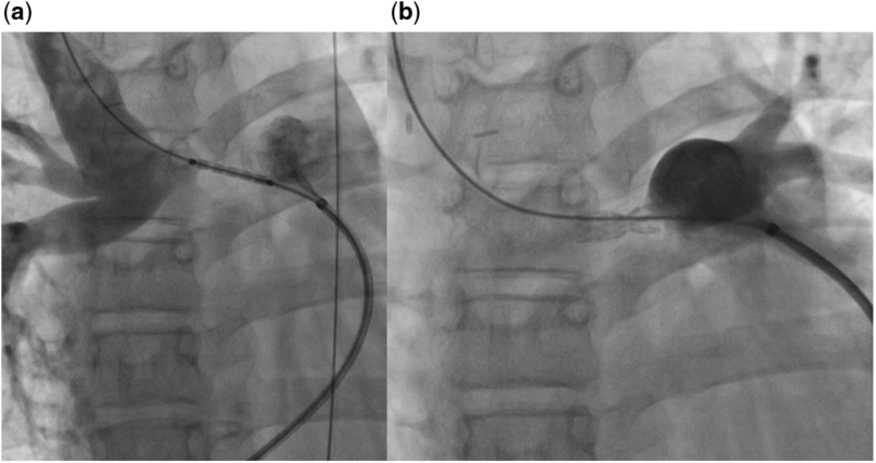
A 0.035-inch Amplatzer Super Stiff guidewire (eV3 Endovascular, Plymouth, Minnesota, United States of America) was introduced from the jugular vein and withdrawn back through the femoral site. A long 6F sheath was introduced through the femoral vein. A 6x16-mm covered LifeStream stent (Bard Peripheral Vascular, Tempe, Arizona, United States of America) was advanced over a 0.035-inch wire and was deployed into the recanalised segment (with 10 atm) (Fig 2a). The narrowest segment of the implanted stent was assessed with 2–3 mm (Fig 2b) which was found optimal due to increased blood pressure in the left pulmonary artery. Control angiography showed no contrast extravasation. The mean pressure in the superior caval vein was 14 mmHg and in the left pulmonary artery it was 37/20/27 mmHg.
(a) A 6x16-mm covered LifeStream stent was advanced into the recanalised segment. (b) Due to increased blood pressure in the LPA, the narrowest segment of the implanted stent was left with the diameter of 2–3 mm. LPA=left pulmonary artery.

After the procedure, the clinical state of the patient has improved significantly. Oxygen saturation 3 months later was measured with 90%. A repeated catheterisation was scheduled 18 months later and the stent was redilated to a diameter of 5 mm (pressures after redilation superior caval vein 1:10846 11 mmHg, left pulmonary artery 32/13/23 mmHg). On angiography, the intrapulmonary shunts had declined significantly.
Discussion
We report on spontaneous closure of arterio-venous fistulas by a successful percutaneous recanalisation of the right pulmonary artery origin after surgical ligation with the use of radiofrequency energy perforation and consecutive covered stent implantation in patient with Ebstein’s anomaly 9 years after a unidirectional Glenn procedure. A few months after the intervention, the arterial blood oxygen saturation has increased to 90% and the girl no longer required oxygen supply. At a repeated catheterisation, 18 months after recanalisation, the stent was redilated to a diameter of 5 mm and only slightly elevated left pulmonary artery pressures were assessed.
The spectrum of clinical manifestation in patients with Ebstein’s anomaly is very wide.Reference Celermajer, Bull and Till2 In some cases, heart failure is diagnosed shortly after birth and these children need surgical or interventional management in the neonatal period to improve pulmonary arterial perfusion. In other patients (like in our patient), the necessity for surgery appears later if any. Today, if surgery is indicated, most patients with Ebstein’s anomaly are managed with a cone operationReference da Silva, Baumgratz and da Fonseca3 at our centre with very good results.Reference Lange, Burri and Eschenbach4
However, in patients in whom tricuspid valve reconstruction results in unacceptable haemodynamics, a cavo-pulmonary shunt, hence a so-called 1 ½ circulation, may be helpful as it was in our patient.Reference Celermajer, Bull and Till2, Reference Sirivella and Gielchinsky5, Reference Kreutzer, Mayorquim and Kreutzer6
Severe intrapulmonary shunts with subsequent increasing arterial cyanosis are a known long-term complication after a classical Glenn operation or a Kawashima procedure, where the hepatic blood does not reach parts of the pulmonary arteries.Reference Hoffman1 The lack of the hypothetical hepatic factor is believed to be responsible for severe arterio-venous fistulas formation which comprises a significant clinical problem. The clinical manifestation of multiple arterio-venous fistulas may include central cyanosis, frequent infections and polycythemia with increased haematocrit value.Reference Papagiannis, Apostolopoulou and Sarris7 Our patient was cyanotic and additionally had increased pressure and pulmonary arterial resistance in the left lung.
Percutaneous right pulmonary artery recanalisation provided redirection of hepatic blood flow into the right lung and this had a positive effect on the fistulas in the right lung. Despite the fact that this intervention was performed in 12-years-old patient even 9 years after Glenn operation, spontaneous closure of arterio-venous fistulas was obtained anyway. The clinical state of the patient improved significantly soon after intervention. Repeated surgery was avoided. Our case report shows that in patients after a unidirectional partial cavo-pulmonary anastomosis and important arterio-venous fistulas in the lung without hepatic blood flow, the attempt of redirection of hepatic blood supply into the affected lung should be made by catheter interventional means or by surgical means, not only in small children but also in older ones who underwent unidirectional Glenn procedure years before. Our case report proves that even these patients may significantly benefit from this approach. In children with functionally univentricular hearts after bidirectional Glenn, diagnosed with significant arterio-venous fistulas, the early completion to total cavo-pulmonary connection proved to be feasible causative treatment.Reference Praus, Eicken and Balling8
The radiofrequency perforation system, which was used in our patient, proved to be safe and effective. It enables percutaneous approaches for recanalisation of ligated or atretic vessels, for example, in patients with aortic isthmus atresiaReference Butera, Heles and Carminati9 or atretic valves.Reference Hausdorf, Schulze-Neick and Lange10 In the future, the indications for the use of radiofrequency in the field of congenital heart diseases can be expanded due to increasing accessibility and experience with this management option.
Conclusion
Redirection of hepatic blood into the pulmonary artery is an important management step in patients who underwent partial cavo-pulmonary connection and developed clinically significant arterio-venous fistulas in the follow-up time. It may cause fistulas to close, and hence improve the clinical state of the patient. Moreover, our case report shows that spontaneous closure of the arterio-venous fistulas may be achieved not only in small but also in older children long after a unidirectional Glenn procedure. In selected patients, an interventional approach is safe and feasible and repeated surgery may be avoided.
Acknowledgements
The authors thank Prof. Gernot Buheitel for the help in preparing the article.
Financial Support
This research received no specific grant from any funding agency, commercial, or not-for-profit sectors.
Conflicts of Interest
None.
Ethical Standards
The authors assert that all procedures contributing to this work comply with the ethical standards of the relevant national guidelines on human experimentation and with the Helsinki Declaration of 1975, as revised in 2008, and has been approved by the institutional committees.