Introduction
Advanced laryngeal carcinoma usually invades the larynx and prelaryngeal strap muscles, and may even involve the cervical skin. Local reconstruction after wide tumour resection is always indicated when advanced laryngeal carcinoma has invaded the overlying skin. Commonly used repair techniques involve the use of a pedicled skin flap (e.g. a pectoralis major myocutaneous flap) or a free skin flap (e.g. an antebrachial flap or an anterolateral thigh flap).Reference Spriano, Pellini and Roselli1, Reference Ninković, Harpf, Gunkel, Schwabegger, Thumfart and Anderl2 However, flap repair cannot be carried out at all surgical facilities because it is technically difficult and requires an experienced surgical team. The risks associated with flap repair include donor site malformation and dysfunction and flap necrosis. A good result was achieved using acellular dermal matrix to simplify the repair of a cervical skin defect in a patient with advanced laryngeal carcinoma that had invaded the skin.
Case report
A 55-year-old man had undergone two operations 17 and 9 months prior to the current procedure. Pathological diagnoses made after these procedures were severe atypical hyperplasia and squamous cell carcinoma, respectively. Five months before the current procedure, the patient's left cervical skin had broken down and the lesion had gradually enlarged. A pathological diagnosis of squamous cell carcinoma was made.
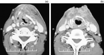
Laryngopharyngeal computed tomography showed that the larynx was abnormally shaped because of a large, irregular soft tissue mass. The mass crossed the anterior commissure and infiltrated the skin. Venous injection of contrast material resulted in clear heterogeneous enhancement of the mass (Figure 1). The mass had destroyed the thyroid cartilage and extended upward to the level of the epiglottis, downward to the trachea, and forward to the cervical skin. No obvious lymphadenectasis was detected in the cervical muscle gap. Colour Doppler ultrasound examination revealed no lymph node enlargement in the cervical region and no tumour metastasis to the sternum and abdomen. The pre-operative diagnosis was recurrent laryngeal carcinoma with invasion into the surrounding tissues and a tumour–node–metastasis classification of T4N0M0. The patient consented to undergo treatment and surgical repair with a single-use acellular dermal matrix, and the ethics committee of our hospital approved the procedure.
Computed tomography scans showing the recurrent laryngeal carcinoma invading the cervical soft tissue and skin.

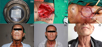
An acellular dermal matrix membrane with a standard size of 6 × 8 cm was used for surgery (Zhenghai Biological Technology, Yantai, China): one side of the membrane is rough (basement membrane side) and the other is smooth (dermal side). The membrane was first soaked and rinsed three times with physiological saline solution before use. Tracheostomy was performed under local anaesthesia, and general anaesthesia was subsequently induced. An oval incision with a 1-cm border was made around the broken down skin, the subcutaneous tissue was opened and the platysma muscle was elevated. The invaded prelaryngeal strap muscles and skin in the middle of the anterior neck were then exposed. Because the tumour stage was advanced and the associated skin defect was large (6 × 4 cm), total laryngotomy and T-type layered suturing of the fistula orifices were performed. The acellular dermal matrix patch was cut to match the area of the defect and sutured to the surrounding skin (Figure 2). Several small holes were made in the membrane to facilitate drainage.
Sequential photographs showing intra-operative repair of an anterior cervical defect with an acellular dermal matrix and the post-operative follow up. (a) Acellular dermal matrix. (b) Neck tissue defect after laryngeal cancer (tumour–node–metastasis staging T4N0M0) resection. (c) Repair of the cervical anterior defect with acellular matrix. (d–f) Cosmetic outcome at 10 days (d), 3 months (e) and 3 years (f) after surgery. M = mandible; NGT = nasogastric tube; PHPW = posterior hypopharyngeal wall; TI = tracheal intubation

Antibiotics were administered for 7 days post-operatively to prevent infection. Negative pressure drainage tubes were placed in the operative site, and a pressure dressing was applied for 7 days. Nasogastric feeding was initially provided: at post-operative day 10, oral feeding became possible and the sutures and nasogastric tube were then removed.
At post-operative day 10, the biological patch and underlying tissue had healed well; no effusion, infection, pharyngeal fistula or dysphagia had developed. Three months after surgery, the cervical wound had completely healed, and no infection, pharyngeal fistula or dysphagia had developed. At the three year follow up, scar tissue was present on the patient's neck but his swallowing function was normal. No local tumour recurrence or metastasis has developed (Figure 2).
Discussion
Acellular dermal matrix is a natural extracellular matrix with a very low risk of immunological rejection; it can be either allogeneic or xenogeneic, depending on its source. Its use and clinical outcomes are well documented.Reference Sbitany and Serletti3–Reference Symbas, McCord and Nahai5 Repair of the skin defect following wide resection of an advanced laryngeal carcinoma always necessitates flap reconstruction. However, there are no reports describing the repair of such skin defects with acellular dermal matrix. The biological membrane used in the present case was bovine skin tissue that had been processed using bioengineering techniques to create an acellular tissue. Removal of cellular components decreases the incidence of host rejection, while preserving the extracellular matrix. Pathogens such as bacteria, viruses and the prion that causes mad cow disease are also removed. The membrane has a three-dimensional structure comprising collagen fibres, with a smooth dermal surface and wide pores. It therefore provides a good environment for host cell migration and growth to facilitate rapid vascularisation and epithelialisation.
In this patient, scar tissue present in the neck at the three-year follow up affected the cosmetic result. However, the patient's swallowing function was normal, and neither local tumour recurrence nor metastasis had developed. This treatment outcome indicates that cervical skin defects can be successfully repaired with acellular dermal matrix. Nevertheless, a few points about the surgical repair of skin defects with acellular dermal matrix should be noted. Firstly, reliable pharyngeal fistula suture techniques are required for successful repair outcomes. In this case, a three-layer T-type suture technique was adopted.Reference Silver, Ferlito and Thomas6 The first layer comprised an inverting mucosal suture pattern, the second comprised a submucosal reinforcement suture pattern and the third involved suturing of the inferior constrictor muscle of the pharynx. If a post-operative pharyngeal fistula develops, the transplanted acellular dermal matrix drops off. Secondly, complete drainage is important. In this patient, two negative pressure drainage tubes were placed at the surgical site, and several small holes were created to facilitate drainage. Thirdly, application of a local pressure dressing is necessary to ensure close adhesion of the acellular dermal matrix to the underlying muscle layer.
• Wide resection is necessary to treat an advanced laryngeal carcinoma that has invaded the skin
• The resulting cervical skin defect is commonly repaired using a pedicled or free skin flap
• Acellular dermal matrix is a natural alternative with a low rejection rate
• Use of this matrix reduces the risks associated with flap repair
This is the first reported use of xenogeneic acellular dermal matrix to repair a cervical skin defect in a patient with advanced laryngeal carcinoma. This surgical method has the following advantages: the acellular dermal matrix lacks epidermal cells that induce host rejection; it has dermal components with no cytotoxicity, good biocompatibility, and a low risk of immune rejection; it can be obtained from a wide range of natural sources; it exhibits good mechanical performance with flexibility, with a soft texture, and can be tailored and sutured; the creation of a donor site wound is avoided; and pain relief is provided. Moreover, this repair technique is simpler than flap reconstruction and can be carried out in most hospitals.
Conclusion
Repair of cervical skin defects using this simplified method can be performed at every medical unit. This procedure reduces the risks associated with flap repair techniques such as donor site malformation and dysfunction and flap necrosis.