Introduction
The differentiation and maturation of ovarian follicles in vertebrates requires multiple coordinated responses of follicle cells and oocytes to appropriate stimuli. One way to achieve tighter cellular control is the regulation of the direct exchange of ions, metabolites and other messenger molecules through the intercellular channels clustered at the gap junctions (Kumar & Gilula, Reference Kumar and Gilula1996; Wei et al., Reference Wei, Xu and Lo2004). These intercellular channels are made up of connexins (Cx), a highly related family of proteins that allow the direct exchange of substances smaller than 1200 Da between adjacent cells (Goodenough et al., Reference Goodenough, Goliger and Paul1996). Homologous (granulosa cell–granulosa cell) and heterologous (granulosa cell–oocyte) gap junctions have been observed in the ovarian follicles of different vertebrate species (Bolamba et al., Reference Bolamba, Patiño, Yoshizaki and Thomas2003).
The participation of gap junctions in the physiology of the ovarian follicles of vertebrates, especially in the maturation process, is supported by numerous studies such as those of Buccione et al., Reference Buccione, Schroeder and Eppig1990, Cerdá et al., 1993, Yoshizaki et al., Reference Yoshizaki, Patiño and Thomas1994, Villecco et al., Reference Villecco, Aybar, Sanchez and Sanchez Riera1996, Villecco et al., Reference Villecco, Aybar, Genta, Sanchez and Sanchez Riera2000 and Carabatsos et al., Reference Carabatsos, Sellitto, Goodenough and Albertini2000. However, there is little information concerning the role of gap junctions in the vitellogenic process. At this stage of oogenesis, the major event is the accumulation of nutrients in the form of yolk proteins inside the oocyte until fertilization and embryogenesis occur (Wallace, Reference Wallace1985). The liver vitellogenin (VTG) synthesis and its hormonal requirements have been well elucidated in Xenopus laevis (Wallace & Dumont, Reference Wallace and Dumont1968; Ho, Reference Ho1987; Varriale et al., Reference Varriale, Pierantoni, Di Matteo, Minucci, Milone and Chieffi1988.). It is known that FSH-like gonadotropins promote vitellogenesis through the estradiol synthesized by follicle cells (Redshaw, Reference Redshaw1972). Gonadotropins also promote the uptake of yolk proteins by vitellogenic oocytes, although this process does not seem to be mediated by estradiol-17β (Wallace and Bergink, Reference Wallace and Bergink1974).
Xenopus laevis oogenesis is a continuous, asynchronous process and oocytes at all stages of development are present in the ovary at all times during adult life (Dumont, Reference Dumont1972). Thus, oocytes at different stages of development are exposed to the same hormonal environment, although only vitellogenic follicles are actively involved in VTG endocytosis. This fact would indicate that the stimulation for oocytes to enter active VTG uptake could be developmentally regulated in some manner and would be mediated by follicle cells through an unknown mechanism.
Moreover, vitellogenic oocytes cultured in vitro without follicle cells grow indefinitely at a rate that is strictly dependent on the external concentration of hepatic vitellogenin added to the medium (Wallace et al., Reference Wallace, Misulovin and Wiley1980, Reference Wallace, Misulovin and Etkin1981). These authors suggested that the follicle cells investing fully grown oocytes might serve to prevent vitellogenin and nutrient access to full-grown oocytes in vivo. This fact supports the idea that follicle cells may be involved in the normal vitellogenic process.
A possibility to be considered is whether the acquisition of endocytic competence could be transferred to the oocyte from the overlying follicle cell epithelium via gap junction contacts. In this context, Waksmonski & Woodruff (Reference Waksmonski and Woodruff2002) demonstrated that epithelial cell–oocyte gap junctional communication is required in six different orders of insects for the uptake of yolk precursors. This mechanism could represent an evolutionary conserved manner of regulating the endocytic competence of follicles.
Landesman et al. (Reference Kumar and Gilula2003) explored the role of gap junction intercellular communication during the development of X. laevis. The analysis of embryonic cDNA revealed maternal expression of two novel connexins, Cx31 and Cx43.4. Thus, the early Xenopus embryo contains at least four maternal connexins: Cx31, Cx38, Cx43 and Cx43.4 (Landesman et al., Reference Landesman, Postma, Goodenough and Paul2003). Moreover, de Boer & van der Heyden (Reference De Boer and Van der Heyden2005) suggested their participation in the development of premidblastula transition (MBT). However, there is no detailed information about the temporal expression pattern of connexins during oogenesis.
In this context, the aim of the present study was to investigate the physiological role and the expression pattern of heterologous gap junctions during X. laevis vitellogenesis.
Materials and methods
Biological material
Adult female X. laevis specimens were kept in dechlorinated fresh water tanks at 18–20 °C on a 12 h light/dark cycle and fed twice weekly with chopped heart meat.
Frogs were anaesthetized on ice and ovarian lobes from human chorionic gonadotropin (HCG – Elea) stimulated females (500 IU) were removed via mini-laparotomy. After the incision was sutured, the animals were allowed to recover at room temperature. The ovarian lobes were dissociated by 0.15% collagenase I (Sigma) in O-R2 solution at 26 °C for 20–60 min. This procedure freed many individual follicles from the ovarian pieces, but did not disrupt the association between the oocytes and their surrounding follicle cells (Fortune, Reference Fortune1983).
The vitellogenic follicles (stages III–IV and V; Dumont, Reference Dumont1972) were dissected manually with forceps and incubated in an O-R2 sterile solution.
Preparation of vitellogenin-containing serum (VTGcS)
For the study of receptor-mediated oocyte endocytic activity it is essential that the receptor ligands (VTG) be present in the incubation medium. We initiated vitellogenin accumulation in the blood by injecting X. laevis females with a dose of estrogen (4 mg estradiol-17β dissolved in 0.4 ml propylene glycol per 100 g body weight). After 2–3 weeks, estrogen-treated animals were anaesthetized and bled exhaustively. The serum contained approximately 100–150 mg vitellogenin/ml (Wallace et al., Reference Wallace, Misulovin and Wiley1980).
Dye transfer experiments
Oocytes were microinjected with 20–40 nl of 1% aqueous solution of the fluorescent dye Lucifer yellow CH MW: 457.2 (Emax λ 535 nm – Molecular Probes). To evaluate dye transfer to follicle cells, oocytes were manually defolliculated 2 h after microinjection with watchmaker forceps and observed with a Zeiss fluorescence microscope.
Downregulation of gap junctions
To block the passage of molecules ≥440 Da, follicles were incubated in octanol dissolved in dimethyl sulphoxide (DMSO) and diluted to 1 mM with sterile O-R2 (final concentration of DMSO < 1 %). It should be noted that 1 mM octanol has been often used to disrupt dye coupling between oocytes and their surrounding epithelial cells (Patiño & Purkiss, Reference Patiño and Purkiss1993; Cerdá et al., 1993).
Endocytic tracers
To detect the pathway of vitellogenin endocytosis, the following tracers were used: biotinylated bovine serum albumin (b-BSA): BSA (Sigma) was allowed to react with biotinyl-ϵ-aminocaproic acid N–hydroxy-succinimide ester (BNHS – Sigma) by addition of 10 μl of a 20 mg/ml solution (in dimethylformamide) for each mg of albumin present in the dialysis bag. After 1 h at room temperature, the reaction mixture was dialyzed at 4 °C overnight against an O-R2 solution. The biotinylated protein was stored at −20 °C until used. Fluorescein dextran (FDX): 7 mg/ml (Mr: 40000; Molecular Probes) was prepared in an O-R2 solution.
Tracer incorporation by oocytes
Control groups (coupled gap junctions): 60 vitellogenic follicles (III, IV and V stages) were incubated in 500 μl of O-R2 solution with 50 μg/ml gentamycin and 50 IU/ml of nystatin for 3 h in a humidified temperature-controlled incubator (22 °C). After this incubation period, 40 μl of VTGcS plus 100 μl of b-BSA (1 mg/ml) were added to 30 follicles and 40 μl of VTGcS plus 50 μl of FDX (7 mg/ml) to the remaining 30.
Experimental groups (downregulated gap junctions): 60 vitellogenic follicles (III, IV and V stages) were incubated in 500 μl of O-R2 solution with 50 μg/ml gentamycin, 50 IU/ml of nystatin and 1 mM octanol for 3 h in a humidified temperature-controlled incubator (22 °C). After this incubation period, 40 μl of VTGcS plus 100 μl of b-BSA (1 mg/ml) were added to 30 follicles and 40 μl of VTGcS plus 50 μl of FDX (7 mg/ml) to the remaining 30. Both experimental and control groups were incubated for 48 h at 22 °C.
Histological procedures and immunohistochemistry
After incubation with b-BSA, follicles were fixed in Ancel & Vintemberger solution (Ancel & Vintemberger, Reference Ancel and Vintemberger1948), dehydrated and embedded in paraffin–celloidin according to Manes & Nieto (Reference Manes and Nieto1983). Sections 10–12 μm thick, serially obtained from blocks, were attached to slides.
The b-BSA was determined by incubation with complex ExtrAvidin–peroxidase conjugate (Sigma). The sections were deparaffined, rinsed with PBS (pH 7.4) and incubated with bleaching solution (1% H2O2) for 24 h to allow the development of stain for easy visualization. Then, they were rinsed with PBS (pH 7.4), incubated with 0.3% H2O2 in methanol for 30 min to inactivate the endogenous peroxidase and treated with 0.1% trypsin (Merck) for 10 min at room temperature to unmask antigenic sites. Slides were incubated with 3% BSA–PBS for 1 h at room temperature to prevent non-specific background staining. After blocking they were treated with a 1:500 dilution of ExtrAvidin–peroxidase conjugate for 2 h at room temperature. Peroxidase activity was detected by incubation with 3,3′-diaminobenzidine–H2O2 at room temperature for 10 min. The reaction was stopped by rinsing the samples with distilled water and slices were mounted and observed with a Nikon microscope.
After incubation with FDX, follicles were fixed in MEMFA (0.1 M MOPS, pH 7.4; 2 mM EGTA, 1 mM MgSO4, 3.7% formaldehyde) for 1 h. Then they were transferred to 100% methanol and washed twice for 5 min, an apparently essential procedure for optimal penetration of the reagents into the follicles. Then follicles were rehydrated and blocked for 2 h with 2% Roche blocking reagent at room temperature before overnight incubation at 4 °C with alkaline phosphatase conjugated antifluorescein Fab fragments (Roche Biochemicals). Alkaline phosphatase activity was detected by incubation with NBT/BCIP (Roche Biochemicals) as a substrate. After staining in whole mount, follicles were fixed in Ancel & Vintemberger solution (Ancel & Vintemberger, Reference Ancel and Vintemberger1948), dehydrated and embedded in paraffin–celloidin according to Manes & Nieto (Reference Manes and Nieto1983). Sections 10–12 μm thick, serially obtained from blocks, were attached to slides and observed with a Nikon microscope.
RNA isolation and RT-PCR analysis
Total RNA was isolated from follicles by the guanidine thiocyanate/phenol/chloroform method (Chomczynski & Sacchi, Reference Chomczynski and Sacchi1987) and cDNAs were synthesized using AMV reverse transcriptase (Roche Biochemicals) and oligo(dT) primer. The primers sequences and PCR conditions were obtained from Landesman et al. (Reference Kumar and Gilula2003). The PCR products were analysed on a 1.5% agarose gel. As a control, PCR was performed with RNA that had not been reverse transcribed in order to check for DNA contamination.
Data analysis
Experimental data are presented as the mean ± SD of the number of experiments indicated in the legends. Statistical analysis of value was performed by Student's t-test with p < 0.05 considered statistically significant.
Results
Vitellogenic oocyte–follicle cell interaction
In order to examine follicle cell–vitellogenic oocyte coupling through gap junctions transfer fluorescent dye experiments were performed.
The fluorescent dye lucifer yellow CH is a widely used to determine gap junction functionality, since it can pass freely through gap junctions but does not readily permeate non-junctional membranes as long as the cell is healthy. On the other hand, incubation with 1 mM octanol is an efficient method for downregulating gap junctional communications in ovarian follicles.
Lucifer yellow CH was microinjected through the follicular layer into 40–50 vitellogenic oocytes in the presence or absence of 1 mM octanol. The injected dye was allowed to diffuse through the ooplasm for 2 h, after which abnormal or damaged follicles (generally fewer than 10%) were discarded. Healthy follicles were then dissected and follicular layers were observed with a fluorescence microscope.
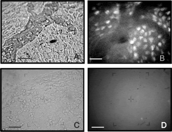
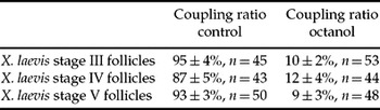
Results showed that follicle cells from vitellogenic follicles incubated without 1 mM octanol fluoresce after dye injection (Fig. 1A, B). In contrast, follicle cells from vitellogenic follicles treated with 1 mM octanol did not show fluorescence. (Fig. 1C, D). The coupling ratio in control follicles of vitellogenic oocytes (stages III, IV and V) averaged 91.7%, which was reduced by octanol to 10.3% (Table 1).
Dye transfer from vitellogenic oocytes to follicle cells in untreated follicles (A, B) and 1 mM octanol treated follicles (C, D). A and C, images of follicle cell epithelia examined for phase contrast; B and D, the same preparations examined for fluorescence. After injection of lucifer yellow into vitellogenic oocytes, dye transfer to the overlying follicle cells can be observed in untreated follicles (B) but not in octanol treated follicles (D). Bars, 45 μm.

Octanol-induced changes in coupling ratios in vitellogenic oocytes

Values are the means ± SD from five different experiments. Each experiment was performed on a different animal.
These results show that during the vitellogenic process there are functional gap junctions at the oocyte/follicle cells interface and that octanol uncouples this intercellular communication.
Dependence of vitellogenesis upon heterologous gap junctions coupling
With the observation of a marked dye coupling between epithelial cells and oocyte during endocytic uptake of VTG, the hypothesis emerges that the gap junctions might provide a route by which a signal that regulates vitellogenesis would be delivered to the oocyte.
We tested this hypothesis by monitoring the formation of nascent yolk platelets during the in vitro incubation of vitellogenic follicles (stages III, IV and V) in O-R2 solution/VTGcS (coupled gap junctions) and in a medium in which gap junctions had been downregulated (O-R2 solution/VTGcS/octanol). In order to label nascent yolk platelets, b-BSA and FDX were added to these culture media (see Materials and methods). Non-specific fluid-phase labeling was a convenient way to follow uptake of yolk precursor proteins.
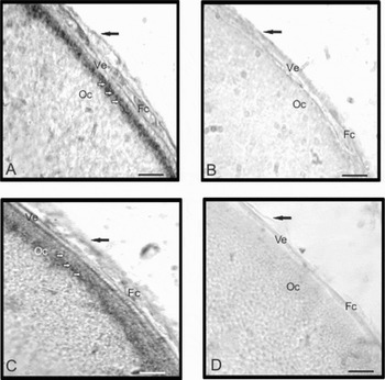
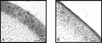
Fixed and sectioned untreated control follicles developed a stratum of newly formed yolk platelets (Fig. 2A, C; Fig. 3A) in the oocyte cortex, while in octanol-treated follicles no sign of nascent yolk sphere formation was observed (Fig. 2B, D; Fig. 3B). The same results were obtained with both endocytic tracers. Examination of previtellogenic oocytes revealed no marked nascent yolk platelets (data not shown).
Effect of gap junction uncoupling on VTG uptake in vitellogenic oocytes. Stage III (A, B) and stage V oocytes (C, D) were incubated in O-R2 solution/VTCcS/b-BSA (A, C) and in O-R2 solution/VTCcS/b-BSA/1 mM octanol (B, D). Light micrographs show newly marked yolk platelets (A, C) corresponding to oocytes with coupled gap junctions; no marked nascent yolk platelets (B, D) corresponding to oocytes with uncoupled gap junctions can be observed. Bars, 25 μm. Fc, follicular cell; Oc, oocyte cortex; Ve, vitelline envelope. Black arrows, theca; white arrows, marked yolk platelets.

Effect of gap junction uncoupling on VTG uptake in vitellogenic oocytes. Stage IV (A, B) oocytes were incubated in O-R2 solution/VTCcS/FDX (A) and in O-R2 solution/VTCcS/FDX/1 mM octanol (B). Light micrographs show newly marked yolk platelets in the cortex (A) corresponding to oocytes with coupled gap junctions; no marked nascent yolk platelets (B) corresponding to oocytes with uncoupled gap junctions can be observed. Bars, 15 μm. Oc, oocyte cortex.

Thus, follicles in which gap junctional communication between epithelial cells and oocyte had been disrupted by octanol ceased vitellogenic uptake of the yolk precursor.
Connexin differential expression during oogenesis
In order to determine what connexin/s are involved in a vitellogenesis, we analysed the temporal expression pattern of three maternal connexins during oogenesis.
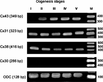
Expression of Cx43, Cx31, Cx38 and zygotic Cx30 were examined by RT-PCR (Fig. 4). Results showed that the mRNA transcripts of Cx31 and Cx38 were present without changes at all oogenesis stages studied. As previously reported (Landesman et al., Reference Landesman, Postma, Goodenough and Paul2003), no maternal expression of Cx30 was observed. On the other hand, Cx43 transcript levels increased from stage II onwards. The major expression of this connexin during the vitellogenic stages suggests a probable role in the VTG endocytic uptake.
Connexin temporal expression patterns. RT-PCR was performed on material from X. laevis oocytes (stages I–V). Cx43 expression was first evident at stage II of oogenesis and increased during the subsequent vitellogenic stages (III, IV and V). No expression changes were detected for Cx31 and Cx38. Maternal expression of Cx30 was not observed. Ornithine decarboxylase (ODC) was used as an internal control, M; ladder 100 bp.

Discussion
In the present paper we showed that a gap junctional intercellular communication plays an important role in the acquisition of endocytic competence by X. laevis oocytes. We also proposed that the connexin Cx43 would form the gap junctions through which the epithelial cell signal would regulate yolk uptake in X. laevis oocytes.
The transformation of oogonia into oocytes is commonly described as oogenesis. In many oviparous vertebrates the growth of oocytes from microscopic to macroscopic dimensions is mainly the result of one of the most exciting examples of cell regulation, namely vitellogenesis. This process, common to all oviparous vertebrate species, is characterized by hepatic production of a glycoprotein, vitellogenin (VTG), under multihormonal control and its transportation via the bloodstream to the ovary, where it enters the oocytes by receptor-mediated endocytosis (Polzonetti-Magni et al., Reference Polzonetti-Magni, Mosconi, Soverchia, Kikuyama and Carnevali2004).
Yolk formation is the oldest and most common feature by which the mother supplies the zygote for embryonic development in most reproductive strategies, with only few exceptions among invertebrates and only Eutheria among vertebrates (Polzonetti-Magni et al., Reference Polzonetti-Magni, Mosconi, Soverchia, Kikuyama and Carnevali2004). On the other hand, ovarian characteristics in lower vertebrates are consistent with oocyte development regulated by hormones through both long-loop feedback and local control mechanisms, in which follicle cells would participate through the gap junctions (Pierantoni et al., Reference Pierantoni, Cobellis, Meccariello and Fasano2002; Fernández & Ramos, Reference Fernández, Ramos and Jamieson2003; Sánchez & Villecco, Reference Sánchez and Villecco2003).
Results from the present study have shown that intercellular communication via gap junctions normally exists between the vitellogenic oocyte and follicle cells in X. laevis ovary. Moreover, X. laevis oocytes, early in their development, establish adhesive contacts and communicate via gap junctions with an investing layer of follicle cells (Browne & Werner, Reference Browne and Werner1984). This was also observed in other amphibians such as Bufo arenarum and Ceratophrys cranwelli, where the analysis of the oocyte–follicle cell interface during oogenesis revealed the development of a complex cell–cell interaction that changes throughout the various oogenetic periods (Sánchez & Villecco, Reference Sánchez and Villecco2003). This observation is in agreement with those reports for other vertebrate follicles that have previously demonstrated metabolic, fluorescent dye and electrical coupling between the oocyte and the investing granulosa cells during maturation (Racowski & Satterlie, 1985; Cerdá et al., 1993; Villecco et al., Reference Villecco, Aybar, Genta, Sanchez and Sanchez Riera2000).
In addition, gap junctional integrity in X. laevis follicles is sensitive to widely used uncouplers of intercellular communication such as long-chain alcohols. We have shown that the presence of octanol produces an inhibition of intercellular communication between oocyte and follicle cells, as revealed by the blockage of dye transfer in 90% of the vitellogenic follicles observed, whichever the stage under consideration (III, IV or V). Octanol has been reported to be a highly effective and specific antagonist of gap junctions in vertebrates (Chu & Treistman, Reference Chu and Treistman1997; Sretarugsa & Wallace, Reference Sretarugsa and Wallace1997) and insects (Woodruff & Tilney, Reference Woodruff and Tilney1998; Adler & Woodruff, Reference Adler and Woodruff2000).
Experiments in which gap junctions were downregulated with octanol show that coupled gap junctions are required for endocytic activity. The examination of octanol-treated vitellogenic oocytes failed to reveal the presence of labelled yolk spheres. Thus, the evidence presented in this report demonstrates that a part of the pathway regulating endocytic activity probably includes the transfer of a diffusible signal from the epithelial cells to the oocyte. These results strongly suggest that a signal molecule small enough to pass through open gap junctions would be involved at least at the onset of vitellogenesis. In the amphibian X. laevis, the ovary simultaneously contain follicles at all developmental stages (Dumont, Reference Dumont1972) that are under the same hormonal environment, so that the signal regulating VTG uptake cannot occur on a global scale but must rather be limited to each individual follicle as a vitellogenesis local control mechanism.
It has long been recognized in vertebrates that gap junctionally transmitted signals from the epithelial (granulosa) cells are involved in the regulation of oogenesis, especially during maturation (Buccione et al., Reference Buccione, Schroeder and Eppig1990; Carabatsos et al., Reference Carabatsos, Sellitto, Goodenough and Albertini2000; Villecco et al., Reference Villecco, Aybar, Genta, Sanchez and Sanchez Riera2000; Kidder & Mhawi, Reference Kidder and Mhawi2002, Bolamba et al., Reference Bolamba, Patiño, Yoshizaki and Thomas2003), but their functions in amphibian vitellogenesis have not been explored. In this paper we report the first direct experimental evidence showing that gap junctions are involved in the regulation of amphibian VTG uptake. Anderson & Woodruff (Reference Anderson and Woodruff2001) showed that a gap junctionally transmitted epithelial cell signal regulates endocytic yolk uptake in the insect Oncopeltus fasciatus, while Brooks & Woodruff (Reference Brooks and Woodruff2004) reported that 17–19 kDa protein calmodulin is involved in the process.
Gap junctional communication is regulated at different levels: transcription, translation, intracellular trafficking, oligomerization, docking and gating (Segretain & Falk, Reference Segretain and Falk2004; Teunissen & Bierhuizen, Reference Teunissen and Bierhuizen2004). Virtually nothing has been described for the latter four mechanisms with respect to Xenopus connexins (De Boer & Van der Heyden, Reference De Boer and Van der Heyden2005). In this sense, only the promoter of Cx43 has been cloned and studied (Van der Heyden et al., Reference Van Der Heyden, Roeleveld, Reneman, Peterson and Destree2001). Translational control provides an excellent mechanism for fast adaptation to rapidly changing demands, as the mRNA is already present (De Boer & Van der Heyden, Reference De Boer and Van der Heyden2005). In this context, we focused our attention on the study of the connexin mRNA temporal expression patterns during X. laevis oogenesis.
Results showed that the expression of Cx43 was first evident at stage II of oogenesis and increased during the subsequent vitellogenic stages (III, IV and V), which could indicate that this Cx is related to the process that regulates yolk accumulation. No expression changes were detected for Cx31 and Cx38 connexins.
To date, 20 and 21 different connexin isoforms have been described in the mouse and human genome, respectively (Willecke et al., Reference Willecke, Eiberger, Degen, Eckardt, Romualdi, Guldenagel, Deutsch and Sohl2002; Sohl & Willecke, Reference Sohl and Willecke2003). In the amphibian X. laevis only seven Cxs have been described, i.e. XlCx30, XlCx31, XlCx38, XlCx40.4, XlCx41, XlCx43 and XlCx43.4 (Gimlich et al., Reference Gimlich, Kumar and Gilula1988, Reference Gimlich, Kumar and Gilula1990; Ebihara et al., Reference Ebihara, Beyer, Swenson, Paul and Goodenough1989; Yoshizaki & Patiño, Reference Yoshizaki and Patiño1995; Landesman et al., Reference Landesman, Postma, Goodenough and Paul2003; De Boer et al., Reference De Boer, Kok, Neuteboom, Spieker, De Graaf, Destree, Rook, Van Veen, Jongsma, Vos, De Bakker and Van Der Heyden2005) and their temporal, developmental pattern and adult tissue distribution were summarized by De Boer & Van der Heyden (Reference De Boer and Van der Heyden2005). However, those analyses are far from complete for many of the Cxs, so that all studies in this connexion are valuable.
In summary, we have demonstrated that direct gap junctional communication is a requirement for endocytic activity since without the appropriate signal from surrounding epithelial cells X. laevis oocytes were unable to endocytose VTG.
Acknowledgements
This research was supported by CONICET and CIUNT grants to S.S.S. We wish to thank Mrs Virginia Méndez for proofreading and Mr Marco Gutiérrez for excellent technical assistance.