INTRODUCTION
Aging is associated with reductions in brain volume and cognitive decline that occur along a spectrum that ranges from “normal aging” to neurodegenerative disease and associated dementia (Armstrong et al., Reference Armstrong, An, Shin, Williams, Doshi, Erus and Resnick2020; Kapasi et al., Reference Kapasi, Yu, Boyle, Barnes, Bennett and Schneider2020). Both neuronal and astroglial pathophysiology have been linked to these changes, including Alzheimer’s disease (AD) (Carter et al., Reference Carter, Herholz, Rosa-Neto, Pellerin, Nordberg and Zimmer2019). Blood-based biomarkers are emerging as readily obtainable non-invasive tools to measure several components of highly dynamic and multifactorial age- and neurodegeneration-associated processes. Neurofilament light (NfL) chain, a sensitive maker of neuronal injury, is consistently elevated in blood as a function of older age and across a range of neurologic conditions like AD and other neurodegenerative diseases (Gaetani et al., Reference Gaetani, Blennow, Calabresi, Di Filippo, Parnetti and Zetterberg2019; Wang et al., Reference Wang, Chen, Xu, Li, Hou, Ou and Tan2019; Zetterberg, Reference Zetterberg2016). Plasma glial fibrillary acidic protein (GFAP) reflects astroglial pathophysiology (Yang & Wang, Reference Yang and Wang2015) and is increasingly recognized as a marker of glial dysfunction in age-associated brain changes (Carter et al., Reference Carter, Herholz, Rosa-Neto, Pellerin, Nordberg and Zimmer2019; Nolan et al., Reference Nolan, De Paula Franca Resende, Petersen, Neylan, Spina, Huang and Grinberg2019).
There is comparatively much more research on plasma NfL than plasma GFAP in aging and neurodegenerative disease populations. Studies show that higher plasma concentrations of NfL are associated with lower gray matter volume, faster gray matter atrophy, and worse cognition (Hu et al., Reference Hu, Chen, Ou, Cao, Chen, Cui and Yu2019; Mattsson, Andreasson, Zetterberg, & Blennow, Reference Mattsson, Andreasson, Zetterberg and Blennow2017; Rajan et al., Reference Rajan, Aggarwal, McAninch, Weuve, Barnes, Wilson and Evans2020). Few studies directly report NfL associations with white matter structural volume (Nyberg et al., Reference Nyberg, Lundquist, Nordin Adolfsson, Andersson, Zetterberg, Blennow and Adolfsson2020), though data suggest older adults with higher blood-based NfL have greater white matter hyperintensity burden (Mattsson et al., Reference Mattsson, Andreasson, Zetterberg and Blennow2017; Nyberg et al., Reference Nyberg, Lundquist, Nordin Adolfsson, Andersson, Zetterberg, Blennow and Adolfsson2020; Sudre et al., Reference Sudre, Bocchetta, Heller, Convery, Neason, Moore and Rohrer2019; Sun et al., Reference Sun, Tan, Xu, Wang, Hu, Li and Yu2020) and worse white matter microstructural integrity (Moore et al., Reference Moore, Hohman, Badami, Pechman, Osborn, Acosta and Jefferson2018; Schultz et al., Reference Schultz, Strain, Adedokun, Wang, Preische, Kuhle and Gordon2020). High plasma GFAP shows similar associations with lower cortical volume, worse cognition (Bettcher et al., Reference Bettcher, Olson, Carlson, McConnell, Boyd, Adame and Thaker2021; Oeckl et al., Reference Oeckl, Halbgebauer, Anderl Straub, Steinacker, Huss, Neugebauer and Kornhuber2019; Rajan et al., Reference Rajan, Aggarwal, McAninch, Weuve, Barnes, Wilson and Evans2020; Verberk et al., Reference Verberk, Thijssen, Koelewijn, Mauroo, Vanbrabant, de Wilde and Teunissen2020), greater white matter hyperintensity burden (Elahi et al., Reference Elahi, Casaletto, La Joie, Walters, Harvey, Wolf and Cobigo2019; Sudre et al., Reference Sudre, Bocchetta, Heller, Convery, Neason, Moore and Rohrer2019), and worse white matter tract integrity (Bettcher et al., Reference Bettcher, Olson, Carlson, McConnell, Boyd, Adame and Thaker2021). A few studies support that GFAP is more sensitive to early brain changes in AD (e.g., cortical amyloid burden (Asken et al., Reference Asken, Elahi, La Joie, Strom, Staffaroni, Lindbergh and Casaletto2020; Verberk et al., Reference Verberk, Thijssen, Koelewijn, Mauroo, Vanbrabant, de Wilde and Teunissen2020)), is more strongly associated with cognitive function (Verberk et al., Reference Verberk, Thijssen, Koelewijn, Mauroo, Vanbrabant, de Wilde and Teunissen2020), and has a similar or better relationship with longitudinal brain atrophy and cognitive decline (Rajan et al., Reference Rajan, Aggarwal, McAninch, Weuve, Barnes, Wilson and Evans2020) compared to NfL. Few studies have investigated brain structure and cognitive function correlates of NfL and GFAP simultaneously (Bettcher et al., Reference Bettcher, Olson, Carlson, McConnell, Boyd, Adame and Thaker2021), so additional characterization of how plasma GFAP relates to brain structure and cognition in older adults is needed.
Here, we compare plasma GFAP to plasma NfL on their relationships to brain volume and cognition when measured simultaneously. We examined associations between plasma GFAP, plasma NfL, brain volume, and cognitive function in two independent cohorts of older adults across the typical aging-to-Alzheimer’s dementia spectrum. As independent samples, we aimed to increase the rigor of our clinical study by testing the consistency of the evaluated relationships. We investigated both gray and white matter volumes and multiple components of cognition including memory, executive function, language, and visuospatial abilities. Based on prior studies of these proteins, plasma NfL and plasma GFAP are expected to reflect axonal degeneration and astrocytic changes, respectively, in aging and neurodegeneration. We therefore hypothesized that GFAP and NfL would show independent associations with brain volume and cognitive function.
METHODS
Study Participants
We cross-sectionally sampled participants at the UCSF Memory and Aging Center from larger ongoing studies of typical aging and AD. All participants provided informed consent prior to enrollment in UCSF IRB-01 approved projects, and the research was completed in accordance with the Helsinki Declaration. Participants received diagnoses via multidisciplinary consensus conference as clinically normal (CN), mild cognitive impairment (MCI) (Albert et al., Reference Albert, DeKosky, Dickson, Dubois, Feldman, Fox and Phelps2011), or dementia (McKhann et al., Reference McKhann, Knopman, Chertkow, Hyman, Jack, Kawas and Phelps2011) based on neurologic exam, cognitive testing, and Clinical Dementia Rating (CDR), which is a clinical measure of disease severity. All participants with MCI or dementia had at least intermediate likelihood of underlying AD (Albert et al., Reference Albert, DeKosky, Dickson, Dubois, Feldman, Fox and Phelps2011; McKhann et al., Reference McKhann, Knopman, Chertkow, Hyman, Jack, Kawas and Phelps2011). The sample was separated into two cohorts (Table 1) based on plasma analytic batches. Plasma from both cohorts was analyzed about 1 year apart on the same analytic platform. However, due to well-described batch and lot effects (Casaletto et al., Reference Casaletto, Elahi, Fitch, Walters, Fox, Staffaroni and Rojas2018), absolute concentrations are not comparable. We therefore studied the samples independently. Models demonstrating similar effect size and directionality across both cohorts may therefore be interpreted as more generalizable and robust.
Sample characteristics stratified by the two independent study cohorts. Cohort comparisons for brain volume controlled for age, sex, total intracranial volume, and scanner type. Cognition comparisons controlled for age, sex, and education. Effect size estimates are presented as Cramer’s V (chi-square), Cohen’s d (independent samples t-test), or partial eta squared (analysis of covariance)

APOE = apolipoprotein E gene, CDR-SB = Clinical Dementia Rating Sum of Boxes score, GFAP = glial fibrillary acidic protein, GM = gray matter, MCI = mild cognitive impairment, MMSE = Mini Mental State Exam, NfL = neurofilament light chain, pg/mL = picograms per milliliter, Sig. = statistical significance.
Cohort Descriptions
Cohort 1 (n = 50) was identified to capture mostly CN older adults and those with mild cognitive changes. Thirty-nine Cohort 1 participants were diagnosed as CN (age 72.7 ± 6.3 years), and 11 were diagnosed with MCI (Albert et al., Reference Albert, DeKosky, Dickson, Dubois, Feldman, Fox and Phelps2011) (age 70.7 ± 8.6 years). Cohort 2 (n = 71) was a non-overlapping sample of older adults diagnosed as CN (n = 32, age 75.4 ± 4.6), MCI (Albert et al., Reference Albert, DeKosky, Dickson, Dubois, Feldman, Fox and Phelps2011) (n = 18, age 70.4 ± 11.2), or dementia (McKhann et al., Reference McKhann, Knopman, Chertkow, Hyman, Jack, Kawas and Phelps2011) (n = 21, age 68.9 ± 11.2). Cohort 2 represented a wider range of cognitive and functional abilities along the typical aging-to-Alzheimer’s dementia spectrum than Cohort 1. Of the 39 participants in Cohort 2 with either MCI or dementia, 18 (46%) were considered “early-onset” cognitive impairment based on reported symptom onset before age 65.
Plasma GFAP and NfL Quantification
Venous blood was collected by trained phlebotomists in lavender-top EDTA tubes, gently inverted 8–10 times, and centrifuged at 4oC for 15 min at 1500 g. Plasma was then pipetted into pre-labeled cryovials and stored at −80oC within 2 hr of blood draw until analysis (1 thawing only). For Cohort 1, GFAP and NfL were measured via multiplex single molecule arrays on an HD-1 analyzer (Simoa, Quanterix Neurology 4-Plex A). For Cohort 2, GFAP and NfL were measured using single analyte Simoa assays on the same HD-1 analyzer. All analyses were performed in duplicate according to the manufacturer’s published protocols. We only included sample concentrations with coefficients of variance (CV) <20% (Cohort 1, n = 0 excluded; Cohort 2, n = 7 GFAP and n = 5 NfL excluded from larger batch prior to dataset aggregation for the current study). Mean ± SD CV% for included samples was 4.1% ± 3.3% (GFAP) and 5.1% ± 4.3% (NfL) for Cohort 1 and 4.6% ± 4.2% (GFAP) and 6.4% ± 6.3% (NfL) for Cohort 2. Lab technicians were blinded to clinical diagnoses.
Structural Neuroimaging
All brain MRIs were performed at the UCSF Neuroscience Imaging Center using either a Siemens Trio 3T or Siemens Prisma 3T scanner and completed within 180 days of blood draw. Magnetization prepared rapid gradient-echo sequences were used to obtained whole brain T1-weighted images (TR/TE/TI = 2300/2.98/900 ms, α = 9°; TR/TE/TI = 2300/2.9/900 ms, α = 9°). The field of view was 240 × 256mm, with 1 × 1 mm in-plane resolution and 1 mm slice thickness and sagittal orientation for both sequences.
Before processing, all T1-weighted images were visually inspected for quality control and those with excessive motion or image artifact were excluded. Magnetic field bias was corrected using the N3 algorithm (Sled, Zijdenbos, & Evans, Reference Sled, Zijdenbos and Evans1998). Tissue segmentation was performed using unified segmentation in SPM12 (Ashburner & Friston, Reference Ashburner and Friston2005). Each subject’s gray matter segmentation was warped to create a study-specific template using Diffeomorphic Anatomical Registration Through Exponentiated Lie algebra (DARTEL) (Ashburner, Reference Ashburner2007). Subject’s native space gray and white matter segmentations were then normalized and modulated to study-specific template space using nonlinear and rigid-body transformation. Images were smoothed using a Gaussian kernel of 4-mm full width half maximum. Each subject’s segmentation was carefully inspected to ensure robustness of the process.
Linear and nonlinear transformations between DARTEL’s space and International Consortium for Brain Mapping (ICBM) space were applied (Mazziotta, Toga, Evans, Fox, & Lancaster, Reference Mazziotta, Toga, Evans, Fox and Lancaster1995). Quantification of volumes in specific brain regions was accomplished by transforming a standard parcellation atlas into ICBM space and summing all modulated gray matter and white matter within each parcellated region of interest (ROI) (Desikan et al., Reference Desikan, Ségonne, Fischl, Quinn, Dickerson, Blacker and Hyman2006). Total intracranial volume was calculated for each subject as the sum of the gray matter, white matter, and cerebrospinal fluid segmentations. For this study, our ROIs were total gray and white matter volume as well as subregions including frontal, temporal, and parietal gray and white matter volumes. The frontal lobe composite included the caudal/rostral anterior cingulate, caudal/rostral middle frontal, medial/lateral orbitofrontal, paracentral, pars opercularis, pars orbitalis, pars triangularis, precentral, superior frontal, frontal pole, and insula regions. The temporal composite included the banks of the superior temporal sulcus, entorhinal, fusiform, inferior temporal, parahippocampal, superior temporal, temporal pole, and transverse temporal regions. The parietal composite included inferior parietal, isthmus cingulate, postcentral, posterior cingulate, precuneus, superior parietal, and supramarginal regions. Frontal, temporal, and parietal composites were chosen based on presumed susceptibility to atrophy seen with aging and AD.
Neuropsychological Testing
Comprehensive neuropsychological testing included assessments of memory, executive functioning, language, and visuospatial abilities (Kramer et al., Reference Kramer, Jurik, Sharon, Rankin, Rosen, Johnson and Miller2003). All raw test scores were converted to Z-scores based on the score distribution of a large sample of CN older adults from the UCSF Hillblom Aging Network (N > 650 per test, 97% with CDR-Sum of Boxes (SB) = 0, age 65 ± 13 years, 60% female, education 16.8 ± 2.4 years). Age, sex, and education adjustments were not applied prior to Z-score conversion but were included in all regression models with cognitive function outcomes (see “Statistical Analysis” section). Composite scores were created to be parsimonious in our analyses and reduce type 1 error inflation associated with multiple comparisons.
Executive Function
The executive function composite score was made up of five tasks (Delis, Kramer, Kaplan, & Holdnack, Reference Delis, Kramer, Kaplan and Holdnack2004; Kramer et al., Reference Kramer, Jurik, Sharon, Rankin, Rosen, Johnson and Miller2003): letter fluency, design fluency, stroop inhibition, digit span backwards, and modified trail making test. Z-scores from the five executive function tests were averaged to compute the executive function composite score. We did not have a priori hypotheses regarding specific aspects of executive functioning that would be associated with plasma biomarker levels and each component score was weighted equally in the composite. This executive composite score has been published on previously by our group (Casaletto et al., Reference Casaletto, Rentería, Pa, Tom, Harrati, Armstrong and Zahodne2020; Elahi et al., Reference Elahi, Casaletto, La Joie, Walters, Harvey, Wolf and Cobigo2019) and demonstrates a strong association with an independently derived executive function composite score that differentially weighted subtests based on confirmatory factor analysis (Staffaroni et al., Reference Staffaroni, Asken, Casaletto, Fonseca, You, Rosen and Kramer2020).
Letter fluency (D-words)
Participants must name as many unique words beginning with the letter “D” as quickly as they can in 1 min. Rule violations include proper names (e.g., David, Doritos), places (e.g., Detroit), and providing the same word with different endings (e.g., drive, drives, driving). Our outcome was the number of correct, unique D-words produced in 1 min.
Design fluency
Design fluency from the Delis-Kaplan Executive Function Scale requires the participant to quickly draw designs using four straight lines that connect dots, with every design being different. Our outcome was the total correct designs on the “filled dots” condition (Trial 1) completed in 1 min.
Stroop inhibition
Participants are shown a page with color words (“RED,” “BLUE,” and “GREEN”) oriented in straight rows of seven words each. Each color worded is printed in an ink color different from the color word itself (e.g., the word “RED” is printed in blue ink). Participants must say out loud the color of the ink that each word is printed in while inhibiting the automatic response of reading the word itself. Our outcome was the total number of correct responses produced in 1 min.
Digit span backwards
Participants are read number strings of increasing length and instructed to repeat the numbers in the reverse order of how the numbers were read to them. Number strings range from 2 to 8 numbers with two trials of each number string length. The test ends when a participant responds incorrectly on both trials of the same string length. Our outcome variable was the longest digit string length that the participant could accurately recite in the reverse order.
Modified trail making test
Modified trail is a mental set-shifting task that requires subjects to serially alternate between numbers and days of the week. The task has a 2-min time limit to complete 14 correct sequences (1, Sunday, 2, Monday, 3…Saturday, 8). Completed lines per minute was used as the outcome measure for this task (e.g., 28 s to complete all 14 lines = 30 lines/min).
Memory
Memory function was based only on the delayed recall trial of the Benson figure task (Possin, Laluz, Alcantar, Miller, & Kramer, Reference Possin, Laluz, Alcantar, Miller and Kramer2011; Weintraub et al., Reference Weintraub, Besser, Dodge, Teylan, Ferris, Goldstein and Morris2018).
Benson figure
Participants are asked to copy a complex geometric figure (maximum 17 points) and then, following a 10-min delay, are asked to draw the figure from memory. Our outcome was the delayed recall score (out of 17) of the Benson figure task.
Language
The language composite score was based on two tasks: animal fluency (Lezak, Howieson, Loring, & Fischer, Reference Lezak, Howieson, Loring and Fischer2004) and the 15-item Boston Naming Test (Mack, Freed, Williams, & Henderson, Reference Mack, Freed, Williams and Henderson1992).
Animal fluency
Participants must name as many different kinds of animals as they can within 1 min. Our outcome was the total number of correctly produced animals.
15-Item Boston Naming Test
Participants are shown line-drawing pictures of objects and asked to name the object. Pictures are arranged hierarchically by obscurity. Our outcome was the total items correct (spontaneous + semantically cued).
Visuospatial
Visuospatial scores were based on two tasks: Benson figure copy score (Possin et al., Reference Possin, Laluz, Alcantar, Miller and Kramer2011; Weintraub et al., Reference Weintraub, Besser, Dodge, Teylan, Ferris, Goldstein and Morris2018) and the Number Location subtest of the Visual Object and Space Perception (VOSP) (Warrington & James, Reference Warrington and James1991).
Benson figure copy
Participants are asked to copy a complex geometric figure. Our outcome was the total number of accurately drawn and correctly placed items from the figure (maximum = 17 points).
VOSP number location
Participants are shown two squares oriented vertically with the top square containing an array of numbers and the bottom square containing a single dot. Participants must indicate which number in the top square corresponds with the position of the dot in the bottom square. Our outcome was the total correct items (maximum = 10).
Statistical Analysis
We compared Cohort 1 and Cohort 2 on demographic and clinical disease factors using independent samples t-tests and chi-square analyses. Associations between plasma GFAP and structural MRI and cognitive outcomes were investigated using linear regression models with all variables entered simultaneously. Models with structural MRI outcomes included plasma GFAP, plasma NfL, age, sex, total intracranial volume, and scanner type as covariates. Covariates for cognitive outcomes included plasma GFAP, plasma NfL, age, sex, and education. A priori alpha level was p < .05. We did not systematically adjust for multiple comparisons but results interpretations heavily weight findings with at least a medium effect size (standardized β > ≈0.3) and results that were consistent across both independent cohorts. Multicollinearity was assessed using the variance inflation factor (VIF) with a conservative cutoff of VIF < 5 considered acceptable (Vatcheva, Lee, McCormick, & Rahbar, Reference Vatcheva, Lee, McCormick and Rahbar2016).
RESULTS
Cohort Characteristics
Cohorts 1 and 2 did not significantly differ in age, sex, education, race/ethnicity, or frequency of APOE e4 carriers (all p’s > .6). As expected, based on the higher frequency of MCI and dementia cases, Cohort 2 had significantly higher CDR-SB, lower Mini Mental State Exam, and worse neuropsychological test scores (all domains) than Cohort 1 (Table 1). In Cohort 1, all participants completed structural MRI and 92–100% completed each cognitive test. In Cohort 2, 43 (61%) participants completed structural MRI and 86-88% completed each cognitive test. Missing structural MRI and cognitive test data within each cohort were assumed missing at random. Cohort 2 participants with and without structural MRI data did not significantly differ in age, sex, education, or CDR-SB. We did not observe statistical evidence of multicollinearity across linear regression model parameters with structural MRI outcomes (maximum VIF = 2.9) or cognitive outcomes (maximum VIF = 1.5).
Plasma GFAP and Structural MRI
Plasma GFAP was consistently associated with white matter volumes but not gray matter volumes across cohorts (Table 2). Higher plasma GFAP was associated with lower total white matter volume in Cohort 1 [β = −0.31, 95% CI (−0.50, −0.11), p = .003] and showed a similar relationship in Cohort 2, though it did not reach statistical significance [β = −0.26, 95% CI (−0.58, 0.05), p = .10]. Examining regionality more closely, higher plasma GFAP was associated with significantly lower white matter volume in both cohorts for temporal [Cohort 1: β = −0.33, 95% CI (−0.53, −0.12), p = .002; Cohort 2: β = −0.36, 95% CI (−0.69, −0.03), p = .03] and parietal ROIs [Cohort 1: β = −0.31, 95% CI (−0.54, −0.07), p = .01; Cohort 2: β = −0.35, 95% CI (−0.68, −0.02), p = .04; Figure 1]. No consistent findings emerged for gray matter volumes. All models included plasma GFAP and plasma NfL simultaneously, and plasma NfL was not significantly associated with gray or white matter ROI volumes in either cohort when controlling for plasma GFAP levels. To evaluate whether clinical disease severity was driving effects (e.g., CN vs. cognitively impaired participants), we further examined interactions between plasma GFAP and CDR-SB scores. No consistent findings emerged within or between study cohorts (Supplemental Figure 1).
Strength of associations between plasma GFAP, plasma NfL, brain volume, and cognition. Values represent standardized beta-weights with 95% confidence intervals shown in parentheses. Bolded values indicate associations that were statistically significant and in the same direction for both Cohort 1 and Cohort 2. Column (a) shows the outcome variable of the regression models. Cells in columns (B) and (D) represent the regression weights for separate models that included either GFAP (B) or NfL (D) as the lone plasma biomarker predictor. Cells in columns (C) and (E) represent the regression weights from a single regression model that included both GFAP and NfL simultaneously; therefore, cells in column (C) show the unique associations of GFAP while adjusting for NfL, and cells in column (E) show the unique associations of NfL while adjusting for GFAP

a Other model covariates: age, sex, total intracranial volume, scanner type.
b Other model covariates: age, sex, education.
*p < .05.
**p < .01.
Scatterplots depicting associations of higher plasma GFAP with lower white matter volumes. Y-axes represent model-predicted standardized (z-score) volumes based on age, sex, total intracranial volume, scanner, plasma GFAP, and plasma NfL. Dashed lines show 95% confidence interval around the linear fit line. Findings were most robust for the temporal and parietal lobe white matter volumes. Only Cohort 1 showed a statistically significant association of higher plasma GFAP with lower total white matter volume. The association in Cohort 2 had a similar effect size and was in the same direction, but did not reach statistical significance. These models showed no significant associations between plasma NfL and gray or white matter volumes in either study cohort.

Plasma GFAP and Cognition
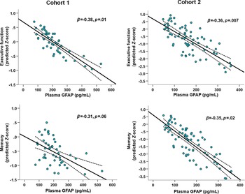
Higher plasma GFAP was associated with worse executive function scores in both Cohort 1 [β = −0.38, 95% CI (−0.67, −0.08), p = .01] and Cohort 2 [β = −0.36, 95% CI (−0.62, −0.10), p = .007; Table 2]. In Cohort 2, higher plasma GFAP was additionally associated with worse visual memory [β = −0.35, 95% CI (−0.64, −0.06), p = .02]. A similar relationship was seen for visual memory in Cohort 1 but did not reach statistical significance [β = −0.31, 95% CI (−0.64, 0.01), p = .06; Figure 2]. In Cohort 2 only, higher plasma GFAP was also associated with lower language [β = −0.30, 95% CI (−0.58, −0.02), p = .03] and visuospatial scores [β = −0.37, 95% CI (−0.67, −0.07), p = .02]. Similar to the brain volume analyses, plasma GFAP associations were significant while adjusting for plasma NfL, but plasma NfL was not significantly associated with cognitive outcomes for either cohort when controlling for plasma GFAP. There were again no consistent findings within or between cohorts when examining plasma GFAP × CDR-SB interaction effects (Supplemental Figure 2).
Scatterplots depicting associations of higher plasma GFAP with lower cognitive scores. Y-axes represent model-predicted standardized (z-score) scores based on age, sex, education, plasma GFAP, and plasma NfL. Dashed lines show 95% confidence interval around the linear fit line. Findings were most robust for executive functioning. There was a statistically significant association of higher plasma GFAP with worse memory in Cohort 2. The memory association in Cohort 1 had a similar effect size and was in the same direction, but did not reach statistical significance. Cohort 2 participants also showed significant associations of higher plasma GFAP with lower language and visuospatial scores (not depicted), either of which approached statistical significance in Cohort 1. These models showed no significant associations between plasma NfL and cognitive scores in either study cohort.

Plasma NfL without Plasma GFAP
Prior research suggests we should expect a relationship between plasma NfL, brain volume, and cognition (Hu et al., Reference Hu, Chen, Ou, Cao, Chen, Cui and Yu2019; Mattsson et al., Reference Mattsson, Andreasson, Zetterberg and Blennow2017; Rajan et al., Reference Rajan, Aggarwal, McAninch, Weuve, Barnes, Wilson and Evans2020), especially in our more cognitively impaired cohort (Cohort 2). We therefore evaluated plasma NfL in isolation without plasma GFAP in the regression models to provide additional context for the unexpected null findings for plasma NfL. In Cohort 1, there were no statistically significant associations between plasma NfL and any white or gray matter ROI volumes, or any cognitive outcomes (Table 2). In Cohort 2, higher plasma NfL was associated with lower parietal GM and worse performance in multiple cognitive domains: executive function, memory, and language.
DISCUSSION
We evaluated associations between plasma GFAP, plasma NfL, brain volume, and cognition among older adults across the AD spectrum. Data were derived from 121 participants comprising two independent cohorts – one predominantly with CN older adults plus a subset with MCI, and one with relatively balanced representation of CN, MCI, and dementia. Cognitively impaired participants met clinical consensus research criteria for MCI or dementia with at least intermediate likelihood of AD (Albert et al., Reference Albert, DeKosky, Dickson, Dubois, Feldman, Fox and Phelps2011; McKhann et al., Reference McKhann, Knopman, Chertkow, Hyman, Jack, Kawas and Phelps2011). In both cohorts, we found the most robust relationships between higher plasma GFAP with lower white matter volume in the temporal and parietal lobes, and lower executive function scores. The association with lower memory scores had a similar effect size and direction in both cohorts, but only reached statistical significance in the second cohort comprising more participants with MCI and dementia. This second cohort also showed significant associations between high GFAP and lower language and visuospatial domains. We found no unique relationship between plasma NfL with brain volume or cognition in any models that also adjusted for plasma GFAP. These data support the added value of measuring astroglial biomarkers in aging cohorts at-risk for neurodegenerative disease in addition to commonly studied biomarkers of neuronal pathophysiology.
GFAP, White Matter, and Executive Function
GFAP is a core intermediate filament protein of the astrocytic cytoskeleton (Yang & Wang, Reference Yang and Wang2015). Upregulation of GFAP may reflect a host of astrocytic functional and structural changes in response to aging and neurodegenerative disease. Astrocytes play a prominent role in blood–brain barrier regulation and glymphatic system maintenance, both of which are intricately tied to the brain’s white matter and cerebrovasculature (Jessen, Munk, Lundgaard, & Nedergaard, Reference Jessen, Munk, Lundgaard and Nedergaard2015). The interchange of cerebrospinal and interstitial fluid occurs through aquaporin-4 channels that densely pack the astrocytic endfeet lining the blood–brain barrier (Jessen et al., Reference Jessen, Munk, Lundgaard and Nedergaard2015). This system plays a key role in metabolite clearance, including amyloid-plaques and tau tangles characteristic of AD (Braun & Iliff, Reference Braun and Iliff2020). It may be that increased GFAP levels in plasma signal abnormal astrocytic responses to increased demand for metabolite clearance, breakdown of the blood–brain barrier, and/or subsequent degeneration of white matter. Data showing that higher plasma GFAP is associated with greater white matter disease burden (hyperintensity volume on T2 MRI) (Elahi et al., Reference Elahi, Casaletto, La Joie, Walters, Harvey, Wolf and Cobigo2019) and worse white matter integrity (Bettcher et al., Reference Bettcher, Olson, Carlson, McConnell, Boyd, Adame and Thaker2021) support these hypotheses.
The link of GFAP to white matter volume is compelling given our finding of a robust association of GFAP with executive functioning. Executive function is an umbrella term capturing multiple higher order cognitive skills such as mental flexibility, inhibition, and updating/working memory, among others (Miyake et al., Reference Miyake, Friedman, Emerson, Witzki, Howerter and Wager2000). These abilities are linked to frontoparietal–subcortical networks. There is growing evidence underscoring the particular importance of healthy white matter structural networks for performing executive functions, above and beyond contributions of frontal lobe gray matter (Bettcher et al., Reference Bettcher, Mungas, Patel, Elofson, Dutt, Wynn and Kramer2016; Kennedy & Raz, Reference Kennedy and Raz2009). We found that higher plasma GFAP was associated with lower white matter volume and worse executive functioning even among CN and mildly cognitively impaired older adults. Associations with other cognitive domains only became apparent in our second cohort that included more cases of MCI and dementia, suggesting that plasma GFAP may be sensitive to white matter and executive functioning changes both early in disease and that persist in later stages of disease. A recent study of older adults along the healthy aging to Alzheimer’s dementia spectrum also found links between plasma GFAP, cognition, and white matter integrity (diffusion metrics) in the medial temporal lobe (Bettcher et al., Reference Bettcher, Olson, Carlson, McConnell, Boyd, Adame and Thaker2021). Bettcher et al. consistently found stronger associations between plasma GFAP and memory than executive function. Study samples differed slightly in the proportion of participants with dementia and there may be other unknown differences in other important underlying pathologies contributing to variation in cognitive domain correlates (e.g., white matter disease burden, severity of AD, or common co-pathologies).
Discordance of Plasma GFAP and Plasma NfL Findings
One limitation of blood-based biomarkers is the potential for nonspecific elevations associated with systemic disease and/or blood–brain barrier breakdown rather than with a particular pathophysiologic process. The simultaneous inclusion of plasma GFAP and NfL in our models allowed us to examine the specificity of these biomarkers. We found several associations between plasma GFAP, white matter volumes, and cognition that did not exist for plasma NfL, which refutes hypotheses of blood-based protein elevations simply reflecting global blood–brain barrier breakdown. In fact, plasma NfL showed some associations with brain volume and cognition only when plasma GFAP was removed from the regression models, and only in our cohort with more severe clinical disease.
Rajan and colleagues studied plasma GFAP and NfL collected in a large sample of older adults and found higher levels of both were independently associated with more severe cortical atrophy and cognitive decline in the years following the blood draw (Rajan et al., Reference Rajan, Aggarwal, McAninch, Weuve, Barnes, Wilson and Evans2020). White matter volume was not analyzed. They also found that both plasma GFAP and NfL were elevated in Alzheimer’s dementia cases (compared to controls), while only plasma NfL differentiated cases with MCI. This runs contrary to our findings of brain structure and cognition correlates for plasma GFAP, but not NfL, even in our less impaired cohort (Cohort 1). Other studies also report a strong signal for plasma biomarkers at the milder end of clinical disease expression (Asken et al., Reference Asken, Elahi, La Joie, Strom, Staffaroni, Lindbergh and Casaletto2020; Verberk et al., Reference Verberk, Thijssen, Koelewijn, Mauroo, Vanbrabant, de Wilde and Teunissen2020). Research that disentangles the complex temporal dynamics of aging and neurodegenerative disease pathophysiology as expressed in plasma biomarkers is in its relative infancy. Additional work requires longitudinal integration of biomarkers capturing multiple disease components – neuronal, glial, and inflammatory changes – among older adults representing the continuum of clinical disease severity.
Limitations
Our study was cross-sectional and had relatively small sample size for each of our two independent cohorts. We therefore were underpowered to investigate possible important interactions, such as sex-specific associations. The sample almost exclusively included White/Caucasian older adults, limiting generalizability to other racial/ethnic groups. The cohorts contained a subset of cases with early-onset AD changes that may have different disease characteristics than late-onset dementia. Plasma GFAP alone may nonspecifically capture a diverse range of astrocytic response to neurologic insults; therefore, we cannot yet link our observed associations to a particular aspect of astrocytic pathophysiology (e.g., astrocyte activation/inflammatory response vs. astrocyte degeneration). Cognitively impaired participants met consensus criteria for MCI or dementia with at least intermediate likelihood of underlying AD pathology; however, co-pathology is common and we cannot determine whether our findings are specific to any particular neurodegenerative disease.
CONCLUSIONS
Higher levels of plasma GFAP are robustly associated with lower white matter volume and worse executive function in older adults with and without cognitive impairment. Plasma GFAP may be more sensitive to white matter and cognitive changes than plasma NfL, particularly in mildly impaired individuals. Biomarkers reflecting astroglial pathophysiology will help advance our ability to capture complex dynamics of aging and neurodegenerative disease.
SUPPLEMENTARY MATERIAL
To view supplementary material for this article, please visit https://doi.org/10.1017/S1355617721000813
ACKNOWLEDGEMENTS
We thank the patients and their families whose help and participation made this work possible. We also thank the National Centralized Repository for Alzheimer’s Disease and Related Dementias (NCRAD) for sample storage, which receives government support under a cooperative agreement grant (U24 AG21886) awarded by the National Institute of Aging (NIA).
Financial Support
We thank contributors who collected samples used in this study, as well as the following funding sources that have supported our work: NIH-NIA ADRC (P30 AG062422) and PPG (P01 AG019724) to BLM, R01(s) AG032289 and AG048234) and Larry L. Hillblom Network Grant (2014-A-004-NET) to JHK, American Academy of Neurology grant to FME, NIH-NIA (K23 AG058752) to KBC, NIH-NIA (K23 AG061253) and Larry L. Hillblom Fellowship (2018-A-025-FEL) to AMS, NIH-NIA (K23AG059888) to JCR, NIH-NIA (U19AG063911, U54NS092089, R01AG038791) to ALB, and Robert W. Katzman Clinical Research Training Scholarship in Alzheimer’s and Dementia Research from the American Academy of Neurology to LV.
CONFLICTS OF INTEREST
JCR reports being a site PI for clinical trials sponsored by Eli Lilly. JHK has provided consultation to Biogen. ALB has served as a consultant for Aeton, Abbvie, AGCT, Amgen, Arkuda, Arvinas, Asceneuron, Eisai, Ionis, Lundbeck, Novartis, Passage BIO, Sangamo, Samumed, Third Rock, Toyama, and UCB. All authors deny conflicts of interest directly pertaining to this study.