Introduction
It has long been known that the diameter of the pupil changes in response to changes in ambient illumination, the purpose being to control the brightness of the light falling on the retina. However, the pupil also responds to changes in color, spatial structure, and movement (Slooter & van Norren, Reference Slooter and van Norren1980; Young & Alpern, Reference Young and Alpern1980; Young et al., Reference Young, Han and Wu1993; Young & Kennish, Reference Young and Kennish1993; Sahraie & Barbur, Reference Sahraie and Barbur1997; Gamlin et al., Reference Gamlin, Zhang, Harlow and Barbur1998). In addition, it is well known that internal states entailing arousal, effort, interest, and emotion are observable through their effect on the diameter of the pupil (Hess & Polt, Reference Hess and Polt1960; Kahneman & Beatty, Reference Kahneman and Beatty1966; Beatty, Reference Beatty1982).
In the experiments reported in this article, it is determined whether the diameter of the pupil can be affected by cognitive mechanisms entailing changes in attention, irrespective of differences in mental effort. More specifically, we investigate whether the diameter of the pupil can be entrained by alternations between broadly spread and narrowly focused attention. If it were found that pupil dilation is greater for broad than narrow attention, it would provide evidence that differences in spherical aberration are associated with the alternating attentional states. That is, greater spherical aberration when the pupil is dilated would blur the retinal image, decreasing the contrast of high spatial frequency information (Campbell & Green, Reference Campbell and Green1965; Campbell & Gubish, Reference Campbell and Gubish1966), and thereby, increasing the relative sensitivity of the large receptive fields that mediate broadly spread attention compared with the smaller receptive fields that mediate more narrowly focused attention (Hock & Balz, Reference Hock and Balz1994; Balz & Hock, Reference Balz and Hock1997; Hock et al., Reference Hock, Balz and Smollon1998; Hochstein & Ahissar, Reference Hochstein and Ahissar2002). It is determined whether pupil-entraining changes in attentional spread can be cued both exogenously (attention elicited by the stimulus) and endogenously (attention controlled by the perceiver) (Jonides, Reference Jonides, Long and Baddeley1981; Corbetta & Shulman, Reference Corbetta and Shulman2002). It also is determined whether differences in pupil diameter can result from sustained broad attention compared with sustained narrow attention. When endogenously cued, changes in attentional spread are intentional, so they must be initiated cortically. Evidence for pupil entrainment with endogenous cueing therefore would provide strong behavioral support for cortical feedback influencing the subcortical control of pupil diameter (Gamlin et al., Reference Gamlin, Zhang, Harlow and Barbur1998; Barbur, Reference Barbur, Chalupa and Werner2004).
Materials and methods
The diameter of the pupil is notoriously variable, even in the absence of stimulation, so differences between broadly spread and narrowly focused attention could not be consistently observed by the examination of pupillary time series. To address this problem, the two attentional states were rhythmically alternated, and the analysis of the data was based on the Fast Fourier transformation (FFT) of the pupillary time series. We then could determine whether the alternations of attentional spread would be accompanied by increased Fourier energy at the frequency of alternation, 0.2 Hz, and its harmonics (temporal frequencies that are integer multiples of 0.2 Hz). To rule out the possibility of spontaneous pupillary oscillations (hippus), which can occur in the 0.05–0.3 Hz range (Stark et al., Reference Stark, Campbell and Atwood1958; Kardon, Reference Kardon and Miller1998; Loewenfeld, Reference Loewenfeld1999), the experiments included trials in which only the fixation dot was present. An increase in Fourier energy at 0.2 Hz for these trials would be indicative of hippus.
Stimuli
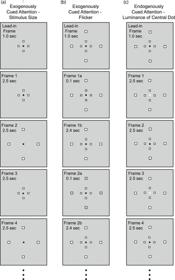
The experimental stimuli were presented on a FlexScan T566 monitor under the control of a Dell Precision 360 computer. Subjects viewed the stimuli while a View Point PC60 EyeTracker (Arrington Research, Inc., Scottsdale, Arizona). recorded their pupillary responses and eye positions. The chin rest was 38.1 cm from the screen, with small adjustments in order to accommodate individual differences in eye location (i.e., deep-set eyes). Four elements were arranged in a diamond configuration, 2.6 deg apart in their central locations and/or 26.0 deg apart in their peripheral locations (from center to center both horizontally and vertically). The elements were centered on a fixation dot that intercepted a visual angle of 5.2 min in the center of the screen. Illustrations of the stimuli in Fig. 1 are not to scale.
The stimuli in Experiment 1 were composed of square luminance-balanced elements located at the four corners of a diamond. (a) Attention–alternation is exogenously cued by changing the size of the stimulus (the four elements are shifted between their central and peripheral locations). (b) All eight elements are simultaneously visible. Attention–alternation is exogenously cued by the momentary removal of the light inner squares of the four central elements or the four peripheral elements. (c) All eight elements are simultaneously visible. Attention–alternation is endogenously cued by changes in the luminance of the central fixation element. The illustrations of the stimuli are not to scale.

The possibility that the appearance or disappearance of the elements would produce luminance effects on the diameter of the pupil was minimized by balancing the luminance of each element such that its overall luminance was approximately equal to the luminance of the background. The luminance-balanced elements, each of which was composed of a light inner square (luminance = 109 cd/m2) surrounded by a dark border (luminance = 0 cd/m2), were presented against an intermediate background luminance of 46.0 cd/m2. In their central locations, the inner light square of each element subtended a visual angle of 10.4 × 10.4 min, and the surrounding dark border around each side of the inner square was 5.2 min thick. In their peripheral locations, the inner light square of each element subtended a visual angle of 15.6 × 15.6 min, and the surrounding dark border around each side of the inner square was 10.4 min thick.
Procedure
Subjects were tested for eye dominance at the start of their first testing session. The eye tracker camera was positioned to record from the dominant eye, while the nondominant eye was covered by an eye patch. Lights in the testing room were dimmed, resulting in an ambient room illumination of 1.9 cd/m2, as determined by reflectance off the wall next to the subject. Prior to each testing session, a calibration procedure for each subject matched gaze locations to a series of screen locations.
During the testing sessions, pupil diameter and eye position were recorded while stimulus cues specifying attention to either the central or peripheral element locations alternated at a rate of 0.2 Hz. The 0.2-Hz rate was selected based on pilot testing indicating that it was comfortable for alternating between broad and narrow attention. Each two-frame broad/narrow alternation cycle (2.5 s per frame, with the exception of Experiment 2) was repeated eight times, so the total duration of each trial was 40 s. Subjects were instructed to maintain fixation on the dot in the center of the screen, which was visible for the entire trial, and to avoid eye movements. Each 40-s trial was preceded by a 1.0-s “lead in” in order to minimize effects due to the onset of the stimulus (see Fig. 1).
Exogenously cueing attention alternation with changes in stimulus size
The four luminance-balanced elements that formed a diamond configuration were located centrally for 2.5 s (cueing narrow attention), and then, its overall size was increased by presenting the elements peripherally for 2.5 s (cueing broad attention), back and forth, as in Fig. 1a.
Exogenously cueing attention alternation with flicker
All eight luminance-balanced elements were stationary and visible for the entire duration of each trial. Narrow attentional spread was cued by removing the inner white squares of the four central luminance-balanced elements for 0.1 s, then restoring them for the next 2.4 s. This alternated with the cueing of broad attentional spread by removing the inner white squares of the four peripheral luminance-balanced elements for 0.1 s, then restoring them for the next 2.4 s (Fig. 1b).
Endogenously cueing attention alternation with changes in central luminance
While all eight luminance-balanced elements were stationary and visible for the entire trial, the luminance of the central fixation dot alternated between 250 cd/m2, which signaled narrow attention for 2.5 s, and 0 cd/m2, which signaled broad attention for 2.5 s (Fig. 1c).
Experiment 1: alternating between broad and narrow attentional spread
In all three attentional cueing conditions, subjects were to either spread their attention to encompass all four peripheral elements or narrow their attention to encompass the four central elements. Included were blocks of fixation-only trials, in which only the fixation dot was presented for 40 s. The fixation dot was black for the fixation-only conditions that were paired with the exogenous attention–alternation conditions (stimulus size and flicker). It alternated between white and black at a 0.2-Hz rate for the fixation-only condition that was paired with the condition, in which attention–alternation was endogenously cued by changes in the luminance of the central dot.
Experiment 2: alternating broad and narrow attention: unpredictable frame durations
This experiment tested stimuli for which attention–alternation was exogenously cued by alternately presenting the luminance-balanced elements in their central and peripheral locations, as in Fig. 1a, but with irregular frame durations. The 5-s broad/narrow cycles were maintained, but each of the eight cycles was composed of frame pairs for which one attentional cue was presented for 2 s and the other for 3 s, randomly determined. Also tested was a corresponding condition in which only the fixation dot was presented.
Experiment 3: sustained broad and sustained narrow attentional spread
All three attention–alternation stimuli were presented but with the instruction that the attention–alternation cues were to be ignored. During separate blocks of trials, subjects were instructed to either sustain broad attention for the entire trial in order to encompass the locations of the four peripheral elements or to sustain narrow attention in order to encompass the locations of the four peripheral elements.
The pupillary light reflex
The luminance of the screen alternated between 150 and 0 cd/m2, while the luminance of the fixation dot alternated between 0 and 150 cd/m2, both at a 0.2-Hz rate.
Design for Experiments 1–3
Seven subjects participated in the first three testing sessions of Experiments 1 and 3; attention–alternation was exogenously cued by changes in stimulus size (Fig. 1a). Five subjects participated in the second three testing sessions; attention–alternation was exogenously cued by flicker (Fig. 1b). Five subjects participated in the final three testing sessions; attention–alternation was endogenously cued by changes in the luminance of the fixation dot (Fig. 1c). Tested during each session, in approximately counterbalanced order, were: 1) narrow/broad attention alternation, 2) fixation only, 3) sustained broad attention, and 4) sustained narrow attention. Blocks of luminance–alternation trials, and blocks of attention–alternation and fixation-only trials for Experiment 2, were intermixed with those tested during the first three sessions. There were nine trials per subject in each of the conditions described above (three trials within each of the three sessions).
Experiment 4: confirmation of sustained attention with a detection task
Sustained attention instructions were as in Experiment 3. As in Fig. 1a, the luminance-balanced elements alternated between their central and peripheral locations in two-frame cycles (2.5 s per frame), but now, there were two, three, or four two-frame cycles per trial (randomly varied from one trial to the next). During the last frame of each trial, the luminance-balanced elements were in their peripheral locations, and the luminance of the border surrounding one of the elements was increased for 0.5 s. It was increased from 0 to either 98, 105, or 120 cd/m2, as determined by pilot testing aimed at minimizing floor and ceiling effects for each subject. Following the luminance increment, the fixation dot changed from white to blue for the final 2.0 s of the trial, indicating that the subject should respond by tapping their finger to indicate which of the four peripheral elements had changed in luminance: one tap if the luminance change occurred for the upper element, two taps for the right element, three taps for the lower element, and four taps for the left element. Subjects were required to guess if they were unsure of their response.
Blocks of 12 trials were generated by the orthogonal combination of the number of central/peripheral alternations of the luminance-balanced elements (two, three, or four) and which of the four peripheral locations had the to-be-detected luminance increment. Each of eight testing sessions included, in counterbalanced order, one block of 12 trials with sustained narrow attention and another block of 12 trials with sustained broad attention. After each trial, six subjects indicated the location of the peripheral element for which there was a luminance increment. There were 96 trials per subject in the sustained broad and sustained narrow conditions.
Experiment 5: alternating attention with eye blinks suppressed
This experiment repeated the Experiment 1 condition, in which attention–alternation was endogenously cued by changing the luminance of the central fixation dot (as in Fig. 1c), but now with the instruction not to blink anytime during the trial. A single naive subject participated in this experiment. There were 18 trials for which there were no eye blinks and 9 trials for which there were eye blinks, despite the instruction not to blink. Data also were obtained for nine subsequent trials during which the subject was told not to be concerned about blinking.
Subjects
A total of 10 subjects voluntarily participated in this study. All except authors L.B.D. and H.S.H. were students at Florida Atlantic University who were naive with respect to the purpose of the experiments.
Data analysis
The eye tracker illuminated the perceiver’s dominant eye with infrared light and recorded its reflection with a video camera. It “captured” the pupil by probing for dark regions within an adjustable search area. The eye tracker’s software was interfaced with a MatLab program that controlled the timing and display of stimuli and marked the start of each trial in the ongoing data stream produced by the eye tracker, which sampled at a rate of 16.7 ms. The data stream included the horizontal pupil diameter, the horizontal and vertical position of the eye, and an indicator of the quality of the pupil measurement. Poor quality samples occurred when the pupil was momentarily “lost,” typically when there was an eye blink, and when the position of the eye was more than 2 s.d. away from the average horizontal and vertical eye positions (the rate of poor quality samples was approximately 3%). One sample preceding and five samples following a poor quality sample were replaced by the average of the minimum and maximum of the horizontal pupil diameter for the trial. Similar results were obtained when these samples were replaced instead by the average of the preceding and following pupil diameter values.
The data stream during the first second of each trial was excluded, and the remainder of the trial’s time series was shortened to 2048 samples, the largest power of 2 that could be accommodated in order for the Fast Fourier Transform (FFT) to be computationally efficient. The resulting time series for the 9 trials in each condition (18 trials in Experiment 5) were averaged for each subject and Fourier transformed into the temporal frequency domain. Data graphs are based on the average of each subject’s FFT. Statistical analyses were based on the reliability, across subjects, of differences in FFT amplitude and correlations between the FFTs for attention alternation and the FFTs for luminance alternation.
Results
Experiment 1: alternating between broad and narrow attentional spread
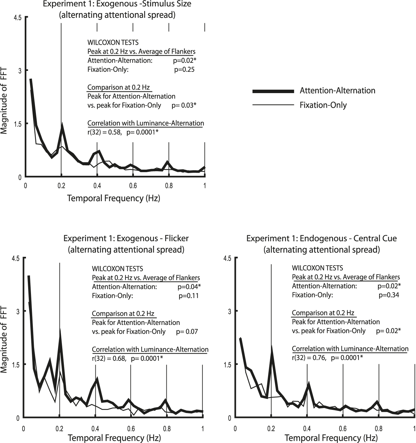
FFTs of the pupillary time series for the attention–alternation and fixation-only conditions of Experiment 1 are presented in Fig. 2. The results of statistical analyses are inset within each FFT graph. The initial analyses focused on the Fourier amplitude at 0.2 Hz, the temporal frequency at which attention was alternated. Local peaks in the FFT amplitude at this frequency can be seen for each of the three attention–alternation conditions; the amplitude at 0.2 Hz was significantly greater than the average FFT amplitude for the flanking 0.17 and 0.23 Hz frequencies.Footnote 1 The same comparisons were not significant for the fixation-only conditions, confirming that a spontaneous hippus response (Kardon, Reference Kardon and Miller1998) was not affecting the results at 0.2 Hz. In addition, FFT amplitudes at 0.2 Hz were significantly larger for the attention–alternation compared with the fixation-only conditions when attention changes were cued by changes in stimulus size or by luminance changes of the central fixation dot. The difference was marginally significant when attention–alternation was cued by flicker (there was a small reversal for one subject).
Experiment 1: FFT results for the attention–alternation and fixation-only conditions when broadly spread and narrowly focused attention alternate at a temporal frequency of 0.2 Hz. Attentional changes were exogenously cued by changes in stimulus size or flicker and endogenously cued by changes in the luminance of the central fixation dot.

Time series for pupil diameter also were obtained for 0.2 Hz alternations in the luminance of the entire screen. The FFT for these data was typical of the pupillary light reflex in that they indicated predominant Fourier amplitude peaks at the alternation rate of 0.2 Hz but also at both the odd and even harmonics of 0.2 Hz. The harmonics reflect the sawtooth character of changes in pupil diameter; that is, fast constrictions in response to luminance increase and slower dilations in response to luminance decrease (Lowenstein & Loewenfeld, Reference Lowenstein and Loewenfeld1950; Ellis, Reference Ellis1981; Heller et al., Reference Heller, Perry, Jewett and Levine1990; Howarth et al., Reference Howarth, Heron and Whittaker2000). A similar pattern of FFT amplitude peaks at 0.2 Hz and its harmonics was observed for the attention–alternation but not for the fixation-only conditions. FFT amplitudes for the attention–alternation conditions were significantly correlated with those for the luminance–alternation condition for all three attention–alternation conditions. (The correlations were based on 34 pairs of FFT amplitudes, 1 pair of attention–alternation and luminance–alternation amplitudes for each temporal frequency between 0.03 and 1.0 Hz.)Footnote 2 Thus, the pattern of FFT harmonics obtained for broad/narrow attention alternations followed the pattern of harmonics that is characteristic of the pupil’s well-established response to changes in luminance. The results of Experiment 1 indicated, therefore, that pupil diameter can be entrained by the alternations in attentional spread, as it is by alternations in luminance, regardless of whether the changes in attention are cued exogenously or endogenously.
Experiment 2: alternating broad and narrow attention: unpredictable frame durations
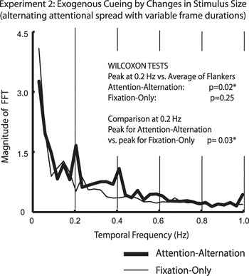
It is possible that the predictability of regularly alternating attentional cues resulted in subjects anticipating the attentional cues and intentionally changing their attentional spread. The changes in attention would then be endogenously controlled, irrespective of the exogenous cues themselves. In order to rule out this possibility, the 5-s (0.2-Hz) broad/narrow cycle was maintained, but for the frame pairs composing each of the eight broad/narrow cycles, one attentional cue was presented for 2 s and the other for 3 s, randomly determined. With changes in attentional spread cued by changes in stimulus size, the results from Experiment 1 were replicated (Fig. 3). That is, the FFT amplitude at 0.2 Hz was significantly greater than the average FFT amplitude for the flanking 0.17 and 0.23 Hz frequencies for the attention–alternation condition, and the same comparisons were not significant for the fixation-only conditions. In addition, FFT amplitudes at 0.2 Hz were significantly larger for the attention–alternation compared with the fixation-only condition. It therefore was confirmed that the effects of attention–alternation on pupil diameter can be cued exogenously, irrespective of endogenous attentional control based on the predictability of attention–alternation cues.
Experiment 2: FFT results when alternations in broadly spread and narrowly focused attention were cued by changes in stimulus size at a temporal frequency of 0.2 Hz but with frame durations of unpredictably varying duration.

This result, however, does not rule out other possible influences of endogenous control when attention changes are cued exogenously. It is likely, for example, that endogenous attentional mechanisms contribute to the maintenance of fixation on the central dot and the symmetrical spreading of attention to encompass all the peripheral or all the central dots. Moreover, the results for sustained broad and sustained narrow attention in the experiment that follows indicate that the presumably obligatory effects of exogenous cueing can be intentionally suppressed. Exogenous cues may automatically elicit changes in attention (Jonides, Reference Jonides, Long and Baddeley1981), but the need to endogenously maintain symmetrical attentional spread around the fixation point may make it possible to suppress further response to the exogenous cues.
Experiment 3: sustained broad and sustained narrow attention
This experiment had two objectives. The first was to determine whether establishing the broad and narrow attentional states entailed differences in mental effort. The second was to confirm that the stimulus changes that cued attention alternation in Experiment 1 were not in themselves sufficient to entrain pupil diameter.
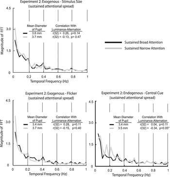
Subjects were instructed to ignore the alternating broad versus narrow attentional cues, and instead, to sustain either broadly spread attention to encompass all four peripheral element locations during the entire trial, or in separate blocks of trials, to sustain more narrowly focused attention to encompass all four central element locations during the entire trial. Regardless of whether the attention–alternation cues were exogenous or endogenous, mean pupil diameters were very similar in the sustained broad and sustained narrow conditions (see insets within each of the Fig. 4 graphs). This indicated that the effect on pupil diameter of alternating broad and narrow attention in Experiment 1 was not attributable to differences between the two attentional states in mental effort or processing load (Hess & Polt, Reference Hess and Polt1960; Kahneman & Beatty, Reference Kahneman and Beatty1966; Beatty, Reference Beatty1982; Steinhauer et al., Reference Steinhauer, Siegle, Condray and Pless2004).
Experiment 3: FFT results and mean pupil diameters for the conditions with sustained broad attention and sustained narrow attention. Subjects were instructed to ignore the 0.2 Hz stimulus changes that cued alternations in attentional spread.

The FFT results in Fig. 4 indicated that changes in the attentional cues without accompanying changes in broad/narrow attention were not sufficient to entrain the pupil. There was little difference between the sustained broad and sustained narrow conditions, and in addition, the Fourier amplitude peaks that were observed in the attention–alternation conditions of Experiment 1 were largely absent in this experiment despite the same 0.2-Hz alternation in attention cues. Correlations with the FFT amplitudes obtained for luminance alternation were not significant for any of the broad attention conditions and were not significant for all but one of the narrow attention conditions (a marginally significant correlation was obtained only in sustained narrow condition with endogenous attentional cueing, possibly because of the white/black alternation in the luminance of the fixation dot). There otherwise was no indication that fluctuations in pupil diameter contained the multiple harmonic components that are characteristic of fast constrictions alternating with slow dilations.
Experiment 4: confirmation of sustained attention with a detection task
Attention alternation was exogenously cued by alternately presenting the luminance-balanced elements in their central and peripheral locations. As in Experiment 3, subjects were instructed to ignore those cues and sustain either broad or narrow attention. The purpose of this experiment was to confirm that they could do so. In addition to sustaining attentional spread for the entire trial, subjects were required to indicate the location of a luminance increment that occurred unpredictably for one of the four peripheral elements. Although they might not have successfully sustained broad or narrow attentional spread during the entirety of every trial, they did so often enough to correctly detect the location of the luminance increment significantly more often when their attention was broadly spread (70.8% correct) compared with when it was more narrowly focused (56.4% correct); t(5) = 3.59, P < 0.02. Thus, the absence of a pupillary effect in Experiment 3 was not due to subjects’ inability to sustain attention in the prescribed manner, reinforcing the conclusion that the entrainment of the pupil requires actual changes in attentional spread rather than changes in the attention-cueing stimulus.
Experiment 5: the effect of eye blinks
The occurrence of eye blinks was not uniform in Experiment 1. In all three attention–alternation conditions, they occurred more often during frames in which attention was narrowly focused compared with frames during which it was broadly spread. FFTs done on the time series of eye blinks indicated the presence of amplitude peaks at 0.2 Hz for the attention–alternation conditions of Experiment 1 but not for the sustained-attention conditions of Experiment 3. Thus, eye blinks occurred most often when attention switched from broad to narrow attention. This was significant because analyses of the pupillary time series from Experiment 1 indicated, consistent with Húpe et al. (Reference Húpe, Lamirel and Lorenceau2009), that eye blinks were consistently followed by pupil constrictions.
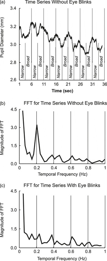
In order to determine whether eye blinks were solely responsible for the pupil entrainment effect, the subject tested in this experiment was instructed to suppress eye blinks (but switch attention) during trials in which attention–alternation was cued endogenously by changes in the luminance of the central fixation dot. When eye blinks were successfully suppressed, it was found that the pupillary time series was sufficiently stable for the time-varying pupil diameter to be directly observed. As expected, the pupil became more dilated after broadly spread attention was cued and more constricted after narrowly focused attention was cued (Fig. 5a), and there was a very large peak in Fourier amplitude at 0.2 Hz and smaller peaks near the harmonics of 0.2 Hz (Fig. 5b). There was, however, no indication that attention–alternation affected pupil diameter for trials during which eye blinks occurred, regardless of whether or not the subject was trying to suppress them (Fig. 5c). This may have been because in contrast with Experiment 1, these eye blinks occurred approximately equally often during frames in which attention was broadly spread and frames in which it was more narrowly focused. The effort to suppress eye blinks may have disrupted their linkage with the narrowing of attention.
Experiment 5: (a) The pupillary time series for trials with endogenously cued alternation between broad and narrow attention at a rate of 0.2 Hz. The data are averaged over 18 trials without eye blinks. The slow drift in the diameter of the pupil is typical. It is responsible for the Fourier energy at low temporal frequencies. (b) FFT results for the 18 trials without eye blinks. (c) FFT results for the pupillary time series averaged over 18 trials with eye blinks, 9 trials in which the subject was trying unsuccessfully to suppress eye blinks and 9 trials in which she was not trying to suppress eye blinks (the data are combined because there was little difference between them).

The results indicate, therefore, that the occurrence of eye blinks is not necessary for the attentional entrainment of pupil diameter, but the entrainment can nonetheless be influenced by pupil-constricting effects of eye blinks, which occur most often when there is a switch in attention from broadly spread to narrowly focused attention.
The near reflex
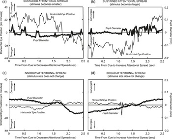
The stimuli in the current study were close enough to the perceiver (38.1 cm), to activate the near reflex, which is composed of vergence eye movements, changes in accommodation, and changes in pupil diameter. Of potential concern is whether the changes in pupil diameter for alternating narrow and broadly spread attention were an indirect result of changes in vergence and accommodation. That is, pupil contraction for more narrowly focused attention might be the indirect result of convergent changes in horizontal eye position and increases in accommodation when the near reflex is activated, and pupil dilation for broadly spread attention might be the indirect result of divergent changes in horizontal eye position and decreases in accommodation when the near reflex is relaxed. This possibility was addressed by measuring changes in eye position and comparing them with changes in pupil diameter for the subject in Experiment 5 who suppressed eye blinks (changes in attentional spread but no change in stimulus size) and for a comparable subject from Experiment 3 with relatively few eye blinks (changes in stimulus size but no change in attentional spread). Analyses were based on the averaged event-locked time series. That is, the time course of changes in eye position and pupil diameter were averaged, both within and between trials (see Fig. 6 for details). One event-locked time series started when the luminance-balanced elements were presented in their central locations (or attention was narrowly focused on these locations). The other started when the luminance-balanced elements were presented in their peripheral locations (or attention was broadly spread over these locations). In both cases, changes in vergence were delayed by approximately 1.5 s, far longer than the 0.2 s that is typically observed in the absence of an attentional task (e.g., Westheimer & Mitchell, Reference Westheimer and Mitchell1956).
The results from Experiment 3 (combining the data from the sustained narrow and sustained broad conditions) were consistent with earlier research in indicating that changes in stimulus size were sufficient to produce changes in vergence eye movements (Erkelens & Regan, Reference Erkelens and Regan1986). When the luminance-balanced elements were shifted into their central locations, changes in horizontal eye position were divergent, consistent with the stimulus moving further away when it became retinally smaller (Fig. 6a). When the luminance-balanced elements were shifted into their peripheral locations, changes in horizontal eye position were convergent, consistent with the stimulus moving closer when it became retinally larger (Fig. 6b). The minimal change in pupil diameter suggests that changes in pupil diameter do not necessarily follow from changes in eye position associated with the near reflex.
Event-locked time series for changes in pupil diameter and horizontal eye position (convergent or divergent) averaged both within and between trials. Prior to creating the event-locked time series from the average of 18 trials, the time course was band-passed filtered such that frequencies below 0.15 Hz were removed, in order to zero-mean the time course and to remove low frequency drift, and frequencies above 2.8 Hz were removed, in order to decrease the contribution of high frequency noise. For a subject from Experiment 3, there were changes in stimulus size but no changes in attentional spread. The stimulus was either: (a) small—the luminance-balanced elements were in their central locations, or (b) large—the luminance-balanced elements were in their peripheral locations (the data for the sustained broad and sustained narrow conditions are averaged). For the subject in Experiment 5, there were changes in attentional spread but no changes in stimulus size. Attention was either: (c) narrowly focused in order to encompass only the centrally located elements or (d) broadly spread in order to encompass the peripherally located elements.

The event-locked time series from Experiment 5 indicated that gradual constriction in pupil diameter began soon after narrow attention was cued (Fig. 6c), and gradual dilations in pupil diameter began soon after broad attention was cued (Fig. 6d). Pupil diameter therefore began changing in accord with endogenously cued changes in attention long before the delayed activation of the near reflex. This indicated that alternating activation and relaxation of the near reflex did not initiate the constrictions and dilations of pupil diameter that occurred with alternations between narrowly focused and broadly spread attention. However, when eye position did change, the ongoing change in pupil diameter was boosted (i.e., further constrictions when attention was being narrowed and further dilations when attention was being broadened). Thus, pupil diameter was affected by the near reflex but long after it began independently changing as a result of feedback from cortical attentional mechanisms. When activated, the near reflex enhanced ongoing pupillary changes but did not directly cause them.
Discussion
Cognitive mechanisms and cortical feedback
There is substantial neurophysiological evidence for neural feedback connections from the cortex to subcortical nuclei involved in the control of pupil diameter (see Gamlin, Reference Gamlin2006). For example, Kardon (Reference Kardon and Miller1998) has shown that pupil dilation can be elicited by the electrical stimulation of several different cortical areas. There is, however, much less behavioral evidence for cortical feedback. One such line of research has entailed the testing of a hemianope with extensive unilateral damage to Area V1 (Barbur & Forsyth, Reference Barbur and Forsyth1986; Sahraie & Barbur, Reference Sahraie and Barbur1997; Barbur, Reference Barbur, Chalupa and Werner2004). It has been found in these experiments that changes in pupil diameter in response to the onset of spatial structure, color, and coherent motion were normal in the hemianope’s intact visual field but were reduced or absent in his impaired visual field. A related effect of cortical damage has been observed as a result of lesions to the inferior temporal cortex of the macaque; they abolish pupillary responses to chromatically modulated gratings (Heywood et al., Reference Heywood, Nicholas, Lemare and Cowey1998). While such results are consistent with feedback from the cerebral cortex to the subcortical Edinger–Westphal nucleus that provides inhibitory control of pupil diameter, they are not definitive. This is because retrograde deterioration of subcortical processing on the side of the brain with unilateral damage (Mihailovic et al., Reference Mihailovic, Cupic and Dekleva1971; Cowey et al., Reference Cowey, Stoerig and Perry1989, Reference Cowey, Johnson and Stoerig2001) could have been responsible for the loss of pupillary responses to changes in spatial structure, color, and movement, not the loss of cortical feedback.
It also is possibly that effects of task difficulty or mental effort on pupil diameter might be indicative of a direct influence on the subcortical nuclei controlling pupil diameter (Hess & Polt, Reference Hess and Polt1960; Kahneman & Beatty, Reference Kahneman and Beatty1966; Beatty, Reference Beatty1982). However, it has been pointed out by McDougal and Gamlin (Reference McDougal and Gamlin2008) that these effects on pupil diameter are likely to be an indirect consequence of differences in arousal effects that are mediated by the locus coeruleus (Aston-Jones & Cohen, Reference Aston-Jones and Cohen2005).
The results obtained in the current study provide definitive behavioral evidence for effects of cognitive processes, and thereby cortical feedback, on pupil diameter. Changes in attentional spread entrain pupil diameter when attention–alternation is cued both exogenously and endogenously (Experiment 1). This entrainment cannot be attributed to differences in the mental effort required to create and maintain the broadly spread and narrowly focused attentional states (in Experiment 3, there were no differences in average pupil diameter between broadly spread and narrowly focused attention when they were sustained for an entire trial). When broad and narrow attentional states were alternated, the pupil was more dilated when attention was broadly spread compared with when it was more narrowly focused. (This is clearly indicated by results obtained in Experiment 5 when eye blinks were suppressed.) Cortical feedback is necessary in order to account for changes in pupil diameter when broad and narrow attention are alternated, particularly when the alternation is cued endogenously (so changes in attention are under volitional control). fMRI studies with human subjects provide converging neural evidence for the cortical origin of endogenous perceiver-controlled attention in a frontal–parietal neural network (Rosen et al., Reference Rosen, Rao, Caffarra, Scaglioni, Bobholz, Woodley, Hammeke, Cunningham, Prieto and Binder1999; Peelen et al., Reference Peelen, Heslenfeld and Theeuwes2004), and neurophysiological studies with macaques show that subcortical areas involved in the control of spatial attention (the superior colliculus and pulvinar nucleus) receive feedback from the parietal and frontal cortex (Ignashchenkova et al., Reference Ignashchenkova, Dicke, Haarmeier and Their2004; Shipp, Reference Shipp2004).
It can be concluded that the effects on pupil diameter of volitional endogenous attention mechanisms were the result of cortical feedback to the subcortical Edinger–Westphal nuclei controlling pupil diameter. This feedback might pass through the pretectal olivary nuclei (Pierson & Carpenter, Reference Pierson and Carpenter1974; Gamlin, Reference Gamlin2006), which also elicits eye blinks and constricts pupil diameter when neurons with receptive fields covering the entire visual field or neurons with macular receptive fields are stimulated (Gamlin et al., Reference Gamlin, Zhang and Clarke1995, Reference Gamlin, Zhang, Harlow and Barbur1998). The activation of the macular receptive fields could be the basis for the pupil’s response when attention is switched from being broadly spread to being narrowly focused (Experiment 1).
Transient versus sustained attention
The effect of sustained broad versus sustained narrow attention on the detection of luminance increments that were observed in Experiment 4 was consistent with previously reported effects of sustained attentional spread for a wide range of perceptual processing tasks (Castiello & Umilta, Reference Castiello and Umilta1990; LaBerge et al., Reference LaBerge, Brown, Carter, Bash and Hartley1991; Hock & Balz, Reference Hock and Balz1994; Balz & Hock, Reference Balz and Hock1997; Hock et al., Reference Hock, Balz and Smollon1998; Goto et al., Reference Goto, Toriu and Tanahashi2001; Poggel et al. Reference Poggel, Treutwein, Calmanti and Strasburger2006). Thus, the evidence in Experiment 3 that broad and narrow attention do not differentially affect the average pupil diameter when each is sustained for an extended period of time suggests that pupil-controlling subcortical nuclei respond to transient changes in cortical feedback (Barbur, Reference Barbur, Chalupa and Werner2004), whereas effects of sustained attentional spread on perceptual processing are based on cortical mechanisms, without pupillary involvement.
The near reflex
Preliminary results indicate that: 1) vergence eye movements can be evoked by changes in attentional spread (with constant stimulus size) as well as by changes in stimulus size (with constant attentional spread); 2) a superimposed attentional task, whether it entails sustained or alternating attentional spread, creates long delays in the onset of vergence eye movements, and presumably, the onset of the near reflex; 3) alternating activation and relaxation of the near reflex is not required for changes in pupil diameter to be entrained by changes in attentional spread; and 4) gradual changes in pupil diameter in response to changes in attentional spread precede vergence changes, but when the latter finally occur, the near reflex facilitates ongoing changes in pupil diameter. In addition to confirming these conclusions, additional research directly measuring accommodation would determine whether it changes in response to changes in attentional spread, as does the pupil diameter and vergence angle, or whether it is more affected by the size of the stimulus than the size of attentional spread.
Spherical aberration, contrast sensitivity, and exaptation
Differences in pupil diameter for alternating broadly spread and narrowly focused attention, approximately 0.2 mm in Fig. 5a, affect spherical aberration. The retinal image is slightly blurrier because of pupil dilation when attention is broadly spread and slightly sharper when attention is more narrowly focused. The linkage between attentional spread and pupil diameter indicates that the resulting change in the contrast of high spatial frequencies, though small in absolute terms (Woodhouse, Reference Woodhouse1975), can nonetheless affect the relative sensitivity of the large receptive fields that mediate broadly spread attention compared with the smaller receptive fields that mediate more narrowly focused attention. The results therefore provide evidence that spherical aberration, an apparent defect in the eyes’ optical system, contributes at least partially to the implementation of a cognitive task entailing the area over which attention is spread. They suggest as well that the control of pupil diameter, which likely evolved in order to modulate the intensity of light reaching the retina, has been co-opted for cognitive functions, one of which is the control of attentional spread (Gould & Vrba, Reference Gould and Vrba1982).
Acknowledgments
We thank Elan Barenholtz for suggesting Experiment 5, Steven Bressler and Larry Leibovitch for their advice regarding the time series analyses, and Liam Mayron, whose technical assistance facilitated the analysis of the data.