Introduction
Studies have consistently demonstrated a relationship between stress and disease activity in people with multiple sclerosis (MS). The majority of these studies have examined clinical exacerbations as an indicator of disease activity (Artemiadis et al. Reference Artemiadis, Anagnostouli and Alexopoulos2011). Although clinical exacerbation is an important outcome to patients, the Therapeutics and Technology Assessment Subcommittee of the American Academy of Neurology, along with the authors of a recent review, have recommended using magnetic resonance imaging (MRI) to evaluate the relationship between stress and disease activity (Goodin et al. Reference Goodin, Ebers, Johnson, Rodriguez, Sibley and Wolinsky1999; Artemiadis et al. Reference Artemiadis, Anagnostouli and Alexopoulos2011). Two neuroimaging markers are commonly used. Gadolinium-enhancing (Gd+) MRI is a marker of the breakdown of the blood–brain barrier, an important pathogenic process in MS. T2 MRI is a marker of more permanent lesions that predict poorer functional outcomes over long-term follow-up (Rudick et al. Reference Rudick, Lee, Simon and Fisher2006). Because neurological evaluation of exacerbation includes symptoms that are subjective (e.g. fatigue) or that can be affected by stress and distress (e.g. ambulation speed), clinical exacerbation could potentially be confounded with measures of stress and distress, whereas MRI is a more objective marker. Additionally, only one out of approximately 5–10 Gd+ lesions is associated with a clinical exacerbation (Grossman, Reference Grossman, Goodkin and Rudick1996), rendering MRI a more sensitive marker of disease activity.
Stressful events or perceived stress may be causally linked to the development of new lesions; indeed, even in healthy people, exposure to an acute stressful event has been associated with white matter changes (Chen et al. Reference Chen, Lui, Wu, Zhang, Zhou, Chen, Huang, Kuang, Chan, Mechelli and Gong2013). A recent randomized controlled trial found that a stress management program reduced the occurrence of Gd+ and T2 lesions in participants with MS (Mohr et al. Reference Mohr, Lovera, Brown, Cohen, Neylan, Henry, Siddique, Jin, Daikh and Pelletier2012). In the present study we examined in more detail the relationship between different types of stress and the subsequent emergence of new brain lesions. To date, two studies have examined this issue. Mohr et al. (Reference Mohr, Goodkin, Bacchetti, Boudewyn, Huang, Marrietta, Cheuk and Dee2000) evaluated the relationship between Gd+ lesions detected on monthly MRI scans and major negative, moderate negative and positive stressful events in 36 patients. Only moderate negative stressors, described as interpersonal conflicts and disruptions in routine, predicted Gd+ lesions 4 to 8 weeks later. In the second study, Yamout et al. (Reference Yamout, Itani, Hourany, Sibaii and Yaghi2010) conducted a review of patient records. Eighteen of the patients completed MRI scans during and 1 month after the July 2006 Israeli–Lebanese war. These 18 patients were compared to 83 patients who completed MRI scans during the same months, but in the years preceding and following the war. Gd+ lesion activity was 2.5 times greater on wartime MRI scans but the statistical significance was marginal because of the small sample size.
Thus, the extant studies differ as to whether moderate or major negative stressful events are associated with increased risk for Gd+ lesions. In addition, both of these seminal studies had methodological weaknesses. The sample studied by Mohr et al. (Reference Mohr, Lovera, Brown, Cohen, Neylan, Henry, Siddique, Jin, Daikh and Pelletier2012) included only 36 patients. The Yamout et al. (Reference Yamout, Itani, Hourany, Sibaii and Yaghi2010) study was cross-sectional and, focusing only on war stress, lacked generalizability to the more common types of stressors encountered by the majority of people with MS. Finally, no studies to our knowledge have examined stress in relation to new or enlarging T2 lesions.
Current study
The present study overcame previous methodological limitations in determining whether stressful events are associated with subsequent brain lesions in MS. We used a prospective design following a larger sample of patients, and MRI scans were used to detect both Gd+ and T2 lesions. We used assessments of negative (major and moderate) and positive stressful events, and also subjectively perceived stress. Events were categorized as positive or negative according to the patient's evaluation, rather than researchers' assumptions regarding the valence of particular life events.
We ensured that stressful events were always measured during a specific time window prior to the MRI scans, such that findings could be more clearly interpreted. We hypothesized that reports of stressful events would predict the presence of Gd+ and T2 lesions on MRI conducted 29–62 days later, above and beyond increases in perceived stress and psychiatric distress that may have accompanied the stressful events. This time window was selected because the average lifespan of a Gd+ lesion is around 4 weeks (Cotton et al. Reference Cotton, Weiner, Jolesz and Guttmann2003); thus, at the time of an MRI scan, any discovered lesions would probably be 0–28 days old. The stressful events would therefore be unlikely to have been assessed, much less to have occurred, after the start of a detected Gd+ lesion, and the temporal order of stressful events prior to lesion formation can be established.
The dearth of literature meant that we did not have pre-existing hypotheses regarding the type of stressful event that would predict Gd+ or T2 lesions.
Method
Study design
This was a secondary analysis of people with MS who were followed for 48 weeks in a Phase II multi-site, randomized controlled trial (Mohr et al. Reference Mohr, Lovera, Brown, Cohen, Neylan, Henry, Siddique, Jin, Daikh and Pelletier2012) comparing stress management therapy for MS (SMT-MS; Mohr, Reference Mohr2010a , Reference Mohr b ) to a waitlist control (WLC). Methods related to the present study are presented here; however, details regarding the trial are available in the primary outcome paper (Mohr et al. Reference Mohr, Lovera, Brown, Cohen, Neylan, Henry, Siddique, Jin, Daikh and Pelletier2012). Participants were informed about the study by their physicians, direct contact, and notices in MS specialty clinics at the University of California, San Francisco, Evergreen Hospital Medical Center in Seattle, and Feinberg School of Medicine at Northwestern University in Chicago, and also through local chapters of the National MS Society. Participants also provided informed consent through these sites. The Institutional Review Boards at all sites approved this study, and a Data Safety Monitoring Board monitored the conduct of the trial and participant safety.
Participants
Individuals interested in the study first completed an initial screening. For those who were potentially eligible, documentation of active MS was then provided by their physician, and a baseline interview and a neurological examination were completed with study staff to fully establish eligibility. Inclusion criteria were: (1) diagnosis of a relapsing form of MS (Polman et al. Reference Polman, Reingold, Edan, Filippi, Hartung, Kappos, Lublin, Metz, McFarland, O'Connor, Sandberg-Wollheim, Thompson, Weinshenker and Wolinsky2005); (2) documentation that MS was active, defined by occurrence of a clinical exacerbation or Gd+ lesion in the past year, occurring ⩾1 month after beginning any currently used interferon drug and ⩾6 months after beginning glatiramer acetate to ensure there was active disease while on disease-modifying medications; (3) age ⩾18 years; (4) ability to speak and read English; and (5) a score of ⩽6.5 on the Expanded Disability Status Scale (EDSS; Kurtzke, Reference Kurtzke1983), indicating that participants could ambulate. Exclusion criteria were: (1) current exacerbation; (2) corticosteroid use in the previous 28 days; (3) use of natalizumab or a cytotoxic drug; (4) non-MS autoimmune or endocrine disorder; (5) inability to undergo MRI procedures; (6) pregnancy or intention to become pregnant; (7) severe psychiatric condition; (8) current psychotherapy or intention to pursue psychotherapy; and (9) dementia (Mohr et al. Reference Mohr, Lovera, Brown, Cohen, Neylan, Henry, Siddique, Jin, Daikh and Pelletier2012).
Measures
MRI
T2/T1-weighted MR images of the brain, with a single dose injection of Gd, were completed every 8 weeks (i.e. at baseline and weeks 8, 16, 24, 32, 40 and 48) using a 3.0-T magnet at each of the sites. For participants taking steroid medication, MRI scans were not conducted until at least 28 days after cessation. MRI scans were read by two masked neurologists using standardized protocols. All scans within each patient were read by the same neurologist. The two neurologists consulted each other and reached consensus in difficult cases. For the purpose of these analyses, we used a conventional and accepted method to identify: (i) the presence of new Gd+ lesions on T1-weighted post-contrast images; and (ii) the presence of new or newly enlarging T2 lesions on T2-weighted images. The presence of such lesions was determined by visual inspection of all serial images co-registered (realigned) to baseline, acquired from a standardized MRI protocol maintained throughout the study. According to accepted definition, the surface area of each lesion had to be >3 mm2 to be counted as a new lesion. No Gd+ lesions were counted twice.
Scans were clinically reviewed by a radiologist for the presence of any potentially dangerous abnormalities that should be brought to the patient's or their physician's attention (e.g. possible tumor, high numbers of lesions).
Interview measures were administered monthly by telephone by evaluators who were masked to participants' treatment assignment. Self-reports were administered in-person at baseline and 48 weeks, and either online or by mail at all the other monthly time points.
Stressful events
The Life Events List (LEL; Cohen et al. Reference Cohen, Tyrrell and Smith1991) was administered every 4 weeks by telephone interview. The LEL was validated in a UK study, where it predicted development of colds after viral exposure and evidenced concurrent validity with perceived stress and negative affect (Cohen et al. Reference Cohen, Tyrrell and Smith1993). Participants indicated whether or not a variety of stressful events occurred during the 3 months prior to the baseline LEL assessment and, for subsequent assessments, during the month prior. For each endorsed event, participants rated their subjective evaluation of the event on a six-point Likert scale. We classified events as negative if they were rated ‘slightly bad’ to ‘very bad’ and we calculated the total positive events by summing the number of events that were evaluated as ‘slightly good’ to ‘very good’. For events missing subjective rating data, the majority evaluation of that event within our sample was used to classify the event as positive or negative.
Negative events
Negative events were classified as ‘major’ if they involved physical threat (e.g. assault, hospitalization for life-threatening illness) to the patient or someone close to them, or if the negative event threatened the structure of the patient's immediate family (e.g. the patient's spouse had had an affair). All other negative stressful events were categorized as ‘moderate’ and assessed interpersonal conflict (e.g. termination of a friendship), disruptions in the patient's routine (e.g. job change) or reduction in resources (e.g. personal finances became worse).
There were four medical stressful events queried on the LEL. Two of these items (one querying ‘physical illness, injury, or disability’, the other querying ‘serious diagnosis of illness’) were modified to exclude MS. The other two items, one querying accidents necessitating emergency medical care, the other querying hospitalization ‘for a serious (life threatening) illness’, were unlikely to be related to MS.
Perceived stress
The Brief Inventory of Perceived Stress (BIPS) is a self-report measure of perceived stress that was validated among MS patients in the USA, most of whom participated in the current study (Lehman et al. Reference Lehman, Burns, Gagen and Mohr2012). The BIPS queries the frequency of various aspects of perceived stress (e.g. feeling criticized or judged, feeling under pressure from deadlines) over the past month.
Psychiatric distress
The Hospital Anxiety and Depression Scale anxiety subscale (HADS-A; Zigmond & Snaith, Reference Zigmond and Snaith1983) and the Center for Epidemiologic Studies Depression Scale (CES-D; Radloff, Reference Radloff1977) were administered to assess self-reported anxiety and depressive symptoms respectively during the past week. The CES-D was initially validated in the USA (Radloff, Reference Radloff1977). We used a six-item subscale of the CES-D to assess somatic depressive symptoms (i.e. restless sleep), and a seven-item subscale for affective depressive symptoms (i.e. sadness; Shafer, Reference Shafer2006). These depressive symptom clusters have evidenced differential relationships with inflammatory processes (Dantzer et al. Reference Dantzer, O'Connor, Freund, Johnson and Kelley2008, Reference Dantzer, O'Connor, Castanon, Lestage, Kelley, Pariante, Nesse, Nutt and Wolpert2009). Although the English version of the HADS-A has been used more extensively in the UK (Herrmann, Reference Herrmann1997), researchers in Canada have validated its use for MS patients (Honarmand & Feinstein, Reference Honarmand and Feinstein2009). The HADS-A consists of seven items querying various anxiety symptoms (e.g. tension, panic, worry).
Treatments
Participants were randomized by the statistician to SMT-MS or a WLC condition. SMT-MS, which features a published therapist manual (Mohr, Reference Mohr2010a ) and patient workbook (Mohr, Reference Mohr2010b ), is a validated stress management intervention for patients with MS. Participants met individually with a licensed clinical psychologist or social worker for 16 sessions each lasting 50 min, spread over 20–24 weeks. Problem solving, relaxation, engagement in positive activities, cognitive restructuring and increasing social support comprised core skills that were taught during the first six sessions. Optional treatment modules that addressed common co-morbidities of depression or MS (e.g. anxiety, sexual dysfunction, cognitive impairments) were then used to tailor the intervention to the patient's needs. Therapists were not allowed to discuss disease-modifying or psychiatric medications; participants who presented with questions regarding such issues were advised to consult their prescribing physician.
The WLC consisted of treatment as usual for the first 40 weeks, followed by an optional 5-h stress management workshop. Over the 24-week SMT-MS treatment period, patients in SMT-MS experienced significantly fewer cumulative Gd+ and T2 lesions than those in the WLC. However, the effect of treatment assignment on cumulative Gd+ and T2 lesions over the 24-week post-treatment follow-up period was non-significant (Mohr et al. Reference Mohr, Lovera, Brown, Cohen, Neylan, Henry, Siddique, Jin, Daikh and Pelletier2012).
Data analysis
We used SAS version 9.2 (SAS Institute Inc., USA) to create generalized estimating equation (GEE) logistic regression models to predict the presence or absence of Gd+ and T2 lesions occurring after baseline. The time-variant variables were assessed 29–62 days before the MRI scan, and comprised stressful events, perceived stress and symptoms of depression and anxiety. Given the follow-back period of each measure, Fig. 1 shows the timeline of these variables in relation to the MRI and formation of a Gd+ lesion. As shown, the maximum possible lag between the occurrence of stressful events and the subsequent MRI scan was 3 months whereas the minimum lag was 1 month (i.e. 29 days).
As the WLC participants had the opportunity to attend a stress management workshop after week 40, their week-48 MRI scans were removed from analysis to ensure that the data were not contaminated by any effects the workshop may have had on the relationship between stressful events and MS disease activity.
Time windows during which stressful events, perceived stress, and distress occurred according to the follow-back periods of each instrument, relative to the start of a gadolinium-enhancing (Gd+) lesion and its detection by magnetic resonance imaging (MRI). Assumes a 4-week maximum lifespan for Gd+ lesions.

We first modeled lesions on time, age, baseline presence or absence of a Gd+ lesion, treatment group and the three types of stressful events. To evaluate whether the results differed between the treatment versus control condition, we then added interactions between each type of stressor and group assignment one at a time to the models and retained any significant interaction terms. Finally, we added the perceived stress and psychiatric distress measures to ensure that the results were not better accounted for by these variables as opposed to the stressful events themselves. For each model, we used the covariance structure that produced the lowest quasi-likelihood information criterion value when run using only the non-imputed (observed) data.
Using IVEware, 20 datasets were created that imputed values for the numbers of stressful events, perceived stress and symptoms of depression and anxiety if any of those variables were not assessed between 29 and 62 days prior to the administration of an MRI scan, based on all other observed data. The results for the 20 datasets were aggregated using SAS proc mianalyze.
Results
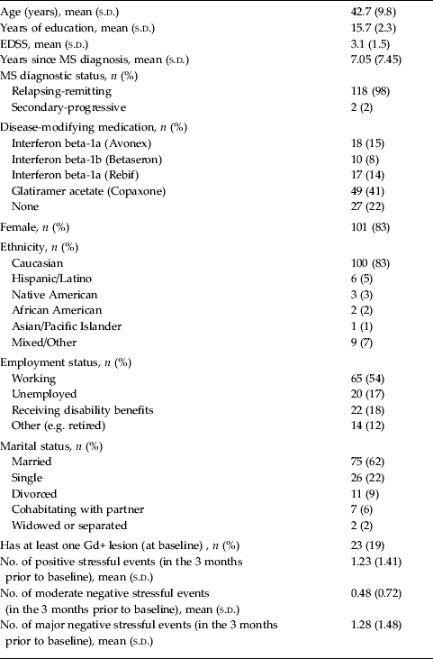
Participants were recruited from 2005 to 2008, and baseline characteristics of the resulting sample (n = 121) are displayed in Table 1. Over the course of 48 weeks, patients self-reported 165 new exacerbations, 77 of which were confirmed. Intravenous and/or oral steroids were prescribed for 27 unique patients during 39 of the confirmed exacerbations.
Demographics and baseline measures of hypothesis-relevant variables

EDSS, Expanded Disability Status Scale; MS, multiple sclerosis; Gd+, gadolinium enhancing; s.d., standard deviation.
Percentages may not total 100 due to rounding.
MRI data
A t test revealed a trend for patients in SMT-MS to have missed a greater proportion of MRI scans (meantreatment = 0.23, meancontrol = 0.12, t 119 = 1.86, p = 0.07). Baseline measures of stressful life events, perceived stress, and depression and anxiety symptoms were not correlated with the number of MRI scans completed in either SMT-MS or WLC (p's > 0.17). There was at least one MRI scan available after baseline for 113 of the 121 participants, and these were the participants included in analyses of our hypotheses (n = 60 in WLC, n = 53 in SMT-MS).
Of the 665 MRI scans possible after baseline, given the assessment schedule and exclusion of week-48 MRI scans in the control group, 527 (79%) were completed and had data available regarding Gd+ lesions; 104 (19.7%) of these 527 scans were positive for Gd+ lesions. For new or enlarging T2 lesions, data were available from 542 (82%) of the 665 possible MRI scans, and 140 (25.8%) of these 542 scans were positive. Data on previous stressful events, perceived stress, anxiety or depressive symptoms were missing, and thus imputed, for 18% of the MRI scans.
Hypotheses
Gd+ lesions
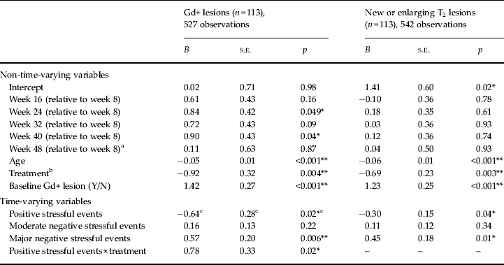
We first considered Gd+ lesions without the perceived stress and distress measures. There was no differential effect of moderate or major negative stressful events by treatment (p's > 0.30). However, as shown in Table 2, there was an overall effect of major negative stressful events [odds ratio (OR) 1.77 for each additional major negative event, 95% confidence interval (CI) 1.18–2.64, p = 0.006]. Furthermore, there was an interaction between positive stressful events and treatment (p = 0.02). Positive stressors were associated with less risk for a subsequent Gd+ lesion in WLC (OR 0.53 for each additional positive stressful event, 95% CI 0.30–0.91, p = 0.02) but not in SMT-MS (OR 1.15, 95% CI 0.76–1.74, p = 0.51).
Parameter estimates for the prediction of presence or absence of lesions

Gd+, Gadolinium enhancing; s.e., standard error.
a Week 48 data were excluded for the control group as it could have been contaminated by the workshop.
b Control = 0, Treatment = 1.
c Given the presence of the interaction effect in the model predicting Gd+ lesions, this parameter constitutes the effect of positive stressful events in the control group.
*p < 0.05, **p < 0.01.
When perceived stress, anxiety and somatic and affective depressive symptoms were added to this model, they were not predictive of Gd+ lesions (p's > 0.66), and the main effect of major negative stressful events (OR 1.76, 95% CI 1.17–2.65, p = 0.007) and the interaction between positive stressful events and treatment (p = 0.02) remained significant. Again, positive stressful events were associated with reduced risk for a Gd+ lesion in WLC (OR 0.53, 95% CI 0.30–0.93, p = 0.03) but not SMT-MS (OR 1.14, 95% CI 0.75–1.75, p = 0.54). Thus, the relationships between positive and major negative stressors and subsequent Gd+ lesions did not seem to be due to psychiatric distress or perceived stress that may have been associated with the stressful events.
T2 lesions
When we examined new or enlarging T2 lesions without the distress measures, no differential effect of the stressful event types by treatment was significant (p's > 0.26). Thus, the model without any interaction terms was used (see Table 2), revealing that positive events were associated with less risk for a subsequent new or enlarging T2 lesion (OR 0.74 for an increase of one positive stressful event, 95% CI 0.55–0.99, p = 0.04). Conversely, the odds of a subsequent new or enlarging T2 lesion increased by a factor of 1.57 for an increase of one major negative stressful event (95% CI 1.11–2.23, p = 0.01).
After adjusting for the distress and perceived stress measures (p's > 0.25), the main effect of positive (OR 0.72, 95% CI 0.54–0.96, p = 0.03) events was still significant. The main effect of major negative events (OR 1.42, 95% CI 0.99–2.04, p = 0.06) retained the same direction as a non-significant trend.
Note that all conclusions remained consistent when only the non-imputed (observed) data were analyzed.
Post-hoc analysis
To investigate whether the significant results could be explained by previous MS disease activity generating the stressful events, we reversed the analyses. We separately modeled positive and major negative stressful events as binary variables on time, age, group assignment, baseline values for that type of stressful event, and the number of Gd+ lesions on the most recent MRI scan completed 29–62 days prior. We then reran these two models using T2 lesions as a predictor rather than Gd+ lesions. We only used MRI data that were included in the analyses for the hypotheses, and no MRI data were used to predict more than one stressful event assessment. We also removed week-40 MRI scans for the control group; these participants may have attended a stress management workshop after week 40 and therefore their subsequent stressful event assessments may have been affected.
Neither Gd+ nor T2 lesions predicted positive events or major negative events (p's > 0.70). We then added interactions between lesion and group assignment to the four models. Group assignment did not significantly modify the relationship between Gd+ or T2 lesions and major negative events, or between Gd+ lesions and positive events (p's > 0.23). By contrast, there was an interaction effect of T2 lesions and group assignment on positive events (p = 0.045); however, the main effect of T2 lesions on positive events was not significant in SMT-MS (p = 0.21) or WLC (p = 0.13). Thus, there was no evidence to suggest that MS disease activity as indicated by neuroimaging was predictive of major negative or positive stressful events. All conclusions remained consistent when only the non-imputed (observed) stressful event data were analyzed.
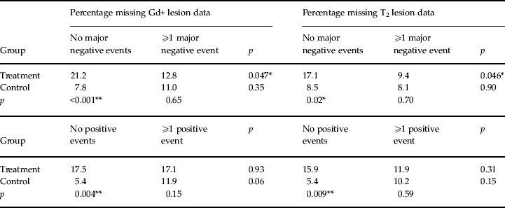
We also examined whether missing data patterns may have influenced the results. Most MRI scans that matched with observed stressful event data were completed at the assessment point 4 or 8 weeks after the stressful event assessment point. Thus, to examine stressful events and missingness of subsequent MRI data, we coded whether or not any major negative or positive stressful events (observed, non-imputed) were reported at the assessments 4 or 8 weeks prior to each MRI assessment point. In Table 3, we present the percentage of missing lesion data by group assignment and prior stressful events. χ 2 tests revealed that participants receiving SMT-MS were more likely than those in the WLC condition to have missing lesion data in the absence of both major negative and positive stressful events (p's < 0.05). Within SMT-MS, major negative events predicted a lower likelihood of missing Gd+ and T2 lesion data (p's < 0.05). There was also a non-significant trend for positive events to predict increased missing Gd+ data in WLC (p = 0.06).
Percentage of lesion data missing, by group assignment and whether the participant reported at least one stressful life event 4 or 8 weeks prior to the scheduled scan

*p < 0.05, **p < 0.01.
Discussion
Patients with MS showed an increased risk of new Gd+ lesions and new or enlarging T2 lesions on MRI scans 29–62 days (4–9 weeks) after the report of major negative life events. During the same time frame, positive stressful events were associated with less risk for subsequent new or enlarging T2 lesions in the overall sample, and with lower risk for Gd+ lesions among MS patients who were not exposed to a stress management intervention. The results were similar when controlling for measures of perceived stress, anxiety, and affective and somatic depressive symptoms; thus, the relationships found between stressful events and subsequent brain lesions do not seem to be driven by psychiatric distress associated with stressful events. The temporal relationship between stressful events and brain lesions was further established by demonstrating that brain lesions did not predict subsequent positive or major negative stressful events.
The current results are consistent with evidence of increased Gd+ lesion activity in MS patients during wartime (Yamout et al. Reference Yamout, Itani, Hourany, Sibaii and Yaghi2010), and also with studies demonstrating a relationship between negative stressful events and MS clinical exacerbations (Artemiadis et al. Reference Artemiadis, Anagnostouli and Alexopoulos2011) and a study showing that the absence of positive events predicted shorter time to MS clinical exacerbation (Brown et al. Reference Brown, Tennant, Sharrock, Hodgkinson, Dunn and Pollard2006). However, our current results are inconsistent those of with Mohr et al. (Reference Mohr, Goodkin, Bacchetti, Boudewyn, Huang, Marrietta, Cheuk and Dee2000), who found that only moderate negative stressful events, and not major negative or positive stressful events, predicted risk for subsequent Gd+ lesions. Differences in methodology may explain these inconsistencies; in the current study the sample size was considerably larger and, rather than relying on classifications of positive or negative events by the researcher, events were classified as positive versus negative according to participants' subjective evaluation.
The latency between MR images and reports of the stressful events was selected to ensure that stressful events occurred prior to the lesions, and thus were not the result of as-yet undetected Gd+ or T2 lesions. Given the 1-month follow-back period of the stressful events interview and assuming a 4-week lifespan for Gd+ lesions, the results suggest that stressful events confer risk for development of lesions immediately to 3 months later (see Fig. 1). However, as changes to normal-appearing white matter have been observed up to 3 months prior to the appearance of Gd+ lesions (Filippi et al. Reference Filippi, Rocca, Martino, Horsfield and Comi1998; Goodkin et al. Reference Goodkin, Rooney, Sloan, Bacchetti, Gee, Vermathen, Waubant, Abundo, Majumdar, Nelson and Weiner1998), our results do not necessarily imply that stressful events trigger the pathogenic factors that result in Gd+ and T2 lesions. Rather, stressful events may be one set of factors among many that determine whether underlying pathogenic processes are successfully regulated or whether they cascade to the point of detectable brain lesions (Mohr & Pelletier, Reference Mohr and Pelletier2006). For example, chronically elevated cortisol levels associated with major negative stressful events may have led to glucocorticoid receptor resistance, thereby decreasing immunoregulatory capacity (Mohr & Pelletier, Reference Mohr and Pelletier2006; Cohen et al. Reference Cohen, Janicki-Deverts, Doyle, Miller, Frank, Rabin and Turner2012). Although the literature on positive stressful events and cortisol is scarce, positive events have been linked to lower cortisol levels in pregnant women (Pluess et al. Reference Pluess, Wurmser, Buske-Kirschbaum, Papousek, Pirke, Hellhammer and Bolten2012); thus, future studies should explore whether positive events may have decreased risk for MS brain lesions by lowering participants' risk for glucocorticoid receptor resistance.
Although the stress management therapy provided to half the participants in this study resulted in fewer Gd+ and T2 lesions and fewer reported negative stressful events (Mohr et al. Reference Mohr, Lovera, Brown, Cohen, Neylan, Henry, Siddique, Jin, Daikh and Pelletier2012), the treatment did not alter the relationship between negative stressful events and the subsequent development of new lesions. Although SMT-MS may have improved patients' strategies for coping with stressors, perhaps its beneficial effect on lesion activity was achieved by helping patients learn to avoid negative stressors altogether. This interpretation is supported by the finding that psychiatric distress and perceived stress, the putative sequelae of negative stressful events, were unrelated to lesion activity. This is also consistent with previous work reporting that new lesions may be predicted by negative stressful events but not by distress (Mohr et al. Reference Mohr, Goodkin, Bacchetti, Boudewyn, Huang, Marrietta, Cheuk and Dee2000). These findings suggest that stress management interventions aimed at reducing the development of new brain lesions should focus more on teaching patients to prevent major negative stressful events and increase positive engagement, even if positive events involve some amount of stress.
The finding that positive stressful events were only related to subsequent Gd+ lesions in the control arm and not to the SMT-MS arm was unexpected. We speculate that the treatment itself may have been a positive stressor that overwhelmed the variability in other positive stressors, thereby decreasing the relationship between measured positive stressors and Gd+ lesions. However, this moderating effect of treatment was not seen for T2 lesions, and further study is needed to clarify these inconsistent results.
Limitations
In a post-hoc analysis, we found patterns of missing data relating to treatment and stressful events. Among participants who did not report any major negative and positive stressful events, those receiving SMT-MS were more likely to subsequently have missing MRI data than WLC participants. As none of the primary results pertained specifically to the SMT-MS group, however, this pattern is unlikely to influence the interpretation of the results. Within SMT-MS, major negative events predicted missing MRI data. However, major negative events predicted lesions across both groups, indicating that this missing data pattern also fails to provide an adequate alternative explanation for the primary results. Finally, there was a trend for positive events in the WLC condition to predict missing Gd+ lesion data. For this missing data pattern to negate the relationship found between positive events and decreased Gd+ lesions in WLC, the WLC participants with positive events who missed their MRI scans would also need to be more likely to be experiencing Gd+ lesions. Thus, it is possible but perhaps unlikely that even this one primary result can be explained by missing data.
There are also limitations to this study in terms of methodology. As participants in this study were enrolled in a trial involving a stress management therapy, their experiences with stress may differ from the general population of individuals with MS. Our instrumentation was also unable to detect the changes to normal-appearing white matter that precede the emergence of Gd+ and T2 lesions, and we were thus unable to determine whether stressful events preceded or followed these early pathogenic processes. More frequent MRI scans and assessments of stressful life events would also have allowed greater precision in determining the time lag between the occurrence of events and changes in risk for lesion development.
It is possible that the relationship between positive and major negative stressful events and subsequent lesion activity may differ based on a patient's stage of MS disease progression. Future studies should examine this possibility, as all participants in the current study were in the relapsing stage. Finally, although neuroimaging markers of MS disease activity were not predictive of stressful events, the experience of MS symptoms, or a subset thereof, may affect the occurrence and/or reporting of stressful events. It was beyond the scope of this study to examine relationships between clinical symptoms of MS and stressful events. However, it should be acknowledged that the progression of MS can in itself generate stressful events, and future studies should tease apart the predictive utility of stressful events versus clinical symptoms of MS on subsequent lesion activity.
Conclusions
This is the largest longitudinal study to date examining the relationship between stressful events and neuroimaging markers of MS. Major negative stressful events were found to be associated with increased risk for subsequent Gd+ and new or enlarging T2 lesions in patients with MS whereas positive stressful events were associated with decreased risk. Future studies should evaluate potential moderators. For example, if positive events occur that are incongruent with the individual's self-concept, they may exert a detrimental rather than a beneficial effect on health (Shimizu & Pelham, Reference Shimizu and Pelham2004), and particular coping styles may ameliorate or aggravate the effect of negative stressful events on Gd+ lesions (Mohr et al. Reference Mohr, Goodkin, Nelson, Cox and Weiner2002). Research that identifies patients who are most vulnerable to the effects of major negative stressful events, and most responsive to the potential benefits of positive life events, can shed light on the pathogenesis of brain lesions in MS and assist with prognosis in clinical settings.
The finding that perceived stress and distress were not statistically associated with the development of new lesions is consistent with a prior study in people with MS (Mohr et al. Reference Mohr, Goodkin, Bacchetti, Boudewyn, Huang, Marrietta, Cheuk and Dee2000), but inconsistent with common models of stress that posit perceived stress and/or negative affect as mediators between stressful events and health outcomes (Lazarus & Folkman, Reference Lazarus and Folkman1984; Cohen et al. Reference Cohen, Kessler, Gordon, Cohen, Kessler and Gordon1995; McEwen & Gianaros, Reference McEwen and Gianaros2010). This indicates the need to develop and evaluate MS-specific biobehavioral models to explain the relationship between stress and neuroimaging markers of MS disease activity.
Acknowledgments
This study was funded by National Institute of Child Health and Human Development grant R01-HD043323 and National Institute of Mental Health grant K08-MH094441-01A1. This study is registered on clinicaltrials.gov (NCT00147446).
Declaration of Interest
None.