




No CrossRef data available.
Article contents
An ultrastructural study of excretory system development in the cercariae of Prosorhynchoides gracilescens (Rudolphi, 1819) and Prosorhynchus squamatus Odhner, 1905 (Digenea, Bucephalidae)
Published online by Cambridge University Press: 06 August 2004
Abstract
The ultrastructure of the developing excretory system of Prosorhynchoides gracilescens and Prosorhynchus squamatus cercariae is described. The development pattern was similar in both species. In early embryos the two main collecting tubes were composed of a layer of cells which were wrapped around the lumen. Later, the tubes fused and the excretory epithelium of the fusion zone and that of the lateral caudal ducts became a syncytium. The collecting tubes in the cercarial body retained their cellular organization. As the tails grew, additional excretory pores were formed in the tail stem where thickened portions of the caudal duct epithelium contacted the surface tegument. Following this, the distal portions of the lateral caudal ducts lost contact with the primary excretory pores and progressively degenerated. Excretory atrium development started with differentiation of secretory active cytons peripheral to the fusion zone. These cells gave rise to cytoplasmic extensions that penetrated the fusion zone wall to eventually form a continuous cytoplasmic layer. This layer eventually replaced some of the fusion zone excretory epithelium and became the lining of the excretory atrium. The anterior end of the fusion zone differentiated into an excretory bladder and a short posterior portion gave rise to the caudal vesicle.
Information
- Type
- Research Article
- Information
- Copyright
- 2004 Cambridge University Press
INTRODUCTION
This study deals with the development of the excretory system in two bucephalid cercariae. Like all cercariae, these organisms initially develop inside an intramolluscan parasite before becoming active free-living larvae that, given the appropriate circumstances, mature into adult parasitic flukes. As a result, it would be expected that the excretory system in mature cercariae should be appropriate for their own requirements and also should possess all the components to deal with the excretory and osmoregulatory problems to be encountered in the next host, in this case a teleost fish. The two bucephalid cercariae chosen for this study initially develop inside branching sporocysts that live in the haemocoel of the pallium, the gonad and digestive gland of marine bivalve hosts. While in that location each cercaria firstly develops a body region that will eventually become the body of an adult fluke. In addition, it also develops a sophisticated and highly muscular tail. The tail, however, is only a temporary feature that is required for a fairly short period of relatively high metabolic activity after the cercaria is released from its sporocyst and escapes from the bivalve. When temporally free-living, the tail is likely to produce considerable amounts of nitrogenous wastes that could constitute a problem if not dealt with. However, as soon as the next host (marine teleosts) is encountered, the tail is immediately discarded taking with it its component of the excretory system without disrupting the remainder which is left behind in the body region. At that point the excretory system must be fully functional and ready to operate to its full capacity as the young fluke develops to maturity.
Transmission electron microscopy was adopted to follow the development of two cercarial excretory systems in this study. The formation of flame cells and the terminal parts of protonephridia is not described as that will be the subject of a separate publication. Instead, this study focuses on the formation and development of the system of main collecting tubes, the excretory bladder and excretory pores. These features of the cercarial excretory system are considered as important criteria in digenean taxonomy. Knowledge of patterns of excretory system development provides for a more accurate interpretation of structures found in fully formed larvae. This has been clearly demonstrated in a number of special comparative embryological investigations of trematode excretory systems (Hussey, 1941, 1943; Kuntz, 1950, 1952; and others) and also by numerous observations on excretory system development included in studies of digenean life-cycles (see reviews by Galaktionov & Dobrovolskij, 1987, 2003). All the studies referred to above were carried out using light microscopy. Relatively little information on the ultrastructure of developing cercarial excretory systems is available and it appears to be limited to that of Krupa, Cousineau & Bal (1969) and Rees (1977) who provided detailed information on this subject for Cryptocotyle lingua. In addition, Rohde & Watson (1992) described the formation of the terminal parts of protonephridia and the ultrastructure of the primary excretory pores of Philophthalmus sp. cercariae. Some data on the later stages of development of the excretory bladder were also provided for cercariae of Ochetosoma aniarum by Powell (1972) and for several microphallid larvae by Malkova & Galaktionov (1989).
The development of the excretory system in bucephalid cercariae was studied for Rhipidocotyle papillosum (syn. Bucephalus papillosus), Bucephalus elegans, R. septpapillata, Prosorhynchus crucibulum and Prosorhynchoides gracilescens (syn. Bucephaloides gracilescens) using light microscopy (Woodhead, 1929; Hussey, 1943; Kniskern, 1952; Matthews, 1973, 1974). The aim of the present study is to describe the ultrastructural aspects of excretory system development in the cercariae of Prosorhynchoides gracilescens (syn. Bucephaloides gracilescens) and Prosorhynchus squamatus as the continuation of earlier studies of Podvyaznaya & Galaktionov (2004) on the fine structure of the excretory system of mature cercariae of the same species.
MATERIALS AND METHODS
Embryonic cercariae of Prosorhynchoides gracilescens and Prosorhynchus squamatus were obtained from sporocysts dissected out of naturally infected bivalves Abra prismatica (Montagu, 1803) and Mytilus edulis L. respectively. Specimens of A. prismatica were collected from the depth of 60–80 m off the southern coast of Iceland in September 2000 and those of M. edulis – from the sublittoral zone of the Onega Bay of the White Sea (Russia) in August 2001. Small pieces of sporocysts, together with surrounding host tissue, were fixed for 7–10 days in 3 or 4% glutaraldehyde in 0·1 M cacodylate buffer (pH=7·4) containing sucrose (760 μosmol in total). After rinsing in buffer the material was post-fixed for 3–4 h at 4 °C in 1% osmium tetroxide in 0·1 M cacodylate buffer, rapidly dehydrated in ethanol and acetone and embedded in Epon–Araldite mixture. While in liquid Epon–Araldite mixture the sporocyst walls were opened with fine needles to release the cercarial embryos. The embryos were sorted according to developmental stages and thereafter were treated separately. Series of thin cross- and longitudinal sections of developing cercariae were cut on LKB III ultramicrotome, double stained with uranyl acetate and lead citrate and examined with Jeol 1200 and/or Leo 900 electron microscopes. In order to trace the developmental sequence of cercariae and that of their excretory system, living specimens were studied using light microscopy and, in the case of P. gracilescens, the literary data (Matthews, 1974) were also used.
RESULTS
The developmental sequence of the excretory system in P. gracilescens and P. squamatus cercariae was traced in all stages of embryos from those with furcae only represented by small buds to those that were almost fully formed. They are presented schematically in Figs 1 and 2. Morphogenesis of the excretory system was very similar in both species and 5 consecutive developmental stages could be recognized. In both P. gracilescens and P. squamatus cercariae the stages appeared to be closely correlated to the formation of the tail.
The first stage
The first elements of the excretory system observed in P. gracilescens and P. squamatus larvae were a pair of primary tubes that opened to the outside at the posterior end of the embryo. The capillaries from a pair of laterally situated flame cells joined the main collecting tubes (a term adopted from Galaktionov & Dobrovolskij, 1987, 2003) just posterior of the pharynx primordium, while the primary excretory pores were located on the inner surface of furcal buds (Figs 1A, 2A). This stage of development persisted until the tail became clearly distinct from the embryo body. Electron microscopy revealed that, throughout their length, these main collecting tubes were composed of separate cells (Figs 1A, 2A, 3A). The number of the cells comprising each tube was not determined, but at least 2 cells were observed in the short region of each furcal primordium. The cells were characterized by well-developed RER, numerous free ribosomes and large distinct mitochondria (Fig. 3A). Occasional small vesicles and microtubules were observed in their cytoplasm and their nuclei contained condensed chromatin at the periphery and small electron-dense bodies. The cells were folded in a ‘c’ shape so that they encircled a narrow lumen. The abutting tips of cells were flattened and attached to one another by a septate junction (Fig. 3A,B). The luminal plasma membrane became progressively folded into ridge-like or lamellated projections which were more abundant in the distal, dilated portions of the tubes. Close to the excretory pores, septate junctions attached the cells of collecting tubes to the cercarial tegument (Fig. 3B). At this point the tegument was thickened slightly and covered by irregular projections.

Fig. 1. Diagram summarizing five consecutive stages of excretory system development in Prosorhynchoides gracilescens cercariae (the number of cells and the number of nuclei in the excretory syncytium are represented arbitrarily). (A) I stage. (B) II stage. (C) III stage. (D,E) IV stage. (F) V stage. act, Anterior collecting tubule; acy, cyton of excretory atrium syncytium; aep, additional excretory pores; as, excretory atrium syncytium; cp, capillary; cv, caudal vesicle; ea, excretory atrium; eb, excretory bladder; fc, flame cell; fz, fusion zone; lcd, lateral caudal duct; mcd, median caudal duct; mct, main collecting tube; pct, posterior collecting tubule; pep, primary excretory pore.

Fig. 2. Diagram summarizing five consecutive stages of excretory system development in Prosorhynchus squamatus cercariae. (A) I stage. (B,C) II stage. (D) III stage. (E,F) IV stage. (G) V stage. For abbreviations see Fig. 1. (From Galaktionov and Dobrovolskij (2003), fig. 65, p. 143, with kind permission of Kluwer Academic Publishers.)

Fig. 3. Excretory system of early embryonic cercariae of Prosorhynchus squamatus and Prosorhynchoides gracilescens. (A) Main collecting tube (MCT) at the first stage of excretory system development in P. squamatus. Cross-section showing septate junction (SJ) where the tips of tube cell abut. L, lumen; MT, mitochondrion; N, nucleus; RER, rough endoplasmic reticulum. (B) Region of primary excretory pore in the same embryo. Note septate junction (SJ) attaching the main collecting tube (MCT) to the tegument (T), a septate junction (SJ) between the tips of tube cell and the increased number of projections on the luminal surface of the main collecting tube. (C–F) The second stage of excretory system development. (C) Cross-section through the posterior portion of the fusion zone in a P. squamatus embryo. Note absence of septate junctions in the duct wall. FZE, fusion zone epithelium; L, lumen; N, nucleus; RER, rough endoplasmic reticulum. (D) Cross-section through the middle portion of lateral caudal duct (LCD) in a P. squamatus embryo. Note narrowing of duct and absence of septate junctions in its wall. L, lumen. (E) Longitudinal section through the fusion zone (FZ) (boundaries are marked by arrowheads) of a P. squamatus embryo showing numerous nuclei in the syncytial lining. L, lumen. (F) Bulge in the proximal portion of a lateral caudal duct (LCD) (boundaries are marked by arrowheads) of a P. gracilescens embryo. In this species the bulge appears to be associated with a slight constriction in each lateral lobe of the tail stem. Note nuclei (N) in excretory syncytium and absence of cell boundaries between adjacent nuclei. L, lumen; T, tegument.
The second stage
As each embryo became larger and more elongated a distinct furrow developed to separate the increasingly large primordial stem and furcal buds from the remainder of the cercarial body. As this happened, the main collecting tubes moved closer to one another and eventually fused to form a short median duct in the posterior region of the body (Figs 1B, 2B). This duct, referred to as the ‘fusion zone’, just extended into the tail primordium where the posterior portions of the main collecting tubes remained separate. Here, these ducts, now referred to as lateral caudal ducts, ran to the tip of each developing furca where they opened to the exterior as they had done previously. By the time the process of fusion was complete, the cells in the fusion zone and those in the lateral caudal ducts were found to have become confluent, thereby forming a syncytium (Fig. 3C,D,F). As development proceeded, the number of nuclei in the fusion zone sharply increased so that they appeared to occupy the bulk of the syncytial lining (Fig. 3E). During this period the furcae continued to grow rapidly resulting in an equivalent elongation of the lateral caudal ducts (Figs 1B, 2C). The walls of the ducts became extremely thin (Fig. 3D). However, this was not the case at the proximal portion of each duct where a distinct symmetrical bulge projected towards the antero-lateral surface of the tail stem primordium (Figs 1B, 2C). When investigated by TEM it was apparent that these bulges were formed by a local thickening of the syncytium and they always contained 1 or 2 nuclei (Fig. 3F).
The unfused portions of the main collecting tubes in the cercarial body (Figs 1B, 2B,C) retained their cellular organization and were connected to the fusion zone syncytium by septate desmosomes. As development proceeded, the flame cell number increased and, accordingly, the network of collecting tubes in the embryo body became more extensive. By the time that fusion of the main collecting tubes was complete, the anterior and posterior collecting tubules had taken on the appearance of branches of the main collecting tubes (Figs 1B, 2B) and the fine structure of each was found to be similar. During subsequent development to fully formed cercariae, the multiplication of the flame cells appeared to be accompanied only by the appearance of new capillaries and small collecting tubules.
The third stage
The third stage of excretory system development coincided with a period in which tail stem lobes became distinct and from these lobes the furcae continued to grow (Figs 1C, 2D). At this time additional excretory pores developed in the tail stem at the points where the previously mentioned bulges in the lateral caudal ducts occurred. The number of nuclei increased (to 3–5) in these small portions of the excretory syncytium and this appeared to result in them becoming thicker (Fig. 4A). In the same region a number of short extensions originating from the syncytium cytoplasm could be seen extending towards the tail stem surface. These extensions, which passed between the developing muscles, were anchored to the surface tegumental layer by septate junctions (Fig. 4A,B). There were 5 or 6 points of contact between the excretory syncytium and the tegument in P. squamatus embryos and at this time cavities could be detected in the thickest of the cytoplasmic extensions (Fig. 4B). It appeared that these cavities extended to connect with the duct lumen on one side and, on the other side, they eventually broke through the tegument to form the additional excretory openings. Favourable serial sections of P. squamatus embryos indicated that several openings were initially formed on each side of the tail (Fig. 4B). In more developed larvae (Fig. 2F) however, only one excretory pore was identifiable on each side of the tail. Although not observed, it is assumed that the single pore was probably formed by fusion of the adjacent openings and associated cytoplasmic extensions of the excretory syncytium. In P. gracilescens cercariae only 1 pair of excretory openings was ever identified during the course of development of the additional excretory pores. This was in spite of the fact that 2 or 3 points of contact between the caudal duct excretory syncytium and the surface tegument were initially observed on either side of the tail (Fig. 1C). In both worm species, when the newly formed excretory openings appeared, the distal portions of the lateral caudal ducts appeared to progressively degenerate from the region of the primary excretory pores towards the bases of the tail furcae (Figs 1C, 2D).

Fig. 4. The third stage of excretory system development in Prosorhynchus squamatus and Prosorhynchoides gracilescens embryonic cercariae. (A,B) Longitudinal sections showing formation of additional excretory pores in P. squamatus. Note several nuclei (N) in a thick portion of duct syncytium, a number of cytoplasmic extensions (arrowheads) projected towards the tail stem surface and septate junctions (SJ) connecting the excretory syncytium to the tegument (T). (B) Note also formation of several excretory openings (EO). Asterisks indicate inner cavities in cytoplasmic extensions. L, lumen; LCD, lateral caudal duct; M, muscles. (C,D) Longitudinal sections through the anterior portion of the fusion zone (AFZ) of P. gracilescens (C) and P. squamatus (D) embryonic cercariae. Note position of junctions (SJ) between the main collecting tubes (MCT) and the fusion zone epithelium. L, lumen; LA, lamellae; MY, developing myocyte. (E) Fusion zone epithelium in P. gracilescens embryonic cercaria. Note Golgi complex (GC), numerous dense mitochondria (MT) and sparse rough endoplasmic reticulum (RER). L, lumen; N, nucleus.
During the period in which the additional excretory pores were forming changes were also observed in the structure of the fusion zone. Light microscopy indicated that in P. gracilescens embryos the fusion zone extended proximally so that the main collecting tubes appeared to join it at about one-third along its length (Figs 1C, 4C). In P. squamatus larvae the septate junctions between the fusion zone syncytium and the cells of the main collecting tubes remained at the anterior terminal position (Figs 2D, 4D) suggesting that, in this case, most growth might have occurred in the middle portion of the duct. In both species the syncytium cytoplasm in the anterior portion of the fusion zone increased in volume and the lumen enlarged (Fig. 4C,D). Transmission electron microscopy showed that, at this stage of development in P. squamatus embryos, the luminal surface possessed folds that formed lamellated projections. These were not observed at the same stage in P. gracilescens and the syncytium cytoplasm contained numerous small mitochondria, Golgi complexes, free ribosomes and some RER (Fig. 4E). A thin basal lamina and developing myocytes were apparent around the fusion zone syncytium. Developing myocytes were more numerous near the posterior portion of the duct close to the body–tail junction (Fig. 5A,B). In P. squamatus embryonic cercariae this portion was relatively constricted and possessed few nuclei (Fig. 5A), while in equivalent P. gracilescens the number of nuclei remained more or less constant and the fusion zone remained relatively wide (Fig. 5B).

Fig. 5. Sections showing details of the late third stage of excretory system development in Prosorhynchus squamatus and Prosorhynchoides gracilescens embryonic cercariae. (A,B) Longitudinal sections through the posterior portion of the fusion zone (PFZ) in P. squamatus (A) and P. gracilescens (B) embryonic cercariae. In P. squamatus this portion is constricted and devoid of nuclei, while in P. gracilescens it is wide and contains nuclei (N). Note developing myocytes (MY) around the excretory syncytium (in P. squamatus they are more immature). L, lumen. (C,D) Collecting tubules of P. gracilescens (C) and P. squamatus (D) embryonic cercariae showing well pronounced Golgi complexes (GC) and various types of vesicles including coated vesicles (CVE) and electron-dense vesicles (VE). L, lumen; MB, multilamellar body; MI, microtubule; N, nucleus; SJ, septate junction.
Cercariae at this stage of development also displayed some changes in the fine structure of the cells of collecting tubules. Here the Golgi apparatus became well pronounced and numerous vesicles appeared in the cytoplasm. These included coated vesicles and small membrane-bound bodies filled with electron-dense material (Fig. 5C,D). Multilamellar bodies were now apparent, extending from the walls of collecting tubes and into the lumen. There appeared to be less RER. The ultrastructure of the collecting tubes appeared to retain this structure throughout subsequent development to fully formed cercariae.
The fourth stage
Progressive development of the excretory atrium was observed in cercariae that were undergoing intensive development of the tail musculature. At the same time, special tegumental cells associated with the tail tegument became differentiated and the portions of the lateral caudal ducts that were distal to the additional excretory pores progressively degenerated (Figs 1D,E, 2E,F). First evidence of development of the excretory atrium epithelium observed was the differentiation of several secretory cells near the posterior portion of the fusion zone, alongside developing myocytes. These cells were identified by their well-developed RER, pronounced Golgi complexes and small secretory inclusions of moderate electron density (Fig. 6A). Eventually, a number of cytoplasmic extensions from these cells were identified projecting towards the fusion zone. The extensions, which were packed with secretory inclusions, passed between the developing muscles to the excretory syncytium where they were attached by septate desmosomes (Fig. 6B). As they penetrated for some distance into the wall of the fusion zone, they extended laterally between the overlying excretory syncytium and underlying basal lamina and eventually appeared to become confluent as a thin continuous cytoplasmic layer (Fig. 6C). As development proceeded this layer appeared to wrap round a portion of the posterior region of the fusion zone. At this stage a few electron-lucent vacuoles were identified in the newly formed cytoplasmic layer (Fig. 6C) and in more mature specimens they appeared to have fused to give rise to larger cavities (Fig. 7A). Our observations suggested that these cavities increased in size and eventually became continuous with the duct lumen but, unfortunately, all details of this process were not observed. However, it was apparent that, as the cytoplasmic layer progressively increased in complexity, the excretory syncytium lost its integrity and disintegrated (Fig. 7B), leaving the cytoplasmic layer as the new lining (Fig. 8A). (As this process was taking place in P. gracilescens larvae, progressively fewer nuclei could be seen in the epithelium of the posterior portion of the fusion zone. Eventually, no nuclei were left in this region.) It should be remembered that the new excretory atrium syncytium lining retained its continuity with nucleated cytons which were now peripheral to a layer of well-pronounced circular muscles and some longitudinal muscle fibres that developed around the basal lamina of the syncytium (Figs 6C, 7A,B, 8A). From the earliest stages observed, the surface cytoplasmic layer of the excretory atrium syncytium was largely filled with secretory inclusions similar to those observed in the cytons. The contents of the inclusions were seemingly continuous with and possibly responsible for the characteristic thick glycocalyx coating the luminal plasma membrane (Fig. 7A,B).

Fig. 6. The fourth stage of excretory system development in Prosorhynchus squamatus and Prosorhynchoides gracilescens embryonic cercariae. (A) A differentiating secretory cyton (ACY) near the posterior portion of the fusion zone in a P. squamatus embryonic cercaria. Note pronounced Golgi complex (GC), rough endoplasmic reticulum (RER) and secretory inclusions (SI). MT, mitochondrion; N, nucleus. (B) Longitudinal section through the posterior portion of the fusion zone of P. squamatus embryonic cercaria showing curved cytoplasmic extension (arrows) of a secretory cyton which is attached to the fusion zone epithelium (FZE) by a septate junction (SJ). Note secretory inclusions (SI) in this cytoplasmic extension. L, lumen; M, muscles. (C) Longitudinal section of P. gracilescens embryonic cercaria showing the posterior portion of the fusion zone which has been invaded by a cytoplasmic layer (CL) formed by fusion of cytoplasmic extensions from secretory cytons. Note developing circular muscles (CM) around the newly formed cytoplasmic layer (CL) and a nucleus (N) in epithelium of the posterior portion of the fusion zone (FZE). Arrowheads indicate electron-lucent vacuoles in cytoplasmic layer. B, cercarial body; BTJ, body–tail junction; L, lumen; TL, tail.

Fig. 7. Longitudinal sections of Prosorhynchus squamatus embryonic cercariae showing consecutive stages of excretory atrium formation. (A) Note large cavity in the cytoplasmic layer (CL) seemingly formed by fusion of electron-lucent vacuoles (arrowheads). ACY, cyton of excretory atrium syncytium; BL, basal lamina; CM, circular muscles; FZE, fusion zone epithelium; GL, glycocalyx; L, lumen; MY, developing myocyte; SJ, septate junction. (B) Note disintegration of fusion zone epithelium (FZE) over new lining. Note also septate junctions (SJ) connecting the atrium lining to the excretory syncytium anteriorly and posteriorly, secretory inclusions (SI) in the cyton (ACY) and surface cytoplasmic layer of excretory atrium syncytium (AS), glycocalyx (GL) covering the luminal surface and well-developed circular muscles (CM). Arrow indicates connection between cyton and surface cytoplasmic layer. MY, developing myocyte; N, nucleus.

Fig. 8. Excretory system of late embryonic cercariae of Prosorhynchus squamatus and Prosorhynchoides gracilescens. (A) Longitudinal (sagittal) section of P. squamatus embryonic cercaria showing the late fourth stage of excretory system development. Primordial excretory bladder (EB), excretory atrium (EA) and median caudal duct (MCD) are well defined. Arrowheads indicate connections between the surface cytoplasmic layer of excretory atrium syncytium (AS) and deeper nucleated cytons (ACY). B, cercarial body; BTJ, body-tail junction; CM, circular muscles; SJ, septate junctions; TL, tail. (B) Longitudinal (frontal) section of P. gracilescens embryonic cercaria showing the early fifth stage of excretory system development. Note well-developed longitudinal muscles (LM) of the excretory atrium (EA) and early stages of enlargement of the excretory atrium (arrows) in the region of body–tail junction (BTJ). The median caudal duct (MCD) is not yet dilated to form the caudal vesicle and is only slightly enlarged at its anterior end. AS, excretory atrium syncytium; CM, circular muscles; EB, excretory bladder; LCD, lateral caudal duct; SJ, septate junctions; TL, tail. (C) Formation of the caudal vesicle (CV) in a P. squamatus cercaria. The anterior portion of the caudal vesicle is lined by an extension of the excretory atrium syncytium (AS). The median caudal duct is clearly enlarged to form the posterior portion of the caudal vesicle, its wall has become thin and devoid of surface lamellae. Note constricted extensions of the excretory syncytium (arrows) containing mitochondria. LCD, lateral caudal duct; SJ, septate junctions.
Observations of cercariae in which the excretory atrium syncytium was developing indicated that the portion of the fusion zone situated anteriorly to the excretory atrium became larger, eventually developing into the excretory bladder (Figs 1E, 2F). At the same time the posterior portion of the fusion zone became reduced to become the short median caudal duct (Figs 1E, 2F, 8A). In P. squamatus cercariae the luminal surface of this duct was covered by numerous ridge-like and lamellate projections that were absent in P. gracilescens larvae.
The fifth stage
During the fifth stage, development of the excretory bladder, the excretory atrium and lateral caudal ducts was completed (Figs 1F, 2G, 8B) and the lining of each acquired all the features seen in mature cercariae and described by Podvyaznaya & Galaktionov (2004). The median caudal duct increased in width to become the caudal vesicle (Figs 1F, 2G). In P. squamatus cercariae the excretory atrium syncytium extended posteriorly as the lining of the anterior portion of this vesicle (Fig. 8C). Completion of the development of the excretory system coincided with the final stage of tegument development which was represented by the appearance of tegument cell secretory products in the tegument surface cytoplasmic layer.
DISCUSSION
When compared with information on other bucephalid cercariae, the general plan of the excretory system development in P. gracilescens and P. squamatus larvae most closely resembles that described by Hussey (1943) for Bucephalus elegans. Kniskern (1952) reported the presence of a commissural vessel between the main collecting tubes in early embryos (blunt furcae stages) of Rhipidocotyle septpapillata. We found no similar structure at the equivalent stage of development in P. gracilescens and P. squamatus, nor did Hussey (1943) in B. elegans or Matthews (1973) in Prosorhynchus crucibulum. It might be supposed, however, that in R. septpapillata larvae fusion of the main collecting tubes begins earlier than in other bucephalid cercariae and that the transversal commissural vessel represents the early stage of this process. Matthews (1973, 1974) described the continuing presence of lateral caudal ducts in the furcae of fully formed P. gracilescens and P. crucibulum cercariae. According to our observations, in P. gracilescens and P. squamatus developing larvae the lateral caudal ducts degenerated in an anterior direction from their posterior limits to the region of the excretory pores in the tail stem, before the cercariae reached maturity. A similar developmental sequence was also recorded by Hussey (1943) in cercariae of B. elegans and by Kniskern (1952) in R. septpapillata larvae.
In spite of the above-mentioned minor discrepancies in the descriptions of members of four bucephalid genera (Bucephalus elegans, Rhipidocotyle septpapillata, Prosorhynchoides gracilescens, Prosorhynchus crucibulum and P. squamatus) the basic developmental pattern of the cercarial excretory system is essentially similar. A radically different type of excretory system morphogenesis was described by Woodhead (1929) for Rhipidocotyle papillosum cercariae. According to Woodhead, the early embryos possessed a single excretory pore positioned between the furca buds. In their later ‘middle furcae stage’ he described and provided a figure of 1 pair of excretory pores at the tips of furcae and 3 pairs of additional excretory pores in the tail stem primordium (Woodhead, 1929, Fig. 7). In the light of more recent investigations it would appear that the pattern of excretory system development in R. papillosum cercariae should be checked.
The fact that the excretory pores in the tail stem of bucephalid cercariae appear as a secondary development in the course of excretory system morphogenesis has been shown previously by Hussey (1943), Kniskern (1952) and Matthews (1973, 1974). Our ultrastructural study of P. gracilescens and P. squamatus embryonic cercariae revealed that these pores are formed by contact of thickened portions of lateral caudal duct excretory syncytium with the tail stem tegument. In P. squamatus larvae this process, when compared with the same process in P. gracilescens cercariae, appeared to progress in small steps over a long period. Perhaps this can be attributed to the inaccessibility of the tail stem tegument due to the early development of a layer of thick muscles under the tegument in this species.
None of the earlier investigators have drawn attention to the fact that the secondarily formed excretory pores of bucephalid cercariae are temporary structures. They substitute the primary excretory pores in early larvae and presumably function until the cercaria discards its tail leaving the unpaired definitive excretory pore to open at the posterior end of the body. Our study indicates that, in bucephalids, excretory pores are formed on 3 occasions during development of the protonephridial system. In all other digeneans there is typically only 1 pair of primary excretory pores and a single definitive excretory pore which is secondarily derived. From an evolutionary point of view, the excretory pores in the tail stem of bucephalid cercariae are most likely relatively recent (tertiary) developments, the origin of which is related to the highly modified nature of the larval tail. This is the reason that we have referred to them as ‘additional’ pores rather than ‘secondary’ pores as accepted in the literature (Hussey, 1943). The tail of bucephalid cercariae possesses extremely long and contractile furcae which start to grow intensively in early embryos. The lateral caudal ducts originally open near the tips of these appendages. If the excretory pores did not change their position until completion of larval development, the lateral caudal ducts that extend the whole length of each furca would become so inefficient that they could not transport the metabolic wastes fast enough. In addition, these ducts appear to be limited in their capacity for growth. It was clear that, in P. gracilescens and P. squamatus embryos, the initial extension of lateral caudal ducts in elongating furcae (II stage) coincided with their walls becoming distinctly thinner and an associated reduction in lumen width. A noticeable increase of excretory syncytium volume was only observed at the points where the additional excretory pores were formed.
The excretory atrium of mature P. gracilescens (syn. Bucephaloides gracilescens) and P. squamatus cercariae was described by Podvyaznaya & Galaktionov (2004) as a short, broad median duct positioned in close proximity to the body–tail junction and lined by a syncytial epithelium which was structurally similar to that of the tegument. In the region of the body–tail junction this epithelium abutted with the outer tegument and this led the authors to conclude that the excretory atrium of P. gracilescens and P. squamatus larvae was a primordial definitive excretory pore (which would open to the exterior after the loss of the tail). The lack of continuity between the atrium lining and the outer tegument in mature cercariae suggested that the excretory atrium might have been formed independently of the body wall. The results of the present investigation have confirmed this assumption. It has shown that the excretory atrium syncytium was derived from several cells which initially differentiated near the posterior portion of the fusion zone as separate secretory cytons. Cytoplasmic extensions from these cytons grew into the wall of the fusion zone, expanded over its basal lamina and fused to form a continuous cytoplasmic layer which gradually substituted a portion of excretory epithelium. The sequence of morphological changes accompanying development of the atrium lining in P. gracilescens and P. squamatus cercariae is comparable with that described for the formation of tegument in mother sporocysts of Fasciola hepatica and Schistosoma mansoni (Southgate, 1970; Meuleman et al. 1978). This fact is noteworthy because a very different pattern of tegument formation is considered to be typical for trematode cercariae (Smyth & Halton, 1983; Halton & McCrae, 1985; Galaktionov & Dobrovolskij, 1987, 2003). In cercariae, an anucleate cytoplasmic layer is formed independently of cytons and the two become connected only in the later stages of tegument morphogenesis.
In all digeneans investigated the fully developed definitive excretory pore is lined by a prolongation of the body tegument (Smyth & Halton, 1983; Galaktionov & Dobrovolskij, 1987, 2003). The only ultrastructural observation on the process of definitive excretory pore formation has been made by Rees (1977) for cercaria of Cryptocotyle lingua. In developing C. lingua larvae the definitive excretory pore results from invagination of the body wall and its lining is formed by an inward extension of the surface cytoplasmic layer. The present study showed that in bucephalid cercariae the tegumental lining of the primordial definitive excretory pore originates as a separate structure. Even these sparse comparative data show that the pattern of definitive excretory pore development may differ greatly in various groups of digeneans. Further investigations are needed to comprehend the value of this feature for digenean phylogeny and taxonomy.
It is quite apparent that the development of the excretory system that we have described in the bodies of these bucephalid cercariae provides all the fundamental components of the adult excretory system. However, perhaps surprisingly, at its completion the tail furcae are devoid of both flame cells and excretory ducts. It would appear therefore that nitrogenous wastes produced by the massive tail musculature have no mechanisms for their expulsion. Possibly it may be stored in the central lumen of the furcal rami or the tail stem. When released from their molluscan host bucephalid cercariae do not swim actively. Their furcal rami appear to twist rather than beat, and their action keeps the cercariae suspended in the water for several days. The tails of numbers of cercariae become knotted together so that they form ‘nets’ that have neutral buoyancy and move freely in the water on microcurrents (Wardle, 1988). This naturally increases the chances of the cercariae encountering the second intermediate hosts, fishes. It has been suggested by Smyth & Halton (1983) and others that the rate of depletion of glycogen reserves in cercarial tails is the limiting factor for their active life-span. This may well be the case in many actively swimming furcocercariae such as the strigeids which have flame cells in their tails. However, in bucephalids, nitrogenous waste accumulation and glycogen depletion could be equally important factors in the limitation of their free-swimming existence. Perhaps both glycogen consumption and nitrogenous waste accumulation are slow processes due to the passive swimming style adopted by these cercariae and, when the cercariae penetrate their hosts and shed their tails, they are disposing of their accumulated metabolic wastes as well as their redundant means of propulsion. On the other hand, the excretory system is also responsible for regulation of body fluid composition (Smyth & Halton, 1983). As a cercaria inside a mollusc, or swimming in the sea, is in an environment that has 3-fold the osmolarity that it will encounter inside a teleost, the developmental processes that we have observed in this study appear to be more concerned with preparation for future existence inside a fish than with maintenance while swimming in the sea.
We wish to thank Dr M. Eydal (University of Iceland, Reykjavik) for the help in obtaining specimens of molluscs A. prismatica infected by P. gracilescens and Mr A. Levit (Oncological Institute, St Petersburg) for excellent technical assistance. This investigation was supported by the Russian Foundation for Basic Research (Grant 01-04-49646) and INTAS (project 01-210).
References
REFERENCES
GALAKTIONOV, K. V. &
DOBROVOLSKIJ, A. A.
(
1987).
Hermaphroditic Generation of Trematodes.
‘Nauka’ Publications,
Leningrad (in Russian).
GALAKTIONOV, K. V. &
DOBROVOLSKIJ, A. A.
(
2003).
The Biology and Evolution of Trematodes.
Kluwer Academic Publishers,
Dordrecht, London.CrossRef
HALTON, D. W. &
McCRAE, J. M.
(
1985).
Development of the tegument and alimentary tract in a digenetic trematode, Fellodistomum fellis.
Parasitology 90, 193–204.CrossRefGoogle Scholar
HUSSEY, K. L.
(
1941).
Comparative embryological development of the excretory system in digenetic trematodes.
Transactions of the American Microscopical Society 60, 171–210.CrossRefGoogle Scholar
HUSSEY, K. L.
(
1943).
Further studies on the comparative embryological development of the excretory system in digenetic trematodes.
Transactions of the American Microscopical Society 62, 271–279.CrossRefGoogle Scholar
KNISKERN, V. B.
(
1952).
Studies on the trematode family Bucephalidae Poche, 1907. Part 2. The life history of Rhipidocotyle septpapillata Krull, 1934.
Transactions of the American Microscopical Society 71, 317–340.Google Scholar
KRUPA, P. L.,
COUSINEAU, G. H. &
BAL, A. K.
(
1969).
Electron microscopy of the excretory vesicle of a trematode cercaria.
The Journal of Parasitology 55, 993–1003.CrossRefGoogle Scholar
KUNTZ, R. E.
(
1950).
Embryonic development of the excretory system in fork-tailed cercariae of the schistosomes and in a blunt-tailed brachylaemid cercaria.
Transactions of the American Microscopical Society 69, 1–20.CrossRefGoogle Scholar
KUNTZ, R. E.
(
1952).
Embryonic development of the excretory system in a pleurolophocercous (Acanthostomid) cercaria, three stylet cercariae (a microcercous cercaria, a brevicaudate and a longicaudate dicrocoelid cercaria) and in a micro-caudate eucotylid cercaria.
Transactions of the American Microscopical Society 71, 45–82.CrossRefGoogle Scholar
MALKOVA, I. I. &
GALAKTIONOV, K. V.
(
1989).
Electron microscope study of microphallid cercariae excretory system (Trematoda: Microphallidae).
Parasitologiya 23, 511–517 (in Russian).Google Scholar
MATTHEWS, R. A.
(
1973).
The life-cycle of Prosorhynchus crucibulum (Rudolphi, 1819) Odhner, 1905, and a comparison of its cercaria with that of Prosorhynchus squamatus Odhner, 1905.
Parasitology 66, 133–164.CrossRefGoogle Scholar
MATTHEWS, R. A.
(
1974).
The life-cycle of Bucephaloides gracilescens (Rudolphi, 1819) Hopkins, 1954 (Digenea: Gasterostomata).
Parasitology 68, 1–12.CrossRefGoogle Scholar
MEULEMAN, E. A.,
LYARUU, D. M.,
KHAN, M. A.,
HOLZMANN, P. J. &
SMINIA, T.
(
1978).
Ultrastructural changes in the body wall of Schistosoma mansoni during the transformation of a miracidium into a mother sporocyst in the snail host Biomphalaria pfeifferi.
Zeitschrift für Parasitenkunde 56, 227–242.CrossRefGoogle Scholar
PODVYAZNAYA, I. M. &
GALAKTIONOV, K. V.
(
2004).
An ultrastructural study of the cercarial excretory system in Bucephaloides gracilescens and Prosorhynchus squamatus.
Journal of Helminthology (in the Press).CrossRefGoogle Scholar
POWELL, E. C.
(
1972).
Optical and electron microscope studies on the excretory bladder of the supposed Epitheliocystid cercaria of Ochetosoma aniarum.
Zeitschrift für Parasitenkunde 40, 19–30.CrossRefGoogle Scholar
REES, F. G.
(
1977).
The development of the tail and the excretory system in the cercaria of Cryptocotyle lingua (Creplin) (Digenea: Heterophyidae) from Littorina littorea (L.).
Proceedings of the Royal Society of London, B 195, 425–452.CrossRefGoogle Scholar
ROHDE, K. &
WATSON, N. A.
(
1992).
Ultrastructure of the developing protonephridial system of the cercaria of Philophthalmus sp. (Trematoda, Digenea).
Parasitology Research 78, 368–375.CrossRefGoogle Scholar
SMYTH, J. D. &
HALTON, D. W.
(
1983).
The Physiology of Trematodes.
Cambridge University Press,
Cambridge.
SOUTHGATE, V. R.
(
1970).
Observations on the epidermis of the miracidium and on the formation of the sporocyst of Fasciola hepatica.
Parasitology 61, 177–190.CrossRefGoogle Scholar
WARDLE, W. J.
(
1988).
A bucephalid larva, Cercaria pleuromerae n. sp. (Trematoda: Digenea), parasitizing a deepwater bivalve from the Gulf of Mexico.
The Journal of Parasitology 74, 692–694.Google Scholar
WOODHEAD, A. E.
(
1929).
Life history studies on the trematode family, Bucephalidae.
Transactions of the American Microscopical Society 48, 256–275.CrossRefGoogle Scholar
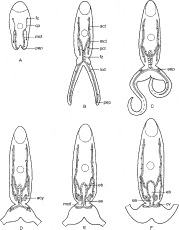
Fig. 1. Diagram summarizing five consecutive stages of excretory system development in Prosorhynchoides gracilescens cercariae (the number of cells and the number of nuclei in the excretory syncytium are represented arbitrarily). (A) I stage. (B) II stage. (C) III stage. (D,E) IV stage. (F) V stage. act, Anterior collecting tubule; acy, cyton of excretory atrium syncytium; aep, additional excretory pores; as, excretory atrium syncytium; cp, capillary; cv, caudal vesicle; ea, excretory atrium; eb, excretory bladder; fc, flame cell; fz, fusion zone; lcd, lateral caudal duct; mcd, median caudal duct; mct, main collecting tube; pct, posterior collecting tubule; pep, primary excretory pore.
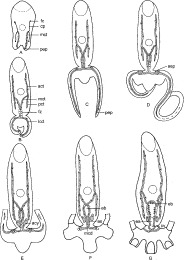
Fig. 2. Diagram summarizing five consecutive stages of excretory system development in Prosorhynchus squamatus cercariae. (A) I stage. (B,C) II stage. (D) III stage. (E,F) IV stage. (G) V stage. For abbreviations see Fig. 1. (From Galaktionov and Dobrovolskij (2003), fig. 65, p. 143, with kind permission of Kluwer Academic Publishers.)
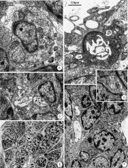
Fig. 3. Excretory system of early embryonic cercariae of Prosorhynchus squamatus and Prosorhynchoides gracilescens. (A) Main collecting tube (MCT) at the first stage of excretory system development in P. squamatus. Cross-section showing septate junction (SJ) where the tips of tube cell abut. L, lumen; MT, mitochondrion; N, nucleus; RER, rough endoplasmic reticulum. (B) Region of primary excretory pore in the same embryo. Note septate junction (SJ) attaching the main collecting tube (MCT) to the tegument (T), a septate junction (SJ) between the tips of tube cell and the increased number of projections on the luminal surface of the main collecting tube. (C–F) The second stage of excretory system development. (C) Cross-section through the posterior portion of the fusion zone in a P. squamatus embryo. Note absence of septate junctions in the duct wall. FZE, fusion zone epithelium; L, lumen; N, nucleus; RER, rough endoplasmic reticulum. (D) Cross-section through the middle portion of lateral caudal duct (LCD) in a P. squamatus embryo. Note narrowing of duct and absence of septate junctions in its wall. L, lumen. (E) Longitudinal section through the fusion zone (FZ) (boundaries are marked by arrowheads) of a P. squamatus embryo showing numerous nuclei in the syncytial lining. L, lumen. (F) Bulge in the proximal portion of a lateral caudal duct (LCD) (boundaries are marked by arrowheads) of a P. gracilescens embryo. In this species the bulge appears to be associated with a slight constriction in each lateral lobe of the tail stem. Note nuclei (N) in excretory syncytium and absence of cell boundaries between adjacent nuclei. L, lumen; T, tegument.
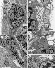
Fig. 4. The third stage of excretory system development in Prosorhynchus squamatus and Prosorhynchoides gracilescens embryonic cercariae. (A,B) Longitudinal sections showing formation of additional excretory pores in P. squamatus. Note several nuclei (N) in a thick portion of duct syncytium, a number of cytoplasmic extensions (arrowheads) projected towards the tail stem surface and septate junctions (SJ) connecting the excretory syncytium to the tegument (T). (B) Note also formation of several excretory openings (EO). Asterisks indicate inner cavities in cytoplasmic extensions. L, lumen; LCD, lateral caudal duct; M, muscles. (C,D) Longitudinal sections through the anterior portion of the fusion zone (AFZ) of P. gracilescens (C) and P. squamatus (D) embryonic cercariae. Note position of junctions (SJ) between the main collecting tubes (MCT) and the fusion zone epithelium. L, lumen; LA, lamellae; MY, developing myocyte. (E) Fusion zone epithelium in P. gracilescens embryonic cercaria. Note Golgi complex (GC), numerous dense mitochondria (MT) and sparse rough endoplasmic reticulum (RER). L, lumen; N, nucleus.
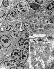
Fig. 5. Sections showing details of the late third stage of excretory system development in Prosorhynchus squamatus and Prosorhynchoides gracilescens embryonic cercariae. (A,B) Longitudinal sections through the posterior portion of the fusion zone (PFZ) in P. squamatus (A) and P. gracilescens (B) embryonic cercariae. In P. squamatus this portion is constricted and devoid of nuclei, while in P. gracilescens it is wide and contains nuclei (N). Note developing myocytes (MY) around the excretory syncytium (in P. squamatus they are more immature). L, lumen. (C,D) Collecting tubules of P. gracilescens (C) and P. squamatus (D) embryonic cercariae showing well pronounced Golgi complexes (GC) and various types of vesicles including coated vesicles (CVE) and electron-dense vesicles (VE). L, lumen; MB, multilamellar body; MI, microtubule; N, nucleus; SJ, septate junction.
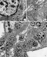
Fig. 6. The fourth stage of excretory system development in Prosorhynchus squamatus and Prosorhynchoides gracilescens embryonic cercariae. (A) A differentiating secretory cyton (ACY) near the posterior portion of the fusion zone in a P. squamatus embryonic cercaria. Note pronounced Golgi complex (GC), rough endoplasmic reticulum (RER) and secretory inclusions (SI). MT, mitochondrion; N, nucleus. (B) Longitudinal section through the posterior portion of the fusion zone of P. squamatus embryonic cercaria showing curved cytoplasmic extension (arrows) of a secretory cyton which is attached to the fusion zone epithelium (FZE) by a septate junction (SJ). Note secretory inclusions (SI) in this cytoplasmic extension. L, lumen; M, muscles. (C) Longitudinal section of P. gracilescens embryonic cercaria showing the posterior portion of the fusion zone which has been invaded by a cytoplasmic layer (CL) formed by fusion of cytoplasmic extensions from secretory cytons. Note developing circular muscles (CM) around the newly formed cytoplasmic layer (CL) and a nucleus (N) in epithelium of the posterior portion of the fusion zone (FZE). Arrowheads indicate electron-lucent vacuoles in cytoplasmic layer. B, cercarial body; BTJ, body–tail junction; L, lumen; TL, tail.
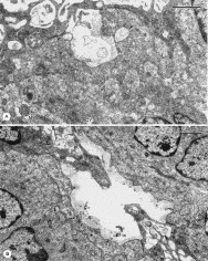
Fig. 7. Longitudinal sections of Prosorhynchus squamatus embryonic cercariae showing consecutive stages of excretory atrium formation. (A) Note large cavity in the cytoplasmic layer (CL) seemingly formed by fusion of electron-lucent vacuoles (arrowheads). ACY, cyton of excretory atrium syncytium; BL, basal lamina; CM, circular muscles; FZE, fusion zone epithelium; GL, glycocalyx; L, lumen; MY, developing myocyte; SJ, septate junction. (B) Note disintegration of fusion zone epithelium (FZE) over new lining. Note also septate junctions (SJ) connecting the atrium lining to the excretory syncytium anteriorly and posteriorly, secretory inclusions (SI) in the cyton (ACY) and surface cytoplasmic layer of excretory atrium syncytium (AS), glycocalyx (GL) covering the luminal surface and well-developed circular muscles (CM). Arrow indicates connection between cyton and surface cytoplasmic layer. MY, developing myocyte; N, nucleus.
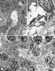
Fig. 8. Excretory system of late embryonic cercariae of Prosorhynchus squamatus and Prosorhynchoides gracilescens. (A) Longitudinal (sagittal) section of P. squamatus embryonic cercaria showing the late fourth stage of excretory system development. Primordial excretory bladder (EB), excretory atrium (EA) and median caudal duct (MCD) are well defined. Arrowheads indicate connections between the surface cytoplasmic layer of excretory atrium syncytium (AS) and deeper nucleated cytons (ACY). B, cercarial body; BTJ, body-tail junction; CM, circular muscles; SJ, septate junctions; TL, tail. (B) Longitudinal (frontal) section of P. gracilescens embryonic cercaria showing the early fifth stage of excretory system development. Note well-developed longitudinal muscles (LM) of the excretory atrium (EA) and early stages of enlargement of the excretory atrium (arrows) in the region of body–tail junction (BTJ). The median caudal duct (MCD) is not yet dilated to form the caudal vesicle and is only slightly enlarged at its anterior end. AS, excretory atrium syncytium; CM, circular muscles; EB, excretory bladder; LCD, lateral caudal duct; SJ, septate junctions; TL, tail. (C) Formation of the caudal vesicle (CV) in a P. squamatus cercaria. The anterior portion of the caudal vesicle is lined by an extension of the excretory atrium syncytium (AS). The median caudal duct is clearly enlarged to form the posterior portion of the caudal vesicle, its wall has become thin and devoid of surface lamellae. Note constricted extensions of the excretory syncytium (arrows) containing mitochondria. LCD, lateral caudal duct; SJ, septate junctions.
 You have
Access
- 2
- Cited by