Introduction
Acute rheumatic fever is an inflammatory disease caused by group A beta-haemolytic streptococcal infection. Arthritis is the most common symptom and occurs in 60–80% of patients.Reference Martins, Veasy and Hill1 Although it is a multi-systemic disease, the most important cause of mortality and morbidity is cardiac involvement which consists of the involvement of either endocardium, myocardium, and/or pericardium.
The exact mechanism of carditis is unclear, although it is known as an inflammatory and autoimmune disease. The most known inflammatory markers are C-reactive protein, erythrocyte sedimentation rate, and white cell count. However, in some autoimmune or inflammatory diseases, the neutrophil and platelet numbers and functions are considered to change.Reference Pitchford and Page2 Moreover, it has been demonstrated that neutrophil-to-lymphocyte ratio and platelet-to-lymphocyte ratio might be the indicators of systemic inflammation.Reference Zahorec3–Reference Biyik, Ucar and Solak5
Sydenham’s chorea, which is one of the major diagnostic criteria of rheumatic fever, is a disorder affecting children and characterised by jerky, uncontrollable movements of the muscles of the face, the arms and legs, and the trunk. It is thought to result from an autoimmune reaction to the streptococcal infection. Besides, tics, emotional changes, behavioural changes, and dystonic disorders can be seen after streptococcal infection. The relationship between psychiatric diseases and monocyte activation have also been studied previously.Reference Mazza, Tringali and Rossetti6
The aims of this study were to compare the ratios of neutrophil-to-lymphocyte to monocyte-to-lymphocyte and platelet-to-lymphocyte of patients with acute rheumatic fever and healthy children, and to correlate them with the other well-known inflammatory parameters.
Material and methods
Patients who were admitted to the paediatric cardiology unit between April 2013 and April 2018 with the diagnosis of acute rheumatic fever were recorded retrospectively. The study included the records of 182 patients who had the data of echocardiographic measurements, complete blood count, erythrocyte sedimentation rate, and C-reactive protein values. All of the admitted patients were in the first acute episode of the disease. The demographic characteristics of the patients were obtained from the patient information system. In the same period, as a control group, 173 healthy children who admitted to the outpatient clinic with the diagnosis of innocent murmur with normal echocardiographic examination and whose records of complete blood count were taken.
Transthoracic echocardiography was performed via Vivid S5 Pro Ultrasound System (GE Medical Systems, Horten, Norway), using 3 and 6 MHz transducers in two-dimensional and M-mode. Cardiac valves were evaluated for regurgitation and prolapse. Mitral and aortic valve regurgitation were assessed by colour Doppler echocardiography in the apical and parasternal long-axis windows. Pathological valve insufficiency was considered for the mitral valve as continuation of regurgitation during systole and for the aortic valve as continuation of regurgitation during diastole in at least in two windows as a jet of mosaic colour 1 cm in length with a peak velocity of 2.5 m/second.Reference Bisno, Butchart and Ganguly7
Exclusion criteria for the patient and the control groups were any infectious or inflammatory diseases, anaemia, thrombocytopenia, or other haematological problems, and usage of anti-inflammatory or anticoagulant drugs in the last month. Local Ethics Committee approval was obtained for the study protocol.
Statistical analysis
Statistical analysis was performed. Descriptive statistics were presented as means and their standard deviations, medians, minimum, and maximum values. The “Mann–Whitney U-Test” was used for neutrophil-to-lymphocyte ratio, monocyte-to-lymphocyte ratio, and platelet-to-lymphocyte ratio, and the “Independent t-test” was used for the other parameters. “Spearman’s Correlation Analysis” was used to determine the relationships between parameters. After evaluation of neutrophil-to-lymphocyte ratio, monocyte-to-lymphocyte ratio, and platelet-to-lymphocyte ratio, erythrocyte sedimentation rate, and C-reactive protein levels one by one (univariate) between subgroups of patients (with or without carditis and chorea) and the control group, analyses of multiple variants were performed (multivariate); p < 0.05 was considered statistically significant.
Results
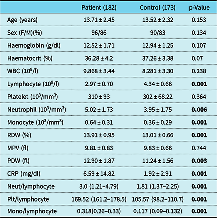
The study included 182 patients with acute rheumatic fever and 173 sex- and age-matched healthy children as the control group. The baseline characteristics are presented in Table 1.
Demographic and laboratory characteristics of the patient and control groups.

CRP = C-reactive protein; ESR = erythrocyte sedimentation rate; F = female; M = male; Mono = monocyte; MPV = mean platelet volume; Neut = neutrophil; PDW = platelet distribution width; Plt = platelet; RDW = red blood cell distribution width; WBC = white blood cell.
p > 0.05 were written in bold.
Carditis was present in the 152 (83.5%) of the patients of whom 52 (28.6%) had mitral regurgitation, 35 (19.2%) had aortic regurgitation, and 65 (35.7%) had both valve involvement. Thirty (16.5%) of the patients had no valve involvement with a history of rheumatic arthritis. Sydenham’s chorea was present in 8 (4.4%) of the patients. Six of the patients with Sydenham’s chorea had no cardiac involvement but had a history of arthralgia/arthritis, and the other two patients had only mitral valve involvement. Erythema marginatum, subcutaneous nodules, and pure fever were not a presenting symptom in any of the patients.
Neutrophil, monocyte levels, and neutrophil-to-lymphocyte, platelet-to-lymphocyte, and monocyte-to-lymphocyte ratios of the patients were significantly higher than controls (p < 0.05) (Table 1).
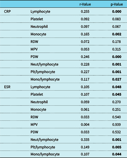
Spearman’s correlation analysis demonstrated low but positive correlations between C-reactive protein levels and neutrophil-to-lymphocyte ratio (r = 0.228, p = 0.001), platelet-to-lymphocyte ratio (r = 0.227, p = 0.01), and monocyte-to-lymphocyte ratio (r = 0.117, p = 0.027) in the patients. Also there was a similar correlation between the erythrocyte sedimentation rate and neutrophil-to-lymphocyte ratio (r = 0.355, p = 0.001), platelet-to-lymphocyte ratio (r = 0.149, p = 0.005), and monocyte-to-lymphocyte ratio (r = 0.107, p = 0.044) in the patients (Table 2).
Correlations between the C-reactive protein and erythrocyte sedimentation rate and other blood count parameters of the patients.

CRP = C-reactive protein; ESR = erythrocyte sedimentation rate; Mono = monocyte; MPV = mean platelet volume; Neut = neutrophil; PDW = platelet distribution width; Plt = platelet; RDW = red blood cell distribution width.
p > 0.05 were written in bold.
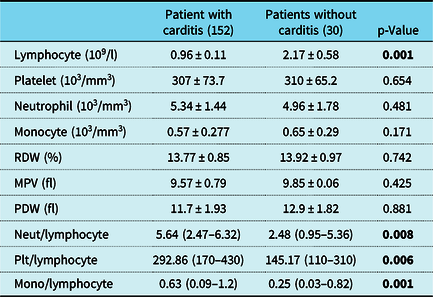
Patients were subdivided into two groups according to the presence of any pathologic rheumatic valve regurgitation (mitral valve, aortic valve, or both) and the presence of isolated arthritis, isolated chorea, or both (without any cardiac involvement). Neutrophil-to-lymphocyte, monocyte-to-lymphocyte, and platelet-to-lymphocyte ratios were significantly higher in patients with carditis than in those without cardiac involvement (p < 0.05) (Table 3). On the other hand, patients with cardiac involvement when grouped according to the presence of mitral valve, aortic valve, or both valve involvement, and compared to the presence of arthritis or chorea, showed no statistically significant difference between these subgroups.
Laboratory characteristics of the patients with and without carditis.

Mono = monocyte; MPV = mean platelet volume; Neut = neutrophil; PDW = platelet distribution width; Plt = platelet; RDW = red blood cell distribution width.
p > 0.05 were written in bold.
Furthermore, in patients with Sydenham’s chorea, neutrophil-to-lymphocyte ratio (4.41 ± 2.25) and monocyte-to-lymphocyte ratio (0.56 ± 0.25) were significantly higher than in patients without chorea (2.94 ± 1.74; 0.28 ± 0.34, respectively) (p = 0.022; p = 0.047, respectively).
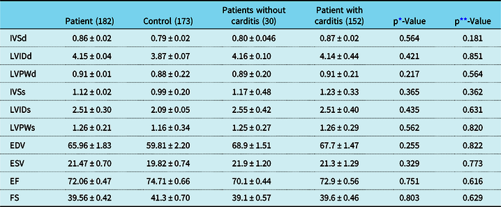
Conventional two-dimensional and M-mode echocardiographic parameters, including end-systolic and end-diastolic volumes, ejection fraction and shortening fraction of the left ventricle, were similar for the patients and the controls (p > 0.05). In addition, echocardiographic parameters were not different in patients with and without heart involvement (Table 4).
Conventional echocardiographic measures of the patient and control groups.

EDV = end-diastolic volume; EF = left ventricular ejection fraction; ESV = end-systolic volume; FS = left ventricular fractional shortening; IVSd = interventricular septal thickness at end diastole; IVSs = interventricular septal thickness at end systole; LVIDd = left ventricular internal diameter at end diastole; LVIDs = left ventricular internal diameter at end systole; LVPWd = left ventricular posterior wall thickness at end diastole; LVPWs = left ventricular posterior wall thickness at end systole;
* For the comparison between control and patient groups;
** For the comparison of patients with carditis and without carditis.
Discussion
In this study, it was found that there were statistically significant correlations between neutrophil-to-lymphocyte, platelet-to-lymphocyte, and monocyte-to-lymphocyte ratios and C-reactive protein level and erythrocyte sedimentation rate, which are well-known minor diagnostic criteria of acute rheumatic fever. Moreover, it was found that the neutrophil-to-lymphocyte, platelet-to-lymphocyte, and monocyte-to-lymphocyte ratios in patients with rheumatic fever were clearly higher in patients with heart disease. Furthermore, the relationship between Sydenham’s chorea and neutrophil-to-lymphocyte and monocyte-to-lymphocyte ratios is here demonstrated for the first time.
The most important major criterion of acute rheumatic fever is undoubtedly cardiac involvement. The development of autoimmunity in sensitive individuals results in inflammation and damage to the heart valves.Reference Guilherme, Cunha-Neto and Coelho8,Reference Stollerman9 The most important markers of this inflammation and also the minor diagnostic criteria of the disease are C-reactive protein and erythrocyte sedimentation rate. It is also known that there may be significant changes in the rates of white blood cells and subtypes such as neutrophils and lymphocytes. In recent studies, it has been stated that when evaluating systemic inflammation with peripheral blood parameters, neutrophil-to-lymphocyte ratio and platelet-to-lymphocyte ratio values can be used as markers. Although the increase in neutrophil and platelet counts during inflammation is known, there is also an increased steroid level due to stress caused by inflammatory disease, resulting in the development of lymphopenia.Reference Onsrud and Thorsby10,Reference Hotchkiss and Karl11 Turak et al. reported in a study that elevation of neutrophil-to-lymphocyte ratio may predict the risk of a cerebrovascular event and mortality in patients with infective endocarditis.Reference Turak, Özcan and Işleyen12 Similarly, it was reported that higher neutrophil-to-lymphocyte ratio was associated with worse clinical outcome and mortality in patients with coronary artery disease.Reference Shen, Chen, Shi and Li13–Reference Kaya, Ertas and Islamoglu15 There are some studies in the literature correlating the increase in neutrophil-to-lymphocyte ratio with the severity of valvular involvement in patients with acute rheumatic carditis, whereas some others comparing the neutrophil-to-lymphocyte and platelet-to-lymphocyte ratios of patients with newly diagnosed acute rheumatic fever before and after treatment had no significant differences.Reference Celik and Celik16–Reference Buyukoflaz and Arslan18 In this study, both neutrophil-to-lymphocyte and platelet-to-lymphocyte ratios were found to be higher in patients with acute rheumatic fever when compared to the control group. These measured markers are accepted as novel indicators of the continuing inflammation in acute rheumatic fever. Not only are these indicators inexpensive, simple, rapid, and readily available markers at nearly any healthcare facility, but also they are less affected by coexisting other infectious diseases – an advantage of these markers over routinely used inflammatory markers, such as C-reactive protein and erythrocyte sedimentation rate.
Furthermore, in this study, monocyte level and monocyte-to-lymphocyte ratio were evaluated in this patient group for the first time and they were significantly higher than the control group. In addition, for the first time, neutrophil-to-lymphocyte ratio, platelet-to-lymphocyte ratio, and monocyte-to-lymphocyte ratio values were correlated with C-reactive protein values and erythrocyte sedimentation rate, demonstrating a positive correlation. Moreover, the neutrophil-to-lymphocyte, platelet-to-lymphocyte, and monocyte-to-lymphocyte ratios were correlated with cardiac involvement of the disease, with a significant difference between the patients with and without cardiac involvement.
Also, red blood cell distribution and platelet distribution width values, additional indicators of inflammation, were found to be significantly higher in our patients compared to the control group, as previously reported.Reference Akboğa, Abacı and Canpolat19
Sydenham’s chorea is well known as the main central nervous system manifestation and a stand-alone major diagnostic criterion of acute rheumatic fever, and it is best known. It has been reported that psychiatric disorders are more common in patients with acute rheumatic fever than the general population even if they have not chorea.Reference Kılıç, Ünüvar and That20 Some other neurological clinical presentations hypothesised to be associated with acute rheumatic fever are paediatric autoimmune neuropsychiatric disorder associated with streptococcal infections (PANDAS), anxiety, behaviour and sleep disorders, obsessive compulsive disorder, attention deficit and hyperactivity disorder.Reference Maia, Teixrira, Cunningham and Cardoso21 There are also some studies investigating the relationship between neutrophil-to-lymphocyte, platelet-to-lymphocyte, and monocyte-to-lymphocyte ratios in psychiatric patients in the literature. Mazza et al. reported that neutrophil-to-lymphocyte ratio, platelet-to-lymphocyte ratio, and monocyte-to-lymphocyte ratio levels were higher in patients with bipolar disorder, major depressive disorder, and schizophrenia when compared to healthy controls, and it was concluded that these ratios might be useful to detect inflammatory activation that occurs in psychiatric disorders.Reference Mazza, Rossetti and Clerici22 In our study, for the first time, the neutrophil-to-lymphocyte ratio and monocyte-to-lymphocyte ratio values of patients with Sydenham’s chorea were compared with healthy control group and were found significantly higher in patients, whereas there was no significant difference in platelet-to-lymphocyte ratio. In addition, neutrophil-to-lymphocyte and monocyte-to-lymphocyte ratios were found higher in patients with chorea than in patients with acute rheumatic fever without chorea. These results of patients with Sydenham’s chorea were similar to those results of other neuropsychiatric diseases in the literature. Although the latent period after the streptococcal infection is longer in patients with chorea, varying from 1 to 7 months, it can also be the only manifestation and the acute phase reactants may be negative in these patients. Neutrophil-to-lymphocyte and monocyte-to-lymphocyte ratios could be used in the long-term follow-up of the disease with serial measurements, leading to treatment as it could be significantly higher despite not being within the acute period of the disease.
Vasan et al. subgrouped patients with acute rheumatic fever in their study. They reported that left ventricular systolic function was normal in all group 1 patients (acute rheumatic fever without clinical evidence of carditis), and in the majority of patients in group 2 (a first attack of acute rheumatic fever and clinical evidence of carditis), and group 3 (previous rheumatic heart disease with acute rheumatic fever and clinical evidence of active carditis). Also, they did not observe any significant differences in the mean values of fractional shortening between the three groups of patients and between their corresponding controls.Reference Vasan, Shrivastava, Vijayakumar, Narang, Lister and Narula23 In this study, there was no difference in conventional echocardiography parameters between the control and the patient groups, as well as patients with rheumatic carditis and those without the cardiac involvement, as in the literature. Our patients were diagnosed at the beginning of their first acute episode when the echocardiogram was performed, and this could explain the absence of dilatation of left heart chambers. Thus, another very important advantage of the measured markers noted in this study is as an add-on for diagnosis and prognosis (with serial follow-up measurements) purposes. As stated, the ratios of neutrophil-to-lymphocyte, platelet-to-lymphocyte, and monocyte-to-lymphocyte were significantly higher in patients with acute rheumatic fever with heart involvement than patients without heart involvement, while the echocardiographic parameters were not different in patients with and without heart involvement. This may be especially helpful for the subgroups of patients with silent carditis (murmur-free) or subclinical rheumatic carditis with trivial or insignificant rheumatic valvular disease, as may be present in acute rheumatic fever patients with chorea or polyarthritis. It is also important to recognise that technical expertise with echocardiography, which is operator-dependent, is necessary to make an accurate diagnosis of subclinical carditis and to avoid over-diagnosis, while neutrophil-to-lymphocyte ratio, platelet-to-lymphocyte ratio, and monocyte-to-lymphocyte ratio markers are not operator-dependent and do not require technical expertise. Moreover, the diagnosis of subclinical rheumatic carditis is extremely important nowadays considering that carditis is now defined as clinical and/or subclinical (echocardiographic valvulitis), fulfilling a major criterion for acute rheumatic fever in all populations.
Limitations
Although, conducted with a relatively large number of patients with acute rheumatic fever, the retrospective design of this study is a major limitation. Also, we only recorded patients with complete blood counts at admission which may limit overall case ascertainment. Moreover, the inflammatory process decreases gradually overtime and is influenced by the use of anti-inflammatory drugs, and the laboratory data here were not collected uniformly and only during the acute phase when the patients were admitted to hospital. Therefore, current results cannot be assumed to be relevant for all patients with acute rheumatic fever. The limited number of the patients with isolated Sydenham’s chorea is another limitation of the study, meaning that patients with chorea could not be subgrouped according to presence or absence of the heart involvement for more accurate comparison. Similarly, patients with chorea who had arthritis in the past or where chorea was a later manifestation of the same acute episode are not included. Moreover, in this study, two of the patients with chorea also had cardiac involvement, resulting in an overlap of the effects of both carditis and chorea, confounding correlations with carditis or chorea alone. Lastly, the presence of a positive throat culture and/or a positive antistreptolysin O titre for group A beta-haemolytic streptococcal infection could not be used in comparison to the other markers used in this study due to incomplete data.
Conclusion
In conclusion, neutrophil-to-lymphocyte, platelet-to-lymphocyte, and monocyte-to-lymphocyte ratios, which can be determined by a complete blood count, may help make the diagnosis of acute rheumatic fever and serial follow-up assessments in children. However, the ratios cannot predict valve involvement or the severity of the disease. In addition, although our novel findings suggest that neutrophil-to-lymphocyte, platelet-to-lymphocyte, and monocyte-to-lymphocyte ratios could be used in the long-term follow-up and management of the disease, more comprehensive prospective studies are needed to confirm this and in order to understand this relationship in detail.
Acknowledgements
None.
Financial support
This research received no specific grant from any funding agency or from commercial or not-for-profit sectors.
Conflicts of interest
None.