Introduction
Eustachian tube dysfunction is a common otolaryngological problem which has multiple aetiologies, including anatomical derangements, chronic sinusitis, allergic rhinitis, adenoid hypertrophy and gastroesophageal reflux.Reference Seibert and Danner 1 Over recent decades, multiple medical and surgical treatments have been explored.
Medical options generally aim to reduce peritubal inflammation and swelling, but fail to relieve symptoms for many.
Surgical options are limited due to the immediate proximity of the eustachian tube to the petrous internal carotid artery and the middle cranial fossa floor, among other critical structures. To date, the only universally accepted surgical treatment has been myringotomy with tympanostomy tube placement. This temporising measure can have its own negative effects on the tympanic membrane and hearing, and sometimes results in more infections. Chronic cases of eustachian tube dysfunction require repeated tympanostomy tube replacement to ensure continued pressure equalisation and symptom control.
A number of surgical techniques have been frustrated by the quandary of eustachian tube dysfunction. Traditionally, it was thought that widening the narrowest part of the eustachian tube, the bony isthmus, would produce symptomatic improvement. Surgeons originally tried techniques such as drilling and stenting the isthmus; however, these methods were abandoned, mainly because of complications, but also because they did not seem to produce long-term results.Reference Wullstein 2 – Reference Zollner 5 Although the bony isthmus is still generally accepted to be the narrowest portion of the eustachian tube, newer procedures addressing the cartilaginous portion of the eustachian tube have been described, since derangements in this latter portion are now thought to be a major source of eustachian tube dysfunction.Reference Poe, Metson and Kujawski 6 – Reference Ockermann, Reineke, Upile, Ebmeyer and Sudhoff 14
Laser dilation, as first described by Kujawski in 1997, removes soft tissue near the nasopharyngeal orifice, and is the most commonly described technique.Reference Kujawski 8 This intervention was most recently evaluated in a pilot study by Poe et al., who used a 980-nm diode (or argon) laser to vaporise mucosa and cartilage on the posterior wall of the eustachian tube lumen.Reference Poe, Metson and Kujawski 6 Out of 10 patients, 70 per cent remained effusion-free at 6 months, with minimal post-procedure complications. A follow-up study showed that laser eustachian tuboplasty was effective in treating some patients, with the highest failure rate in those with reflux or allergic disease, suggesting that concurrent medical therapy may still be necessary.Reference Poe, Grimmer and Metson 9 Laser eustachian tuboplasty was also investigated by Caffier et al., who found improvements in middle-ear ventilation in 66 per cent of patients at one year, and no complications.Reference Caffier, Sedlmaier, Haupt, Goktas, Scherer and Mazurek 10 More recently, Metson et al. used a microdebrider to remove hypertrophied mucosa from the posterior eustachian tube, with subjective symptomatic improvement in 70 per cent of patients over a 13-month mean follow-up period.Reference Metson, Pletcher and Poe 11
These promising results have encouraged some surgeons to continue to explore new techniques, of which balloon eustachian tuboplasty is the newest. Based on studies of balloon dilatation of narrowed sinus ostia in patients with symptoms of chronic sinusitis, balloon dilatation has been similarly applied to patients with chronic eustachian tube dysfunction and presumed derangements within the cartilaginous and bony eustachian tube.Reference Poe and Hanna 12 – Reference Bolger and Vaughan 15 Poe et al. performed a cadaver study investigating transnasal balloon dilatation of the cartilaginous eustachian tube orifice, and found statistically significant dilation of all eustachian tubes.Reference Poe and Hanna 12 The only adverse outcome was minor mucosal tears thought to be of minimal significance.Reference Poe and Hanna 12
A hybrid method of dilating both the cartilaginous and bony portions of the eustachian tube, using a balloon, has recently been introduced by Ockermann et al. Reference Ockermann, Reineke, Upile, Ebmeyer and Sudhoff 13 They tested the safety of transnasal balloon eustachian tuboplasty in a cadaver study, and followed with a human study in which eight patients were treated.Reference Ockermann, Reineke, Upile, Ebmeyer and Sudhoff 13 , Reference Ockermann, Reineke, Upile, Ebmeyer and Sudhoff 14 In both reports, the balloons were inserted far enough to dilate the bony as well as the cartilaginous eustachian tube. There were promising patient satisfaction results, without any complications. However, long-term follow up results have not been reported, so the incidence of recurrence and complications (e.g. patulous eustachian tube) is unknown.Reference Ockermann, Reineke, Upile, Ebmeyer and Sudhoff 14
We conducted the present study to determine whether balloon catheter dilatation via a transtympanic route could be considered as a supplement to other otological procedures. It is rational to consider approaching the eustachian tube from the ear, since the orifice is readily seen during most ear surgical procedures. This approach would only be considered in selected patients who already required transtympanic or transmastoid surgery due to complications of chronic eustachian tube dysfunction such as retraction pockets or cholesteatoma. If deemed successful, this approach could be useful to otologists as a supplement to existing surgical treatments, enabling treatment of the underlying cause of ear disease in addition to the removal of existing pathology. Our study aimed to determine whether eustachian tube balloon dilatation via a transtympanic route was safe and feasible in selected patients undergoing other otological procedures. It was not designed to determine the best part of the eustachian tube to dilate, or to evaluate the functional results of dilatation.
Materials and methods
Approval was obtained from the Walter Reed Army Medical Center institutional review board.
Six fresh-frozen, whole cadaver heads were purchased from Science Care (Phoenix, Arizona, USA). There was no known history of ear or skull base disease in any specimen. The heads were stored securely in our temporal bone laboratory, and were maintained under the supervision of one of the principal investigators.
The tympanic membrane was examined for any abnormalities. The eustachian tube was then approached via an anterior tympanotomy (with an anteriorly based tympanomeatal flap). In several specimens, it was difficult to see the eustachian tube orifice well enough to insert a catheter. The posterior external auditory canal bone was drilled away in 6 of 10 ears until there was a more favourable angle, even if it meant exposing mastoid air cells. A 2-mm Lacricath balloon dilator catheter (model LDC213; Quest Medical, Allen, Texas, USA) was then inserted into each eustachian tube orifice. Whenever there was resistance to catheter placement, we confirmed that the tip was in the eustachian tube orifice, double-checked the insertion angle, and then inserted with more force. The goal of the insertional technique was not to dilate solely the bony isthmus, but rather to insert the catheter as far as resistance would allow in order to also dilate the cartilaginous eustachian tube, if possible. Adequate placement was defined as placement of the tip beyond the bony isthmus. The external position of the catheter was noted on each head prior to transport.
The heads were sequentially transported by the principal investigators to the radiology suite, within the same building, for high-resolution computed tomography (CT) scanning. Scanning was undertaken using a 64 multi-detector spiral CT scanner (GE Light Speed 64; Schenectady, New York, USA) with 0.6 mm section widths. Care was taken to position each head symmetrically in the supine position to enable adequate post-imaging analysis. Prior to scanning, each catheter was inspected to confirm that no dislodgement had occurred during transportation. All catheters were noted to be secure in their pre-transportation positions. A pre-dilatation CT scan was taken. One catheter balloon (per head) was then inflated by a co-investigator. The primary investigator (a neurotologist who would later review the images) was blinded to which side had been inflated. The other balloon catheter was left uninflated within the eustachian tube as a control, and to assure that subsequent evaluation of eustachian tube diameter was not biased by the presence or absence of the catheter. The balloon was inflated using water to a pressure of 13 atmospheres for 90 seconds. This was the maximum operating pressure of the Lacricath catheter, and thus the pressure most likely to cause dilatation. Thirty minutes later, a post-dilatation CT scan was taken. This arbitrary interval was intended to allow soft tissue compression or distraction to resolve, preventing confusion between bony and soft tissue dilatation.
A co-investigator removed identifying information, coded and randomly rearranged each image series, and then compiled them for later evaluation.
Once all imaging was complete, the CT scans were reviewed by a neurotologist (PL) and a board-certified neuroradiologist (JS). They viewed each image series together and agreed on each finding prior to proceeding to the next series. Features evaluated included: catheter position, pre- and post-dilatation eustachian tube diameter, carotid canal integrity, and middle cranial fossa floor integrity. Any abnormalities of surrounding neurovascular structures were identified, including the foramen lacerum, foramen ovale, foramen spinosum, geniculate ganglion, and the bony canal of the greater superficial petrosal nerve. Finally, the minimum diameter of the bony isthmus was measured from the axial images.
Results and analysis
Each pre-dilatation CT scan was evaluated for adequacy and safety of balloon catheter placement.
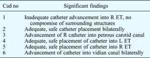
Cadaver one had an inadequate placement on the right side because the catheter tip did not extend beyond the bony isthmus. This was also the designated side of balloon inflation, which had consequently been prevented. Placement in the opposite ear was adequate. The exact location of the isthmus was hard to delineate on both sides.
Cadaver two had adequate catheter placement in both eustachian tubes, each past a clearly defined isthmus. There was no evidence of damage to surrounding structures.
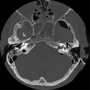
The pre-dilatation CT scan for cadaver three showed a right-sided, mixed temporal bone fracture with an intact otic capsule. Furthermore, the right catheter had entered the petrous carotid canal (Figure 1). The left catheter was adequately placed within the eustachian tube, alongside an intact but thinned carotid canal.
Axial, non-contrast computed tomography temporal bone scan in cadaver three, showing incursion of the catheter balloon into the right petrous carotid artery.

Cadavers four and five were considered as a single specimen, since both had sustained pre-study trauma that did not allow us to insert catheters into each eustachian tube. Thus, a catheter was inserted into the left side of cadaver four and the right side of cadaver five. Both these catheters were placed sufficiently beyond the isthmus, without any evidence of damage to surrounding structures.
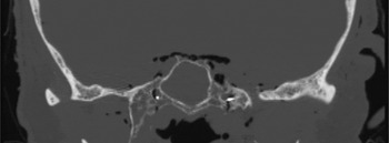
Cadaver six had a pre-existing, right, mixed temporal bone fracture. Both catheter balloons had entered the vidian canal (Figure 2).
Coronal, non-contrast computed tomography scan of bilateral catheter balloon placement in cadaver six, showing catheters entering the vidian canal bilaterally.

Table I outlines the significant observational findings for each head prior to balloon catheter dilatation, regarding the adequacy and complications of catheter placement. All scans showed well-developed, aerated mastoids with no evidence of chronic ear disease.
Pre-dilatation observational findings

Cad no = cadaver number; R = right; ET = eustachian tube; L = left
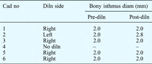
Table II outlines the results of blinded analysis of the post-dilatation CT scans. Only cadaver two showed any difference between the pre- and post-dilatation CT scans: the left eustachian tube had been dilated from a diameter of 2.0 to a diameter of 2.8 mm, while the uninflated catheter was 1.0 mm in diameter (which was the most commonly measured isthmus diameter).
Pre- and post-dilatation CT findings

CT = computed tomography; Cad no = cadaver number; diln = dilatation; diam = diameter
Discussion
The functional anatomy of the eustachian tube is well documented. Its anteromedial portion extending to the nasopharynx is primarily composed of cartilage and soft tissue, and measures 24–26 mm in length. The narrowest portion of the eustachian tube, the bony isthmus, is about 12 mm long.Reference Poe, Metson and Kujawski 6 , Reference Ockermann, Reineke, Upile, Ebmeyer and Sudhoff 14 Muscular contributions from the tensor veli palatini, levator veli palatini, salpingopharyngeus and tensor tympani muscles assist in normal functioning of the eustachian tube. Derangements in any aspect of the eustachian tube anatomy and function can impair its primary functions of pressure equalisation, clearing of middle-ear secretions, and protecting the middle ear from nasopharyngeal secretions.Reference Seibert and Danner 1 , Reference Poe, Metson and Kujawski 6
Many studies have addressed the various anatomical flaws believed to contribute to eustachian tube dysfunction; these have historically been associated with the narrow bony isthmus, but newer reports suggest that the cartilaginous portion of the eustachian tube is the primary culprit. Balloon dilatation eustachian tuboplasty is a promising new modality which has the potential to address both the cartilaginous and bony portions of the eustachian tube within a single procedure. Despite the dual focus of this new technique, it is important to remember that eustachian tube dysfunction may at times be due solely to constriction of the bony portion of the tube.Reference Licameli 7
Three previous publications have reported successful dilatation of the cartilaginous eustachian tube using the transnasal technique, without adverse effects.Reference Poe and Hanna 12 – Reference Ockermann, Reineke, Upile, Ebmeyer and Sudhoff 14 In two of those studies, there were also attempts to dilate the bony eustachian tube by partially placing the catheter into the bony portion, but no significant effects were seen.Reference Ockermann, Reineke, Upile, Ebmeyer and Sudhoff 13 , Reference Ockermann, Reineke, Upile, Ebmeyer and Sudhoff 14
The current study further investigated eustachian tube dilatation using a transtympanic approach, with attempted balloon catheter placement beyond the bony isthmus. We aimed to determine whether this technique might serve as a feasible adjunct to other otological procedures performed to address the complications of chronic eustachian tube dysfunction.
Our findings differed markedly from those of previous investigators, probably because of our approach. Obtaining access to the eustachian tube via a transtympanic approach was more difficult than expected. It is easy to see the eustachian tube orifice, but intubating it with a rigid catheter is a different matter. It was not possible in 6 of 10 ears without some drilling of the posterior ear canal bone. Therefore, this technique should perhaps be considered to require routine performance of a mastoidectomy in order to obtain the appropriate entrance angle to the eustachian tube. Without such drilling, the angle of the catheter was too medial, being directed towards, rather than past, the carotid and parallel to the eustachian tube lumen. The catheter we used would not be practical for transcanal tympanoplasty, or for most canal wall up mastoidectomy procedures. This is unfortunate because eustachian tuboplasty would seem a most logical adjunct to these procedures, performed as they are in the setting of continued eustachian tube dysfunction, which often leads to further disease and surgery.
Our CT findings show that surgical misadventure is a definite possibility during transtympanic balloon catheterisation of the eustachian tube. There are obvious neurovascular concerns when considering the transtympanic route; however, we had thought that balloon catheterisation may be a safer option than the drilling and stenting techniques used in the past. During the catheterisation procedures, we believed the catheter to be progressing down the eustachian tube in all cases. It was therefore highly surprising that one catheter had actually lodged in the carotid canal, and even more surprising that two had extended into the vidian canal. It should be mentioned that we met some resistance during approximately half of the insertion procedures. In such cases, we confirmed that the tip of the catheter was in the eustachian tube orifice, double-checked the insertion angle, and then proceeded with more force. This would be ill advised when treating a patient, but in our cadaver study we were more interested in the ‘worst-case scenario’. It was therefore desirable to observe the results of forced insertion, as long as it proceeded in the appropriate direction. We did not record which eustachian tubes were difficult to intubate, so we cannot know whether the difficult cases went on to have adverse catheter placement.
All specimens had normal tympanic membranes and mastoids, so there was no evidence of any eustachian tube dysfunction. Thus, anatomical conditions should have been ideal, so it was remarkable that we often had difficulty passing a catheter that was 1 mm in diameter when deflated. It may be even more difficult to pass catheters in patients in whom bony constriction is the cause of eustachian tube dysfunction. It might be more informative to study eustachian tube dilatation in cadavers with actual middle-ear disease or eustachian tube dysfunction; however, it would be extremely difficult to assemble specimens that fit criteria. It would also be an unnecessary endeavour, until such time as catheter dilatation appears feasible with normal human heads.
The balloon catheter could probably be modified in order to make eustachian tube insertion easier and safer; however, the anatomy of the eustachian tube limits the possible changes. The Lacricath catheter used in this study was narrow and rigid, as it was designed for the lacrimal duct. We selected the Lacricath from amongst the several types of balloon catheter available, because its rigidity and narrow profile seemed most conducive to navigating the eustachian tube. Any catheter used for eustachian tube catheterisation should have a tip diameter of 1 mm or less, since this is the dimension of the eustachian tube isthmus. However, even a blunt, 1 mm tip can puncture thin bone if it is coupled to a rigid introducer.
It deserves mention that the heads in which the catheter balloon lodged in the carotid artery or vidian canal had pre-existing temporal bone fractures (probably sustained during handling and transport). These fractures may have contributed to directing the balloons erroneously into vital structures, but it was not apparent from the CT scans if the balloons were ever along fracture lines.
Aside from the inherent safety concerns raised by our study, we were unable to show that the catheter balloon actually dilated the bony isthmus. Only one head showed a measurable change in eustachian tube diameter. Two-millimetre balloons were used in this study, whereas previous studies have used 3 mm or 6–7 mm balloons.Reference Poe and Hanna 12 – Reference Ockermann, Reineke, Upile, Ebmeyer and Sudhoff 14 It is possible that a wider calibre, longer balloon is necessary to achieve dilatation of the bony isthmus and cartilaginous eustachian tube. However, it is also possible that the bone is just too robust to be manipulated with balloons – even though we inflated to the maximum operating pressure of the device.
-
• Eustachian tube dysfunction is common, with few medical or surgical treatments
-
• Drilling, stenting, and laser and microdebrider-assisted tuboplasty have previously been studied
-
• This cadaveric study assessed balloon eustachian tuboplasty via a transtympanic approach
-
• This new technique has been proposed as an adjunct to other otologic surgical techniques
-
• However, findings indicated serious safety concerns
The results of this study cannot be interpreted without a discussion of limitations. Our study's inherent flaw was the use of a cadaveric head model, which left us unable to extrapolate all of our results to a clinical model. It was impossible for us to control the handling of the cadavers prior to our use, and this may have resulted in fractures that could have affected balloon catheter placement. In addition, the heads did not have the pathology that we were attempting to treat. Furthermore, given our small sample size, it is possible that our experiment simply did not have enough statistical power to show a difference in post-dilatational bony isthmus diameter. Regardless of these limitations, the aforementioned safety concerns would have made any positive results appear irrelevant. Although our cadaveric model prevented assessment of clinical improvement, such assessment was never our aim, and this topic would have required further research if our findings had indicated feasibility. Ideally, our CT scans would have been evaluated by expert personnel other than the principal investigators of the study. However, we believe that removing any identifying data from the scans, and having the scans read in a random fashion as directed by the associate investigator, minimised bias, which certainly may have been more of a confounding factor had our results shown a post-dilatational increase in bony isthmus diameter.
We were well aware of the problems with the transtympanic approach to the eustachian tube, as reported by other authors. Previous studies investigating drilling and stenting had less than favourable clinical results, even including carotid artery injuries. Nonetheless, we were interested in re-exploring the transtympanic approach, since there was the possibility that a new technology (i.e. balloon catheters) would enhance the safety of the procedure.
Conclusion
Eustachian tube dysfunction is a common problem in the need of more medical or surgical options. Surgeries addressing both the bony and cartilaginous portions of the eustachian tube have included drilling, stenting, and laser and microdebrider-assisted tuboplasty. Balloon eustachian tuboplasty via a transtympanic approach is a new possibility, based on the initial success of balloon sinus surgery. Patients with complications from eustachian tube dysfunction may require transtympanic or transmastoid surgery to address those complications. It would be ideal if dilation could be used as an adjunct to other otological surgical procedures. However, the current cadaveric study found serious safety concerns with the transtympanic approach to eustachian tube balloon catheter dilatation. Otologists should not consider this approach at this time.