A 29-year-old gentleman presented with a history of exertional dyspnoea for the last 10 years and bulbous enlargement of his fingertips for the last 7 months. His clinical examination was unremarkable except for the presence of grade 3 generalised clubbing.
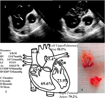
An electrocardiogram revealed features of right atrial enlargement. A chest radiograph revealed an absent pulmonary bay. His haemoglobin level was 24.5 g/dl. Transthoracic echocardiography showed dropout in the interatrial septum, but Doppler study was non-contributory owing to poor windows. The right ventricle and pulmonary arteries appeared small. There was no ventricular septal defect or patent ductus arteriosus. Transoesophageal echocardiography revealed a 27-mm ostium secundum atrial septal defect with a large, floppy Eustachian valve projecting from the right atrium and inferior caval vein junction (Fig 1). Bubble contrast imaging performed from a lower limb injection revealed that the Eustachian valve was diverting right atrial blood across the atrial septal defect into the left atrium (Fig 2) (Video).
Fig 1: transesophageal ECHO showing floppy large eustachian valve; Fig 2: contrast ECHO showing streaming of blood from inferior vena cava to left atrium; Fig 3: saturation study; Fig 4: eustachian valve specimen.

A cardiac catheterisation revealed systemic desaturation consistent with a right-to-left shunt (Fig 3). His total pulmonary blood flow was 2.7 L/minute, and total systemic blood flow was 7.7 L/minute, indicating net right-to-left shunt of 5 L/minute. His mean pulmonary arterial pressure was 11 mmHg, thus excluding pulmonary arterial hypertension as the cause of the right-to-left shunt. Angiograms were performed to rule out a persistent left superior caval vein to the left atrium, unroofing of the coronary sinus, and a pulmonary artery-to-left atrium fistula. The prominent Eustachian valve (Fig 4) was removed, and the atrial septal defect was closed surgically.
Discussion
During early embryological development, the right and left venous valves separate the systemic sinus venosus from the primary part of the right atrium. The left venous valve subsequently fuses with the developing atrial septum. As the systemic sinus venosus becomes incorporated into the right atrium, the right venous valve diminishes. Its rudiments usually become the crista terminalis, the valve of the inferior caval vein, Eustachian valve, and the valve of the coronary sinus, Thebesian valve; however, abnormal regression of the right valve of the sinus venosus may be present occasionally. In our case, muscularisation instead of the normal absorption possibly occurred at the portion of the right valve of the sinus venosus, resulting in the large Eustachian valve diverting the blood stream from the inferior vena cava into the left atrium through the atrial septal defect.
Gallaher et alReference Gallaher, Sperling, Gwinn, Meyer and Fyler 1 reported three patients, aged 26, 14, and 8 years, in whom a large Eustachian valve directed inferior caval vein inflow into the left atrium. Thomas et alReference Thomas, Tabakin and Ittleman 2 described an elderly woman in whom a large Eustachian valve and a redundant septum secundum caused preferential drainage of the inferior caval vein into the left atrium. Only two anatomical anomalies can contribute to this shunting. The first is a large Eustachian valve that directs inferior caval vein flow to the interatrial septum, the second is a redundant septum secundum that billows outward into the right atrium, intercepting inferior caval vein blood, and shunting it through the widely patent foramen ovale into the left atrium. The second one can occur even without elevated right atrial pressures. A high level of clinical suspicion in otherwise unexplained cyanosis and detailed imaging of the abnormal drainage anatomy can lead to accurate diagnosis and successful surgical treatment.
Acknowledgements
The authors wish to thank Dr. Viji Samuel Thompson.
Financial Support
This research received no specific grant from any funding agency or from commercial or not-for-profit sectors.
Conflicts of Interest
None.
Supplementary material
To view supplementary material for this article, please visit https://doi.org/10.1017/S1047951116002754