Tetralogy of Fallot is the most common cyanotic congenital heart condition with an incidence of ~1 in 2500.Reference Child 1 Following the advent of surgical repair in the 1950s, patients with tetralogy of Fallot now typically survive into adulthood and represent a significant proportion of the adult CHD population. The presence of incisional scar and ongoing altered haemodynamics create the ideal substrate for atrial (and ventricular) arrhythmias, both associated with an increased risk of sudden cardiac death.Reference Gatzoulis, Balaji and Webber 2 – Reference Yap, Harris, Chauhan, Oechslin and Silversides 4 The prevalence of “late” atrial tachyarrhythmias is believed to be ~20%.Reference Khairy, Aboulhosn and Gurvitz 5 In younger patients (<45 years), it has been observed that intra-atrial re-entrant tachycardia is more prevalent than atrial fibrillation; however, after the age of 55 years, this situation is reversed, and the prevalence of atrial fibrillation is believed to exceed that of intra-atrial re-entrant tachycardia, occurring in at least 30% of patients.Reference Khairy, Aboulhosn and Gurvitz 5 It is now clear that in addition to the risk of sudden cardiac death, atrial tachyarrhythmias represent a major source of morbidity in the tetralogy of Fallot population.Reference Roos-Hesselink, Perlroth, McGhie and Spitaels 6 – Reference Walsh and Cecchin 8 Despite this, there are little published data on predictors of atrial arrhythmias in patients with tetralogy of Fallot.
The Heart Hospital (University College Hospital, London) provides care for one of the largest number of patients with adult CHD in the world, including ~450 patients with tetralogy of Fallot. We performed a matched case–control study to examine the characteristics and predictors of atrial tachyarrhythmias in our patients with tetralogy of Fallot, describe the mechanisms of these atrial tachyarrhythmias, and discuss the outcome of radiofrequency ablation in this group. We present a comprehensive review of our experience of the development and management of atrial arrhythmias in patients with tetralogy of Fallot.
Methods
Study design, population, and data collection
This is a single-centre, retrospective, case–control study of adults with tetralogy of Fallot and late, postoperative atrial tachyarrhythmias. All patients with tetralogy of Fallot and atrial tachyarrhythmias undergoing radiofrequency ablation at the Heart Hospital, University College Hospital, London, between 2004 and 2013 were included, identified through the Cardiovascular Information Management System (Philips Healthcare, Best, the Netherlands). Ablation was indicated for sustained atrial tachyarrhythmias, either current or previously documented, who had failed medical treatment and had been referred to the electrophysiology service. Patients with significant additional valvular or congenital defects (excepting pulmonary valve lesions) were excluded. For each patient who had undergone ablation for atrial tachyarrhythmias, two controls with tetralogy of Fallot who had not undergone radiofrequency ablation and were not known to have atrial tachyarrhythmias were matched to date of birth (±3 years), gender, and age at the time of index echocardiogram (±2 years) where possible. Index TTE for the cases was one that had been performed shortly before radiofrequency ablation. Patients identified during screening for controls, who had not undergone radiofrequency ablation but were found to have evidence of atrial tachyarrhythmias, were excluded from the case–control comparison, but an additional subgroup analysis was performed on these patients. Baseline and follow-up data, including NYHA functional class, for all patients were collected through a retrospective review of the electronic and archived paper hospital records and subsequently entered into a dedicated database. NYHA class was determined either from clinic letters explicitly stating NYHA class or summarised from a description of daily living activities. Arrhythmia surveillance was performed by a combination of 12-lead ECG – documented at every outpatient clinic visit – and supplemented by additional information when available – for example, Holter monitoring or cardiac rhythm management device interrogation if one were in situ.
Echocardiography
All patients had transthoracic echocardiogram performed by a specialist adult CHD echocardiographer. Biventricular long-axis function on M-mode echocardiography – mitral and tricuspid annular plane systolic excursion – chamber dimensions, left ventricular ejection fraction, right ventricular fractional area change and Doppler calculations of pulmonary valve peak systolic velocity, tricuspid regurgitation, pulmonary regurgitation, and right ventricular systolic pressure were measured according to current recommendations.Reference Wharton, Steeds and Allen 9 All images were digitally stored and reviewed blinded to whether the patient was a case or control.
Mapping and ablation
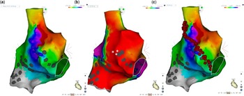
Mapping was guided by three-dimensional navigation systems; either CARTOTM (Biosense Webster, Diamond Bar, California, United States of America) or NavX (St. Jude Medical Inc., St. Paul, Minnesota, United States of America) systems were used. Where possible, bipolar voltage maps in sinus rhythm were created, abnormal endocardium defined as either scar (bipolar amplitude ⩽0.05 mV) or low-voltage (bipolar amplitude of ⩽0.5 mV) (Fig 1a). If atrial tachyarrhythmias were not present at the start of the procedure, induction was attempted with programmed stimulation and repeated with isoproterenol at the discretion of the operator. Intracardiac electrograms were stored on a digital recording system (LabSystemTMPRO, Boston Scientific, Bard, Boston Scientific or EP Medsystems, St. Jude Medical). Tachycardia mechanisms were diagnosed based on electrogram examination, activation, and entrainment mapping (Fig 1b). Atrial tachyarrhythmias were categorised into five types as follows: cavo-tricuspid isthmus-dependent intra-atrial re-entrant tachycardia, in which electroanatomic mapping and/or surface P-wave morphology suggested a macro-reentrant circuit and entrainment mapping demonstrated the cavo-tricuspid isthmus to be critical to the circuit; non-cavo-tricuspid isthmus-dependent intra-atrial re-entrant tachycardia involving scar tissue, suture lines, or prosthetic materials (“SCAR”); atrial fibrillation characterised by indistinct surface P-waves with chaotic atrial electrograms; or “FOCAL”, non-automatic focal/micro-reentrant atrial tachyarrhythmias, defined as originating from a small discrete region with centrifugal activation. If the tachycardia clearly originated from the left atrium, it was separately classified as “LEFT” (excluding atrial fibrillation). In cases where atrial tachyarrhythmias were non-inducible, multiple, or frequently changing, a decision was made at the discretion of the operator to perform an empirical linear or substrate-based ablation if appropriate. The vast majority of these ablations were for non-inducible patients with previous ECG evidence strongly suggestive of typical atrial cavo-tricuspid flutter.
Electroanatomic mapping. Activation map ( a ) of the right atrium from the right anterior oblique aspect showing an intra-atrial re-entrant tachycardia, demonstrated by entrainment mapping to be dependent on a critical isthmus between the superior caval vein and tricuspid valve annulus. Fractionated potentials were seen in this area before ablation, as indicated by the asterisks. An extensive area of low-voltage electrograms are seen on bipolar mapping ( b ) after ablation, in sinus rhythm (colour scale indicates local peak-to-peak electrogram amplitude; scar depicted grey, defined as no identifiable electrogram or bipolar amplitude <0.05 mV, normal endocardium purple, and low-voltage endocardium red to purple, 0.05–0.5 mV). The lesion set (superior caval vein to anterior tricuspid valve annulus) is shown in ( c ).

Ablation “lesion sets” were defined as follows: “cavo-tricuspid isthmus”, tricuspid valve annulus to inferior caval vein; “SCAR”, intercaval, inferior caval vein to atriotomy, superior caval vein to atriotomy, and atriotomy to tricuspid valve annulus; “FOCAL”, right; “LEFT LINEAR”, mitral isthmus, roof line, or septal line, right upper pulmonary vein to mitral valve annulus; “WACA”, wide area circumferential ablation of the pulmonary veins; or “CFE”, complex fractionated electrogram ablation and left.
Ablation was performed using a 4-mm irrigated tip catheter (Navistar Thermocool, Biosense Webster or Therapy Cool Path, Cool Path Duo or Coolflex; St. Jude Medical Inc., St. Paul) with a power of 30–40 W and a flow rate of 17–30 ml/minute. Success was defined as termination of tachycardia during ablation, demonstration of bidirectional block where applicable, and inability to re-induce tachycardia with programmed stimulation. If additional atrial tachyarrhythmias were induced, these were ablated until atrial tachyarrhythmias were rendered non-inducible.
Statistical analysis
Categorical variables are presented as numbers of occurrences and percentages. Continuous variables are shown as means and standard deviations. The association of baseline variables with cases and controls was assessed using conditional logistic regression; where conditional logistic regression could not calculate an odds ratio because of insufficient exposure, an exact binomial test was used.
Kaplan–Meier survival curves were used to examine the relationship between baseline variables and the risk for repeat ablation. Differences in the Kaplan–Meier survival curves were assessed by the log-rank test. A multivariable model was constructed to elicit the most predictive risk factors for recurrence. A Cox model (proportional hazards) was used to estimate the associations of the hazard of recurrence with case patient baseline variables. All univariable associations were examined, and variables with a p-value <0.1 were included in the multivariable modelling.
The comparison of NYHA status at baseline versus follow-up was conducted using the Wilcoxon signed rank test. Fisher’s exact test was used to evaluate the associations of rhythm at long term follow-up, atrial fibrillation ever, and death with categorical baseline variables. Logistic regression was used to calculate the associations of these outcomes with continuous variables.
Data analyses were performed using Stata version 11.2 (StataCorp LP, College station, Texas, United States of America).
Results
Patient characteristics associated with atrial tachyarrhythmias
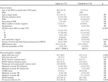
The study population consisted of 37 cases, tetralogy of Fallot patients who had undergone radiofrequency ablation for treatment of atrial tachyarrhythmias, and 53 age- and gender-matched controls, tetralogy of Fallot patients without atrial arrhythmias. It was not possible to identify any appropriately matched control for six of the case patients, and only one matched control was possible for a further nine. All but four patients – two cases and two controls – had undergone complete corrective repair. By the end of the study period, there were four deaths in the atrial tachyarrhythmias group and none in the control group. Mean age at time of death was 57 years, and all four deaths were attributable to biventricular heart failure, one patient having never undergone reparative surgery and none procedure related. At final follow-up, two were in sinus rhythm, one in atrial fibrillation, and one in atrial tachycardia. At baseline, all four patients had NYHA class 3 status: one had severely impaired left ventricular function, two had mildly impaired left ventricular function, and two had cardiac resynchronisation therapy devices in situ. All four had moderate or severe right ventricular dilatation and dysfunction. Full patient characteristics of ablated cases and arrhythmia-free controls are shown in Table 1. Compared with controls, cases were more likely to be on anti-arrhythmic drugs, which included β-blockers (81.1 versus 15.1%, p=0.001), have a cardiac rhythm management device in situ (24.3 versus 1.9%, p=0.008), and be in a worse NYHA symptomatic class (1.95 versus 1.32, p<0.001). Both right and left atrial size by area were associated with the presence of atrial tachyarrhythmias requiring ablation, whereas left ventricular ejection fraction (%) was inversely related (Table 1). Of the 37 cases, 23 (62%) were on long-term warfarin compared with 1/53 (1.9%) of controls.
Baseline characteristics of atrial tachyarrhythmias cases and controls.

ICD=implantable cardioverter defibrillator; LV=left ventricle; NSVT=non-sustained ventricular tachycardia; PR=pulmonary regurgitation; PVR=pulmonary valve replacement; RFA=radiofrequency ablation; RV=right ventricle; RVIT=right ventricular inflow tract; RVOT=right ventricular outflow tract; RVSP=right ventricular systolic pressure; TAPSE=tricuspid annular plane systolic excursion; TR=tricuspid regurgitation; TTE=transthoracic echocardiogram
Data are expressed as mean±standard deviation and number (%) as appropriate
* To include β-blockers
p Values <0.05 are shown
Procedural outcome
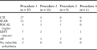
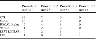
There were 55 radiofrequency ablation procedures performed on 37 atrial tachyarrhythmia cases. The number of cases recorded as being performed under general anaesthesia was 16, under local anaesthetic and conscious sedation – using midazolam and diamorphine –24, and the details of anaesthesia were not recorded for 15 procedures. The presenting or induced arrhythmias during the electrophysiological study are shown in Table 2 and ablation sets are shown in Table 3. Cavo-tricuspid-dependent atrial tachyarrhythmia was the most commonly observed arrhythmia (27/37) at first procedure and cavo-tricuspid isthmus ablation was performed in almost all cases (35/37). Tachycardia cycle length for intra-atrial re-entrant tachycardia ranged from 150 to 500 ms and for focal atrial tachyarrhythmias 210–500 ms. Acute success was achieved in 54/55 procedures (98%) with a mean of 1.56 lesion sets required per procedure. The mean procedure time was 185±67 minutes. Further procedural details are shown in Table 4. Left atrial ablation was required in 3% of first, 21% of second, and 50% of third and fourth procedures. Procedural complications included complete heart block in two patients, one requiring a pacemaker, one with a previously implanted implantable cardioverter defibrillator, and a retroperitoneal bleed, managed conservatively, in one patient. During the study period, four patients additionally underwent right surgical Maze procedures, performed at the time of concomitant cardiac surgery, predominantly pulmonary valve replacement. The data were censored at this point. In three of these four patients, the Maze was a terminal procedure; one patient required a further radiofrequency ablation.
Arrhythmia at procedure.

AF=atrial fibrillation; CTI=cavo-tricuspid isthmus
Ablation at procedure.

CFE=complex fractionated electrogram; CTI=cavo-tricuspid isthmus; WACA=wide area circumferential ablation
Procedural details.

Predictors of recurrence
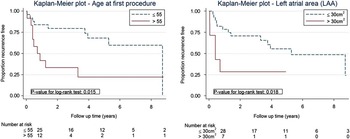
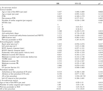
Age at the time of first procedure, presence of cardiac rhythm management device, left atrial area, and presence of atrial fibrillation at first procedure were significant predictors of recurrence after first procedure on univariate Cox analysis (Table 5a). On multivariate regression, age at the time of first procedure, number of cardiac surgeries, presence of cardiac rhythm management devices, and atrial fibrillation at first procedure were the strongest predictors (Table 5b). Age ⩽55 years and left atrial area ⩽30 cm2 predicted recurrence-free survival (log-rank p=0.015 and 0.018, respectively) (Fig 2).
Freedom from atrial tachyarrhythmia recurrence.

Predictors of recurrence of atrial tachycardia requiring further ablation after first procedure.

AF=atrial fibrillation; AT=atrial tachycardia; CTI=cavo-tricuspid isthmus; ICD=implantable cardioverter defibrillator; LV=left ventricle; NSVT=non-sustained ventricular tachycardia; PR=pulmonary regurgitation; PVR=pulmonary valve replacement; RFA=radiofrequency ablation; RV=right ventricle; RVIT=right ventricular inflow tract; RVOT=right ventricular outflow tract; RVSP=right ventricular systolic pressure; TAPSE=tricuspid annular plane systolic excursion; TR=tricuspid regurgitation *p<0.05 considered to be statistically significant.
Long-term outcome
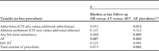
The mean follow-up was 41 months (range 2–87, interquartile range 14–67), after which 29/37 (78%) were arrhythmia-free. NYHA status improved (p<0.001) with the number of patients in NYHA class 1 increasing from 11/36 (31%) to 25/36 (69%); there was one patient for whom post-procedural NYHA data were not available. In addition, 13/37 (35%) did not require anti-arrhythmic drugs at the most recent follow-up. Of the patients on anti-arrhythmic drugs, 22/37 were on β-blockers, one of whom was also on a calcium-channel blocker; four of these patients were in an atrial arrhythmia at the end of follow-up (two atrial fibrillations), and the remainder in sinus rhythm. Moreover, five patients were on amiodarone and four of these five in sinus rhythm; 3/5 were also on a β-blocker (all in sinus rhythm). In addition, two patients were on digoxin and on amiodarone – one of whom was in atrial fibrillation and the other also on a β-blocker and in sinus rhythm. NYHA status at the time of first procedure predicted death (p=0.002). On univariate analysis, there was an association between rhythm at first procedure and the outcome variables: (1) rhythm at final follow-up, sinus rhythm (best) compared with atrial tachyarrhythmia compared with atrial fibrillation (worst); and (2) documentation of atrial fibrillation at any time (Table 6).
Predictors of long-term outcomes.

AF=atrial fibrillation; AT=atrial tachycardia; CTI=cavo-tricuspid isthmus; SR=sinus rhythm
* SR (n=30), AT (n=1), AF (n=6)
** AF ever (n=9), no AF (n=28)
Univariable p-values
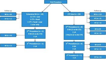
At the first procedure, the observation of cavo-tricuspid isthmus-dependent intra-atrial re-entrant tachycardia alone was associated with a significant decrease in the risk of subsequent atrial fibrillation (p=0.013), and a trend towards the maintenance of sinus rhythm long term, (p=0.053) with an 83% long-term procedural success – that is, freedom from recurrence of any atrial tachyarrhythmia. If a left atrial tachyarrhythmia or atrial fibrillation were observed, this predicted a poorer rhythm status at follow-up. If cavo-tricuspid isthmus ablation alone was performed, regardless of arrhythmia, there was a 52% single, long-term procedural success. Long-term outcome classified by ablation at first procedure is shown in Figure 3. Overall, 20/37 (54%) patients maintained sinus rhythm during follow-up after a single procedure.
Outcome classified by ablation required at first procedure.

Adverse outcomes in non-ablated atrial fibrillation patients
A preliminary screen to identify age- and gender-matched controls with tetralogy of Fallot but without history of radiofrequency ablation for atrial tachyarrhythmias yielded 67 patients. However, a subsequent review of the case records found that 14 of these had to be removed as controls because of evidence of atrial tachyarrhythmias, predominantly atrial fibrillation, highlighting the prevalence of atrial arrhythmias in patients with tetralogy of Fallot. Of these 14 patients who were excluded as controls in our study, four had undergone radiofrequency ablation for atrial tachyarrhythmias at other institutions, whereas nine of the remaining 10 patients had never been ablated and were in atrial fibrillation – that is, 13.4% (9/67) of the total originally matched non-ablated tetralogy of Fallot control group – a figure in keeping with previously published dataReference Khairy, Aboulhosn and Gurvitz 5 . These nine patients had originally been matched to seven ablated cases as two of the cases had two matched controls. The mean age of these nine patients at the time of matched TTE was 62 years and six of them (67%) had died at the end of the follow-up period. In contrast, of the seven ablated cases, with the same mean age, all were alive: four (57%) remained in sinus rhythm and three in atrial tachyarrhythmias (including 1 in atrial fibrillation) at long-term follow-up.
Discussion
This study characterises in detail the largest single-centre series of 37 patients with tetralogy of Fallot and atrial tachyarrhythmias treated with radiofrequency ablation and compares them with arrhythmia-free, non-ablated controls. We have demonstrated that radiofrequency ablation for atrial tachyarrhythmias in patients with tetralogy of Fallot has acute success rates of 98% and in the longer term improves functional status. The observation of lone cavo-tricuspid isthmus-dependent intra-atrial re-entrant tachycardia at first procedure was associated with a lower risk of subsequent atrial fibrillation and an 83% long-term procedural success – that is, freedom from recurrence of any atrial tachyarrhythmia. If left-sided atrial tachyarrhythmias or atrial fibrillation were observed, this predicted a poorer rhythm status at follow-up. Our study also provides important incidental information on atrial arrhythmia burden in non-ablated patients with tetralogy of Fallot compared with those who have undergone radiofrequency ablation.
Patients with tetralogy of Fallot who develop atrial tachyarrhythmias requiring radiofrequency ablation have significantly more biatrial dilatation those who do not. Bonello et alReference Bonello, Kempny and Uebing 10 have previously described the morphological and haemodynamic characteristics of tetralogy of Fallot patients with atrial arrhythmias; of 154 patients, atrial tachyarrhythmias – sustained atrial tachyarrhythmias or atrial fibrillation – were observed in 11, a slightly lower incidence than has previously been reported, and were predicted by right atrial area on cardiac magnetic resonance imaging. Another studyReference Rosianu, Paprika, Osztheimer, Temesvari and Szili-Torok 11 examined the echocardiographic profiles of tetralogy of Fallot patients with palpitations but no documented tachycardia and also noted that the only significant difference in echocardiographic parameters between those with and without a history of palpitations was right atrial size. Our study in contrast highlights that atrial tachyarrhythmias are not only associated with right atrial enlargement but also with an increase in left atrial size. Patients requiring ablation for atrial tachyarrhythmias also have a higher likelihood of having a cardiac rhythm management device in situ – predominantly implantable cardioverter defibrillators – and a higher mortality rate than controls, which reflects the more advanced substrate present in this patient group. NYHA class at presentation was the only variable on univariate analysis that predicted death.
This series has demonstrated that radiofrequency ablation for atrial tachyarrhythmias in patients with tetralogy of Fallot has high acute success rates and improves long-term functional status, in keeping with data from others, although recurrences are common and a significant challenge in ablating patients with adult CHD. In our cohort, the recurrence rate of cavo-tricuspid isthmus-dependent flutter was ~19%, which is higher than that expected in “normal heart” isthmus-dependent flutter and may reflect the more complex nature of the substrate.
Mechanisms and outcomes have only been described in a small number of previous studies.Reference de Groot, Lukac and Schalij 12 – Reference Biviano, Garan, Hickey, Whang, Dizon and Rosenbaum 14 De Groot et alReference de Groot, Lukac and Schalij 12 described a comparable large proportion of cavo-tricuspid isthmus-dependent tachycardia with scar-dependent intra-atrial re-entrant tachycardia, and similarly reported acute success rates of 98% with sinus rhythm maintained in ~85% at long-term follow-up. They also observed a similar incidence of atrial fibrillation during follow-up but did not describe left atrial ablation in their patients. In our study, we observed that the proportion of left atrial tachyarrhythmias increases with time and that left atrial ablation is therefore increasingly likely to be needed with repeat procedures. This must be balanced, however, against the possibility of creating further arrhythmic substrate through ablation. As can be seen in Tables 2 and 3, although there were seven cases where a left-sided macro-reeentrant atrial tachycardia was observed at the first procedure and four cases where atrial fibrillation was observed, only two left-sided ablations were actually performed at the first procedure – both of these patients also had right-sided ablations at the same time – that is, our practice has been to ablate predominantly on the right side initially if possible. That said, given the otherwise relatively good prognosis of many of these patients, it may be reasonable to consider ablation on the left side as well, given the potentially significant side-effects of medical treatment.
Age at the time of radiofrequency ablation, left atrial size, and presence of atrial fibrillation at first procedure were all predictive of recurrence of atrial tachyarrhythmias on univariate analysis in our study population. This is consistent with the critical mass hypothesis of atrial fibrillation proposed in patients without CHDReference Moe 15 and that duration of arrhythmia – that is, time to ablation – may be an important predictor of recurrence.Reference McCready, Smedley and Lambiase 16 On multivariate analysis, age at the time of procedure, number of cardiac surgeries, and presence of atrial fibrillation at first procedure were all independent predictors of recurrence, consistent with data reported by Khairy et al on prevalence of intra-atrial re-entrant tachycardia and atrial fibrillation in the surgically corrected tetralogy of Fallot population.Reference Khairy, Aboulhosn and Gurvitz 5 Older age at ablation and complex atrial surgery have been associated with higher risk of atrial tachyarrhythmia recurrence in the combined adult CHD population;Reference Yap, Harris, Silversides, Downar and Chauhan 17 however, ours is the first study to specifically describe patients with tetralogy of Fallot. These data support a policy of early intervention in patients with atrial tachyarrhythmias and tetralogy of Fallot, and may help clinicians give more informed consent to patients with a higher risk factor profile. Yap et alReference Yap, Harris, Silversides, Downar and Chauhan 17 also examined possible associations between risk of recurrence after radiofrequency ablation and history of atrial fibrillation or procedural complexity and found none. In that study, however, the population was younger and slightly less contemporary, as well as comprised a significant proportion of patients with Fontan circulations, in addition to other adult CHD diagnoses. With respect to long-term outcome in our population, presence of cavo-tricuspid isthmus-dependent flutter alone predicted a better long-term likelihood of freedom from atrial fibrillation and maintenance of sinus rhythm. There are a number of potential explanations for this observation but primarily there tends to be a less complex arrhythmogenic substrate in patients when cavo-tricuspid isthmus-dependent flutter alone is observed – for example, related to surgical scar or volume overload – and that patients with a more complex substrate who require extensive ablation may also inherently, by nature of the ablation itself, have a lower rate of freedom from recurrence. Although this study was not designed to address the effect of ablation on future arrhythmia, it suggests that by intervening on cavo-tricuspid isthmus-dependent flutter at an earlier time in the natural history of tetralogy of Fallot, it may be possible to protect against adverse re-modelling as a result of sustained arrhythmias, which may subsequently lead to the development of a more complex substrate perpetuating further multiple tachyarrhythmias.
Finally, we also observed that the incidence of atrial fibrillation was higher in patients with tetralogy of Fallot who had never undergone radiofrequency ablation for atrial tachyarrhythmias. During screening for matched controls – that is, patients who had not undergone radiofrequency ablation – we noted on case record review that >60% of those over the age of 60 years who had not undergone ablation and had been assumed to be arrhythmia-free in fact had evidence of atrial tachyarrhythmias, predominantly atrial fibrillation. Although these patients were excluded as controls from the case–control analysis, it is interesting to observe that of the 9 non-ablated patients with atrial fibrillation, the majority had died by the end of the follow-up period, whereas, in contrast, of the seven matched ablated cases all were alive and the majority were in sinus rhythm. Older age has been consistently observed to be a risk factor for atrial tachyarrhythmias, specifically atrial fibrillation, in previous studiesReference Gatzoulis, Balaji and Webber 2 , Reference Yap, Harris, Chauhan, Oechslin and Silversides 4 ; however, these new findings suggest that active early surveillance for and ablation of atrial tachyarrhythmias in patients with tetralogy of Fallot should be undertaken in order to prevent the subsequent development of atrial fibrillation. In their cross-sectional study, Khairy et alReference Khairy, Aboulhosn and Gurvitz 5 observed that atrial fibrillation was far less prevalent than intra-atrial re-entrant tachycardia in patients <45 years but that atrial fibrillation prevalence exceeded that of intra-atrial re-entrant tachycardia after 55 years of age, an exaggeration of the increased prevalence of atrial fibrillation seen with age in the general population. They also noted an association between hypertension and intra-atrial re-entrant tachycardia, not observed in our study, and suggested that determinants of intra-atrial re-entrant tachycardia and atrial fibrillation may differ. In the present study, we have observed that the incidence of atrial fibrillation in patients who have undergone atrial radiofrequency ablation at our centre is lower than that expected in a comparable cohort and that electrophysiological intervention with radiofrequency ablation may influence the future development of atrial fibrillation. Further studies on the long-term effects of radiofrequency ablation of atrial tachyarrhythmias in the Fallot population are needed to examine this.
Study limitations
This study has limitations inherent to any retrospective study, although a complete data set was available for the majority of cases and controls. It was not possible to match two controls per case as specified in the original study design, after careful review of control cases revealed evidence of arrhythmias. These “controls” were, however, removed from the case–control analysis. Some analyses are exploratory in nature because of the small sample size, particularly those of longer-term outcomes where there are a few events and a large range of follow-up periods, although 29/37 patients did have a follow-up period >12 months. The cases were evaluated by four different electrophysiologists during the study period, but with a standardised approach, and the vast majority were performed by the same operator. Data recorded on recurrence of atrial fibrillation were based on 12-lead ECG or Holter recordings of variable duration according to availability, and therefore may be an underestimate.
Conclusion
Catheter ablation is a highly effective treatment for atrial tachyarrhythmias in patients with tetralogy of Fallot. Atrial size predicts the development of atrial tachyarrhythmias in this population, and the type of arrhythmia at first presentation may predict long-term outcome. Left atrial arrhythmias and need for left atrial ablation increase over time. Early radiofrequency ablation may have a protective effect against the development of atrial tachyarrhythmias, particularly atrial fibrillation.
Acknowledgements
None.
Conflicts of Interest
None.