Introduction
Total anomalous pulmonary venous return is a rare CHD, with an incidence between 5.9 and 7.1 per 100,000 live births. Reference Seale, Uemura and Webber1–Reference Correa-Villasenor, Ferencz, Boughman and Neill3 Infradiaphragmatic total anomalous pulmonary venous return is often associated with obstruction—either from extrinsic compression or internal narrowing of the venous pathway. Sites of obstruction include the ductus venosus, stenosis of the vertical vein as it crosses the oesophageal hiatus in the diaphragm, or the junction of the vertical vein and the portal vein. Reference Ward, Mullins, Garson, Bricker, Fisher and Neish4 Significant obstruction can result in hemodynamic instability and death if not intervened on in an expedited way. The standard of care for these patients is prompt surgical correction. Stenting of the ductus venosus or vertical vein has been described in patients who are not considered surgical candidates due to size, prematurity, or unstable clinical status. Reference Rothman, Galindo and Evans5–Reference George, Lozier and Bocks8 We present a case of a premature infant who required stenting of the ductus venosus followed by vertical vein stenting via transhepatic portal venous access with closure of the hepatic tract using the VASCADE Vascular Closure System who was successfully bridged to surgical correction.
Case report
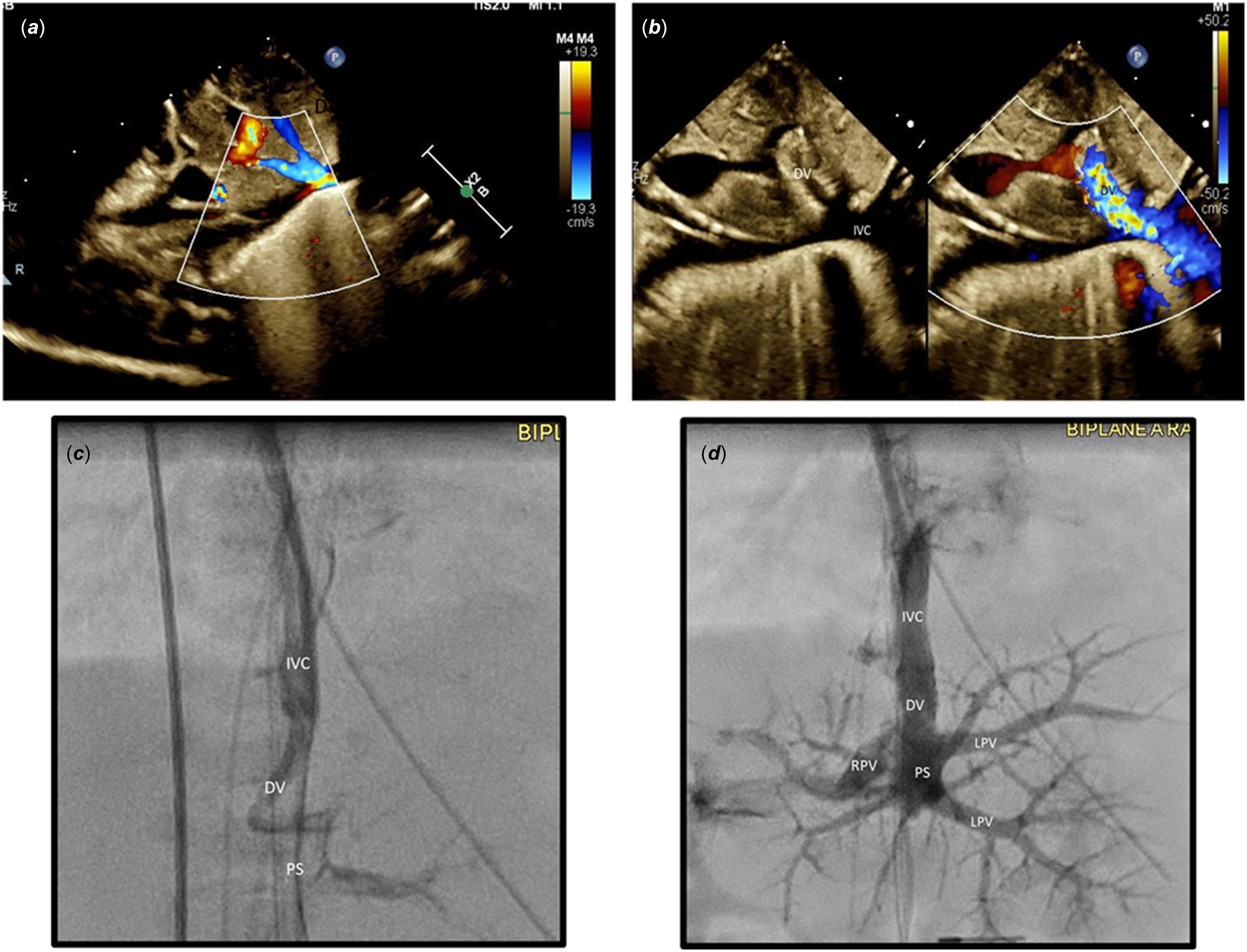
A 29-week premature infant weighing 1.29 kg was postnatally diagnosed with infradiaphragmatic total anomalous pulmonary venous return. He was transferred from an outside institution on day of life 2. He had respiratory failure requiring high ventilatory support with chest X-ray showing diffuse opacification of the lung fields. Transthoracic echocardiogram showed infradiaphragmatic total anomalous pulmonary venous return with a pulmonary venous confluence posterior to the left atrium and a vertical vein draining connecting to the left portal vein. His clinical status worsened overnight with PaO2 in the 20–30s on high oscillator settings with an echocardiogram showing obstruction of the ductus venosus (Figure 1 a ). The echocardiogram showed no significant gradient across the vertical vein. CT scan demonstrated obstruction at the ductus venosus as well as mild calibre change of the vertical vein at the level of the diaphragm (Supplemental Figure 1).
Transthoracic echocardiogram and angiogram in the IVC showing significant narrowing at the DV ( a , c ) that improves after stent placement ( b , d ). IVC = inferior vena cava; DV = ductus venosus; PS = portal sinus; RPV = right portal vein; LPV = left portal vein.

He presented to the cardiac catheterisation laboratory on day of life 3. As the CT scan and echocardiogram suggested only mild narrowing in a large-calibre vertical vein, targeted ductal venosus stenting was planned to not prolong the procedure on a critically ill premature neonate. Access across the ductus venosus was achieved using right internal jugular venous access. Balloon angioplasty of the ductus venosus was performed using a 3 mm × 2 cm NC Emerge balloon (Boston Scientific, Marlborough, MA) to open the significantly narrowed vessel (Figure 1 c ), followed by stent placement using a 4 mm × 12 mm Synergy Megatron (Boston Scientific, Marlborough, MA) stent (Figure 1 b and d ). Angiography showed the vertical vein draining to the portal venous system with mild narrowing at the vertical vein insertion site.
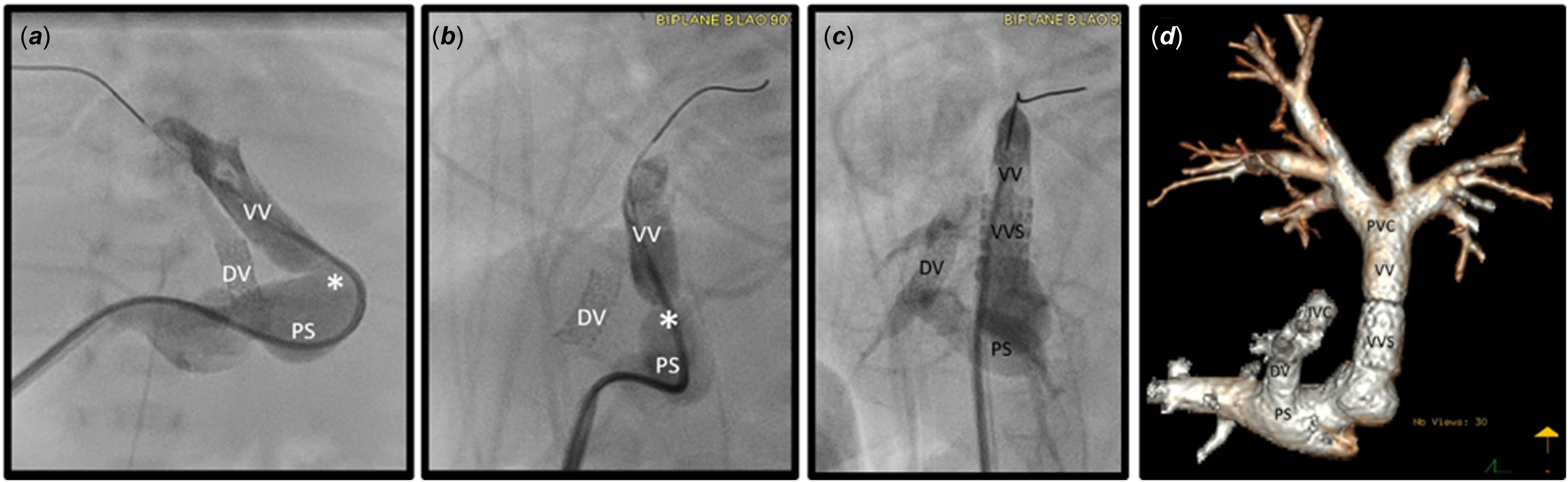
Following initial clinical improvement, he developed a worsened respiratory status with progressive vertical vein narrowing noted on echocardiogram by 2D imaging and increased Doppler mean gradient to 15 mmHg. He returned to the cardiac catheterisation laboratory 11 days after initial catheterisation for stent placement in the vertical vein. Ultrasound guidance was used to image the vertical vein and track the vessel to the portal sinus and to a dilated portal vein. Percutaneous portal venous access was obtained, and the wire position was confirmed on ultrasound and on fluoroscopy. The previously placed stent was a valuable landmark, and the wire interaction with the stent confirmed appropriate positioning. A 6-Fr Pinnacle Introducer sheath (Terumo Interventional Systems, Somerset, NJ) was advanced over the wire. The appropriate position was confirmed with angiography. A 4-Fr sheath had been placed in the right internal jugular vein for angiographic guidance if needed; however, this was not necessary. There was a 14-mmHg mean gradient across the vertical vein narrowing. Angiography showed the superior portion of the vertical vein that measured 7 mm × 6.1 mm with a narrowing to 3.7 mm × 2.7 mm (Figure 2 a and b ). A 0.018″ SV5 wire (Cordis, Miami Lakes, FL) was advanced through the vertical vein into the right upper pulmonary vein. A 7 mm × 12 mm Formula 418 stent (Cook Medical LLC, Bloomington, IN) was advanced over the wire and deployed across the vertical vein narrowing with no residual vertical vein stenosis post-stent placement (Figure 2 c ). The portal venous tract was closed using a VASCADE VCS. Using ultrasound guidance, the VASCADE VCS was inserted into the sheath, and the nitinol disc was deployed into the lumen of the vessel. The sheath was removed over the device, and the disc was brought to the vessel wall using gentle tension. The collagen patch was exposed, and after approximately 30 s, the disc was retracted and removed, leaving the collagen within the tract. Ultrasound confirmed good flow through the portal vein and collagen within the tract with no evidence of haematoma.
Straight AP ( a ) and lateral ( b ) angiogram showing significant obstruction (*) of the vertical vein at the entry into the portal venous system. After stent placement, lateral angiogram ( c ) and preoperative CT 3D reconstruction ( d ) shows no residual VV obstruction.

The patient clinically improved after stent placement but required prolonged support on the oscillator ventilator. His continued tenuous respiratory status was attributed to his premature lungs with some degree of residual pulmonary venous obstruction through the portal venous system through the capillary network. Preoperative CT scan showed widely patent ductus venosus and vertical vein stents (Figure 2 d ). He underwent primary sutureless TAPVR repair with creation of a 3.5 mm atrial septal defect and ligation of the vertical vein at 8 weeks of age and 2.79 kg. He underwent delayed sternal closure 2 days later, was successfully extubated on postoperative day 10, and was weaned to room air on postoperative day 32. He was discharged home on postoperative day 35. Echocardiogram at discharge showed an unobstructed pulmonary venous confluence. The ductus venosus stent remained patent with progressively less flow across it, and liver function testing remained normal. At the most recent follow-up at 8 months of age, he was clinically doing well with no residual flow noted through the ductus venosus stent and no pulmonary venous obstruction on echocardiography.
Discussion
Catheter-based palliation utilising stent placement in the ductus venous and in the vertical vein for obstructed TAPVR has been previously described. Reference Rothman, Galindo and Evans5–Reference George, Lozier and Bocks8 This patient required stenting of the ductus venosus as an initial palliation, followed by stenting of the vertical vein two weeks later due to progressive obstruction. At the time of vertical vein stent placement, umbilical access was not available, and the previously placed ductus venosus stent and angulation of the vertical vein into the portal venous system limited the utility of the transjugular approach. We opted for transhepatic portal venous access to avoid manipulation through the side cells of the previously placed stent and for a more direct approach into the vertical vein.
Transhepatic venous access has been utilised for cardiac catheterisation in patients with CHD. To our knowledge, this is the first case report of a transhepatic portal venous access approach to perform vertical vein stenting. This approach is relatively straightforward pathway utilising ultrasound and fluoroscopic guidance and offers direct access into a larger calibre vessel for implantation of larger diameter stents when necessary. Closure of the hepatic tract has been performed with vascular coils, Gelfoam, Amplatzer Vascular Plugs (Abbott, Chicago, IL), and Mynxgrip (Cordis, Hialeah, FL) to reduce the risk of retroperitoneal bleeding and pericapsular haematoma. Reference Qureshi, Prieto, Bradley-Skelton, Bradley-Skelton and Latson9–Reference Eilers, Bansal and Shan10 Eilers et al. described their experience utilising the VASCADE Vascular Closure System (Cardiva Medical Inc., Santa Barbara, CA) for 16 procedures in 10 patients with excellent results. Reference Eilers, Bansal and Shan10 The technique they describe in detail proved useful in our much smaller patient. We utilised this closure system in our patient to avoid placing a permanent device in a premature infant and mitigate bleeding risk, which could be catastrophic in a patient this size. To our knowledge, this case reports the smallest patient in whom VASCADE VCS has been utilised for hepatic tract closure.
Following surgical repair, the ductus venosus stent remained patent. There are reports of clinically significant portosystemic shunts related to persistent flow through ductal venosus stents requiring transcatheter device closure. Reference Imani, Otsuki and Kondo11 Given the risk for hepatic failure related to a portosystemic shunt, we monitored the patient with echocardiography and laboratory evaluation. The patency of the stent was monitored by echocardiography, and the flow through the stent gradually decreased. There was minimal residual flow through the stent by the time of the patient’s discharge from the hospital, and no residual flow was noted by 8 months of age. Liver function tests were performed periodically postoperatively to evaluate hepatic dysfunction related to steal of hepatic blood flow, and they remained normal.
Conclusion
While surgical correction of obstructed infradiaphragmatic total anomalous pulmonary venous return remains the standard of care for most patients, transcatheter palliation utilising stenting of the ductus venous and/or vertical vein has resulted in successful outcomes for patients who are poor surgical candidates initially. Our case offers a stepwise approach to multilevel obstruction in a small, premature patient with favourable transcatheter palliation followed by successful full surgical repair.
Supplementary material
The supplementary material for this article can be found at https://doi.org/10.1017/S1047951126113134.
Financial support
This research received no specific grant from any funding agency, commercial, or not-for-profit sectors.
Competing interests
None.
Ethical standard
Not applicable.