INTRODUCTION
Celleporina Gray, Reference Gray1848 is a specious genus of Cheilostomate Bryozoa, with about 80 Recent species distributed all around the world (Bock & Hayward, Reference Bock, Hayward, Bock and Gordon2013), 14 of which were described in the last 20 years. In the Atlantic–Mediterranean region 15 Recent species are known (Bock, Reference Bock2014), but only two of them were newly described during the last 20 years.
The genus Celleporina is characterized by the small, encrusting, multilaminar colonies, pisiform, nodular or cylindrical; the suberect autozooids, with cryptocystidean frontal shield with few marginal pores; the sinuate orifice flanked by columnar adventitious avicularia, typically paired; the frequent presence of vicarious avicularia; and by the prominent ovicell, acleithral, with a central perforated area called tabula. The tabula is usually considered as an area of uncalcified ectooecium exposing the pseudoporous entooecium (see e.g. Hayward & Ryland, Reference Hayward and Ryland1999), but this assumption may be due perhaps to a wrong interpretation, as explained in the discussion below.
In the present paper we present a re-description of Celleporina parvula (Canu & Bassler, Reference Canu and Bassler1928) based on the original material, and the description of two new species of the genus, collected in the NW Mediterranean and in the south of Portugal, respectively. Moreover, Celleporina fragilis Arístegui Ruiz, Reference Arístegui Ruiz1989 and Celleporina derungsi Souto et al., Reference Souto, Reverter-Gil and Fernández-Pulpeiro2010, only known until now from the Canary Islands and the south of Portugal respectively, are newly reported from the Gorringe Bank and the NW Iberian Peninsula respectively. The ancestrula of C. fragilis is re-described as a new ancestrular type for which a new name is introduced. The structure of the frontal wall in several species of the genus Celleporina is revised.
MATERIALS AND METHODS
The material studied has different origins. Historical specimens are preserved in the collections of the Muséum National d'Histoire Naturelle in Paris (MNHN), and in the Zoologisk Museum in Copenhagen (ZM). Additional material was collected in fishing nets in the Algarve (S Portugal) by H.D.B.; although the depth is unknown, the boat was small enough as to assure that material was collected in shallow waters near the coastline. Material from the NW of the Iberian Peninsula was collected by scuba diving by J.S. Specimens from the Menorca Channel were collected during the sampling survey ‘Canal0811’. Finally, samples from the Gorringe Bank were collected during the sampling surveys LusoExpedição 2006 and 2007, and the campaign EMEPC/Luso/2009.
The samples were examined with a stereomicroscope and uncoated material was photographed with different SEMs: a Zeiss EVO LS15 at the University of Santiago de Compostela, a Tescan VEGA at the MNHN and an Inspect S50 at the University of Vienna. All of them were used with a backscattered electron detector in low vacuum mode. Ultrastructure of the frontal wall was studied from coated and uncoated material with a Zeiss FESEM Ultra-Plus at the University of Santiago de Compostela. Measurements were taken with the software ImageJ® on the SEM photographs.
The new specimens collected during this work have been deposited in the collections of the Museo Nacional de Ciencias Naturales, Madrid (MNCN) and in the Museu Nacional de História Natural e da Ciencia, Lisbon (MB).
SYSTEMATICS
Superfamily Celleporoidea Johnston, Reference Johnston1838
Family Celleporidae Johnston, Reference Johnston1838
Genus Celleporina Gray, Reference Gray1848
Celleporina parvula (Canu & Bassler, Reference Canu and Bassler1928)
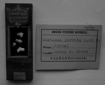
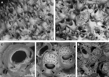
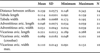
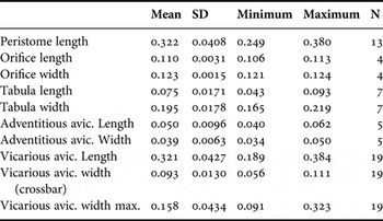
(Figures 1 & 2; Table 1)
Original sample of Celleporina parvula (lectotype and paralectotypes).

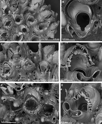
Celleporina parvula: (A) paralectotype; note the abundant columnar avicularia and regeneration processes in vicarious avicularia; (B) ovicellate zooids and vicarious avicularia (lectotype); (C) primary orifice (lectotype); (D) ovicells (paralectotype); (E) ovicells and vicarious avicularia (Atlantide Station 146).

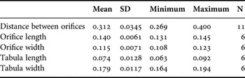
Measurements (in mm) of Celleporina parvula (lectotype + paralectotypes).

SD, Standard deviation; N, number of measurements.
Costazzia parvula Canu & Bassler, Reference Canu and Bassler1928, p. 60, pl. 8, figures 6–8.
Celleporina parvula (Canu & Bassler): Cook, Reference Cook1968, p. 221; Cook, Reference Cook1985, p. 182.
MATERIAL EXAMINED
Lectotype (designated here): MNHN.F.A51237 Atlantic Morocco, Vanneau Station. XLVII, 33°59′N 07°50′W, 17 June 1924, 53 m, on Smittina cervicornis. Canu coll. The ovicellate colony in the top of the slide (see Figure 1).
Paralectotypes (designated here): rest of the colonies in the same slide as lectotype.
Other material: Zoologisk Museum: Atlantide Station 145, 9°20′N 14°15′W, 32 m, 13 April 1946, Cook coll. Atlantide Station 146, 9°27′N 14°48′W, 50–51 m, 13 April 1946, Cook coll. Atlantide Station 151, 10°40′N 16°22′W, 65 m, 16 April 1946, Cook coll. Galathea Station 4, 22°19′N 17°05′W, 62 m, 02 November 1950, Cook coll. (two samples).
DESCRIPTION
Colony multilaminar, multiserial, forming small domed patches. Autozooids recumbent at colony margins, suberect and chaotically arranged elsewhere, with finely granular frontal wall and few, distinct round marginal pores. Primary orifice difficult to see, transversely oval, without distinct sinus or condyles. Peristome thick, well developed proximally and laterally, jointed with the basal wall and forming a pseudotube, incorporating on each side a columnar avicularium with oval rostrum, and complete crossbar, and sometimes finely toothed distal rim; acute to frontal plane and directed obliquely distally. Additional columnar avicularia may appear elsewhere, so each zooidal orifice may be surrounded by up to four avicularia. Vicarious avicularia frequent, with rostrum spatulate and deeply cupped distally; crossbar complete, without columella, palate with a large, frequently lanceolate foramen. Multiple regeneration by intramural buds may occur, with up to four vicarious avicularia growing one within each other. Ovicell hyperstomial; hemispherical ooecium formed by the flattened distal kenozooid budded from the maternal autozooid. Tabula large, crescent, occupying half or more of the ooecium, with large numerous pores in a roughly reticular design in younger ooecia. Pores strongly decrease in size with age during tabula's calcification. Proximally the tabula is restricted by a raised rim, peaked in some zooids. Proximal part of ooecium in the form of a well-developed funnel with its opening proximally surrounded by the peristome and not visible in a frontal view. Ancestrula not seen.
REMARKS
Celleporina parvula was briefly described by Canu & Bassler (Reference Canu and Bassler1928, as Costazia parvula) from several colonies collected between Rabat and Casablanca (W Morocco) at 53 m depth, growing on Smittina cervicornis (Pallas, Reference Pallas1766). The original material is now preserved in the Palaeontological Section of the MNHN mounted in a wooden slide (Figure 1). Celleporina parvula is easily distinguished from any other species of the genus in the Atlantic–Mediterranean region by the abundant columnar avicularia growing elsewhere and the broad tabula on the ovicell.
The species was later considered by Gautier (Reference Gautier1962) as a junior synonym of Lekythopora lucida (Hincks, Reference Hincks1880a) but without further discussion. However, it is unlikely if Gautier saw the original material of Canu & Bassler. As already stated by Cook (Reference Cook1968), C. lucida differs from C. parvula mainly by having a single oral avicularium, and by the perforations of the tabula, with a simple crescent of slit-like pores.
Cook (Reference Cook1968) initially transferred the species to the genus Celleporina, and found it in several localities along NW Africa. This author pointed out its similarity with Lekythopora laciniosa Calvet, Reference Calvet1906, a species described from the Azores (Figure 3). However, this species differs from C. parvula by the orifice, longer than wide with an open U-shaped sinus, the larger oral avicularia, placed close together, the smaller vicarious avicularia, and a smaller tabula with few, small pores. Calvet (Reference Calvet1931, p. 115) considered L. laciniosa as a synonym of Costazzia costazzi (Audouin, Reference Audouin and Audouin1826) but without further discussion. The material reported by Cook (Reference Cook1968, Reference Cook1985) as C. parvula, partly studied here (Figure 2E), seems to differ from the type material only by the larger pores of the tabula.
Lekythopora laciniosa: two views of the type material (Photos courtesy of B. Berning).

Several other species of the genus have been cited from the nearby Canary Islands. Celleporina canariensis Arístegui Ruiz, Reference Arístegui Ruiz1989 differs, among other characters, by its orifice, longer than wide and with a marked sinus, and by the characteristic V-shape of the secondary orifice, flanked by a pair of flat, triangular avicularia. Celleporina fragilis Arístegui Ruiz, Reference Arístegui Ruiz1989 (see below) presents a U-shaped sinus and the pair of small oral avicularia are placed in the inner side of the suboral umbones, nearly facing each other. Finally, C. labiata Arístegui Ruiz, Reference Arístegui Ruiz1989 differs by the orifice with a wide U-shaped sinus, the distinctly labiate peristome, the smaller tabula and the vicarious avicularia with variable shape.
Celleporina caminata (Waters, Reference Waters1879), a Mediterranean species, differs by its large, globular autozooids, and by its tubular peristome with three cylindrical umbones bearing avicularia.
Two other Atlantic species may develop a laminar peristome somewhat similar to C. parvula. Celleporina tubulosa (Hincks, Reference Hincks1880b) differs by its longer peristome, with avicularia projecting upwards, the broadly spatulate vicarious avicularia and the comparatively narrow tabula. Celleporina derungsi Souto et al., Reference Souto, Reverter-Gil and Fernández-Pulpeiro2010 (see below) differs by the orbicular orifice with a somewhat quadrate sinus, the peristome slightly flared apically and frequently with a median triangular projection, and the broadly spatulate vicarious avicularia.
Celleporina parvula is known from the Atlantic Morocco at 53 m depth (Canu & Bassler, Reference Canu and Bassler1928) and from the south of Western Sahara at 62 m depth and Senegal and Guinea-Bissau between 32 and 65 m depth (Cook, Reference Cook1968, Reference Cook1985), so the species is present in shallow waters along the NW coast of Africa. Cook (Reference Cook1968, Reference Cook1985) also reported C. parvula from Azores, but this material was not revised.
Celleporina fragilis Arístegui Ruiz, Reference Arístegui Ruiz1989
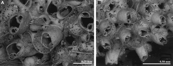
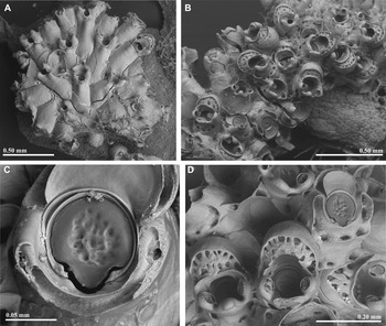
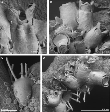
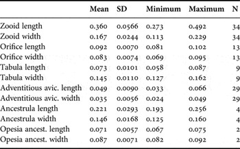
(Figures 4, 5 & 10; Table 2)
Celleporina fragilis: (A) a young unilaminar colony (MB37-000045); (B) a multilaminar colony (MB37-000046); (C) primary orifice (MB37-000047); (D) ovicells (MB37-000047).

Celleporina fragilis: (A) a young zooid; note the small uncalcified frontal window, the two frontal umbones and the marginal pores spared by the gymnocystal calcification (MB37-000044); (B) marginal zooids in a multilaminar colony; note the peristome projecting medially, the larger frontal window and the marginal pores spared by the gymnocystal calcification; pores also open in the uncalcified window (MB37-000044); (C) lateral view of the ancestrula (MB37-000044); (D) ancestrulae and first autozooids (MB37-000044).

Measurements (in mm) of Celleporina fragilis.

SD, Standard deviation; N, number of measurements.
Celleporina fragilis Arístegui Ruiz, Reference Arístegui Ruiz1989, p. 152, figures 4, 12–16.
MATERIAL EXAMINED
MB37-000044: Celleporina fragilis, Gettysburg (Gorringe Bank), 36.30°N 11.36°W. 35 m, 05 June 2006.
MB37-000045: Celleporina fragilis, Ormonde (Gorringe Bank), 36.40°N 11.17°W, 38 m, 04 June 2006.
MB37-000046: Celleporina fragilis, Gettysburg (Gorringe Bank), 36.52°N 11.57°W, 34 m, 11 June 2008.
MB37-000047: Celleporina fragilis, Ormonde (Gorringe Bank), 36.72°N 11.17°W, 38 m, 10 June 2008.
DESCRIPTION
Colony unilaminar, forming small patches on seaweeds, or multilaminar, forming nodular colonies on filiform substrates like hydroids. Autozooids recumbent in unilaminar colonies, radially arranged; tubular, with few proximo-lateral marginal pores; frontal wall gymnocystal, smooth, vitreous, with fine transverse lines and usually one or two frontal umbones. Two lateral, stout, columnar avicularia, separated by a circular pseudosinus with a small proximal uncalcified window; avicularia with rostrum semielliptical, small and finely serrated directed upwards and outwards. Autozooids in multilaminar colonies suberect, chaotically arranged. Peristome more developed, lacking the circular pseudosinus, but projecting medially in a low tubercle. Uncalcified window usually larger. Avicularia similar as in unilaminar colonies. Primary orifice subcircular, slightly longer than wide, with a wide U-shaped sinus, occupying more than a half of the proximal margin, flanked by small, rhomboid condyles. Vicarious avicularium not present in available material. Ovicell hyperstomial, hemispherical. Ooecium formed by the flattened distal kenozooid budded from the maternal autozooid. Tabula narrow, perforated by a single series of irregular, peripheral pores. Proximal part of ectooecium forms a vertical ‘lamina’ and in some case a thickened upper rim and medial peak. Ancestrula celleporiniform, i.e. oval, with a large gymnocyst and a subterminal orifice, roughly D-shaped with straight proximal border, surrounded by three proximal spines and two or four lateral spines. The ancestrula produces one distal and two distolateral autozooids.
REMARKS
Celleporina fragilis was described 25 years ago by Arístegui Ruiz (Reference Arístegui Ruiz1989) from specimens collected in shallow waters around the Canary Islands, encrusting seaweeds. The species is not known outside the Canaries, although, as the author said, it would not be surprising if it were found in nearby regions because of its affinity for growing on Sargassum, algae which, thanks to their vesicles, can disperse easily. The specimens of C. fragilis studied in the present work were collected on algae and hydroids at the Gorringe Bank, an isolated seamount placed 200 km off the south-west of the Iberian Peninsula, and the summit of which reaches 30 m depth.
As stated in the original description, the morphology of the autozooids of C. fragilis is different in colonies growing as unilaminar patches or as multilaminar nodules. In the examined material, colonies on laminar algae were unilaminar, while colonies growing on filiform substrates, like hydroids, were multilaminar. In both cases, our material seems to fit the original description by Arístegui Ruiz (Reference Arístegui Ruiz1989). Nevertheless, the specimens from the Gorringe Bank seem slightly smaller than the measurements given in the original description, but as the proportions are the same, we consider these differences not significant. On the other hand, we have not observed the vicarious avicularia, which anyway seems to be rather scarce.
Ancestrular morphology has been reported in few Celleporina species. Ikezawa & Mawatari (Reference Ikezawa and Mawatari1993) stated that some ancestrulae of Celleporina are schizoporelloid (i.e. they somewhat resemble a daughter zooid) and others have a modified type of tatiform ancestrula, with a subterminal opesium usually surrounded by spines. The ancestrula of C. fragilis was originally described as tatiform, as other species of the genus (see e.g. Hayward & Ryland, Reference Hayward and Ryland1999). The ancestrulae observed by us fully match the original description of the species, although Arístegui Ruiz (Reference Arístegui Ruiz1989) reported 5 spines while in our material the number varies between 5 and 7. The ancestrula has an extensive gymnocyst and a subterminal opesia coincident with the operculum, so there is no membranous frontal wall (Figure 5C, D). This ancestrula cannot be called ‘tatiform’, nor ‘modified tatiform’ insofar as the spines surround not a membranous frontal wall but an operculum. We define here this type of ancestrula as ‘celleporiniform’. Further studies will be necessary to prove if other species of the genus, or even of different genera, also present this type of ancestrula.
Celleporina derungsi Souto et al., Reference Souto, Reverter-Gil and Fernández-Pulpeiro2010
(Figure 6)
Celleporina derungsi (As Laixiñas): (A) detail of a colony; note the cryptocystal frontal wall and the strips of exterior wall (gymnocyst) along the zooidal margins; (B) peristomes with extremely developed triangular projection; (C) primary orifice.

Celleporina derungsi Souto et al., Reference Souto, Reverter-Gil and Fernández-Pulpeiro2010, p. 1425, figure 6.
MATERIAL EXAMINED
MNCN- 25.03/3874: Celleporina derungsi, As Laixiñas (Golfo Ártabro), 43.4601°N 008.4236°W, 31 July 2011, 43 m, on Cellaria fistulosa.
REMARKS
Celleporina derungsi was until now only known from its type locality, at 19–20 m depth in Armaçao de Pêra (Algarve, S Portugal), collected on calcareous algae and shells (Souto et al., Reference Souto, Reverter-Gil and Fernández-Pulpeiro2010). The material collected in the NW Iberian Peninsula only differs from the type material by the smaller size, the lack of vicarious avicularia, and by the median triangular projection on the peristome, that in some autozooids may be extremely developed (Figure 6B). The specimens newly collected were found growing on Cellaria fistulosa (Linnaeus, Reference Linnaeus1758). The different substrate and the small size of the colonies may perhaps explain the differences observed.
Taking into account that C. derungsi is present in the NW and the SW of the Iberian Peninsula, it is not impossible that some of the previous records of the similar C. hassallii in Atlantic Iberian waters (see Reverter-Gil & Fernández-Pulpeiro, Reference Reverter-Gil and Fernández-Pulpeiro2001; Reverter-Gil et al., Reference Reverter-Gil, Souto and Fernández-Pulpeiro2014) actually correspond to C. derungsi.
Celleporina algarvensis n. sp.
(Figure 7; Table 3)
Celleporina algarvensis n. sp. (holotype): (A) ovicellate zooids with different developed peristomes; (B) primary orifice; (C) different peristomes; note the frontal umbo, the regeneration of the peristome and the marginal pores spared by the gymnocystal calcification; (D and E) two developing ooecia; (F) uncleaned colony showing subcleithral ovicells, adventitious avicularia, and tabula covered by a membrane; note the regeneration of the peristome in the central zooid.

Measurements (in mm) of Celleporina algarvensi s n. sp. (Holotype).

SD, Standard deviation; N, number of measurements.
MATERIAL EXAMINED
Holotype: MNCN- 25.03/3875: Armaçao de Pêra, Algarve (S Portugal), March 2004. One ovicellate colony collected in fishing net at the beach.
ETYMOLOGY
Alluding to the geographic origin of the studied material.
DESCRIPTION
Colony multilaminar, forming a small domed patch. Autozooids small, suberect, with distinct marginal pores. Primary orifice orbicular, longer than wide, with a deep U-shaped sinus occupying one third of the proximal margin; condyles rhomboid, sloping towards the sinus but hardly reaching its margins. Peristome thick, developed suborally. Gymnocystal wall folded laterally to cover two stout, columnar avicularia, and extending also onto the frontal-basal area of the peristome, leaving a central membranous (non-calcified) window, partially covered proximally by a conical mucro. Rostrum of avicularia small, semi-elliptical, nearly facing each other and directed upwards. Avicularia frequently broken and substituted by a central prolongation formed by intramural budding, not closed at the apex in the material studied. Ovicell hyperstomial, subcleithral. Hemispherical ooecium formed by the flattened distal kenozooid budded from the maternal autozooid. Tabula narrow, perforated by a single series of peripheral pores, irregular in shape. Proximal part of ectooecium surrounding the ovicell opening forms a vertical ‘lamina’ with a thickened upper rim and medial peak. Ovicell opening visible in frontal view. Vicarious avicularium and ancestrula not seen.
REMARKS
Celleporina algarvensis n. sp. is easily distinguished from all other species present in the Atlantic–Mediterranean region by the shape of the orbicular primary orifice, longer than wide, with a U-shaped sinus emphasized at the binocular by the large condyles, the thick suboral peristome, and by the shape of the ovicell, with a short tabula and pointed rim.
Celleporina derungsi, recently described from the same locality (see above), differs by its orbicular orifice with a broad shallow sinus, the tubular peristome with a median triangular projection and a pair of denticulate avicularia directed outwards, the wider tabula on the ovicell, and the presence of broadly spatulate vicarious avicularia.
Celleporina decipiens Hayward, Reference Hayward1976, also present in the same locality (see Souto et al., Reference Souto, Reverter-Gil and Fernández-Pulpeiro2010) differs by its small sinus and smaller condyles, and the different peristome and tabula.
Celleporina fragilis also presents a gymnocystal peristome, but differs from C. algarvensis mainly by the circular orifice, with a wider sinus occupying more than a half of the proximal border of the orifice; moreover, the oral avicularia of C. algarvensis are smaller, lack a serrate border, and do not reach the top of the columnar cystid (see Figures 4D, 5A, B & 7A, F).
Other European species of the genus also develop a tall peristome: C. parvula, C. pygmaea (Norman, Reference Norman1868), C. siphuncula Hayward & McKinney, Reference Hayward and McKinney2002 and C. tubulosa. None of them has an orifice longer than wide, with a narrow U-shaped sinus and the particular morphology of the peristome.
Most of the avicularia of the colony of C. algarvensis n. sp. are broken, losing the rostrum. The two columnar avicularian chambers regenerate by intramural budding, joining to form a single, medial prolongation. None of these are closed in the tip, so we do not know if a new rostrum is formed later.
The single colony examined was collected at the beach of Armaçao de Pêra, Algarve (S Portugal) in fishing net. However, the boat was small enough as to assure that material was collected in shallow waters near the coastline.
Celleporina mediterranea n. sp.
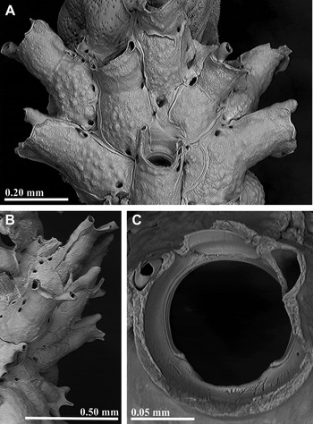
(Figures 8 & 9; Table 4)
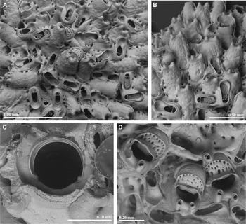
Celleporina mediterranea n. sp. (Holotype) (A) detail showing abundant vicarious avicularia; (B) frontal view of the peristomes and vicarious avicularia; (C) primary orifice; (D) ovicells and avicularia.

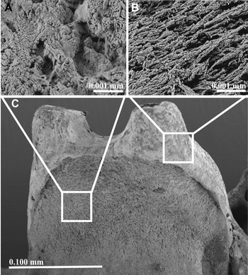
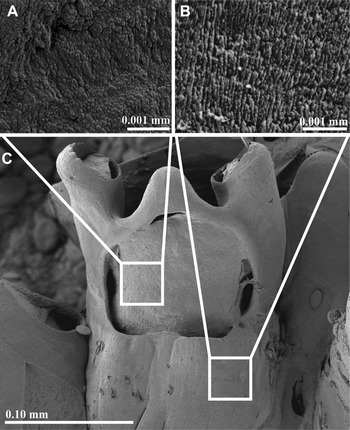
Ultrastructure of the frontal wall in C. mediterranea n. sp. (paratype): (A) cryptocystidean calcification; (B) planar spherulitic ultrastructure of the gymnocystidean calcification; (C) detail of the peristome of a zooid.

Measurements (in mm) of Celleporina mediterranea n. sp. (Holotype + Paratype).

SD, Standard deviation; N, number of measurements.
?Celleporina hassallii var. tubulosa: Gautier, Reference Gautier1962, p. 247.
Celleporina sp.: Souto et al., Reference Souto, Reverter-Gil and Fernández-Pulpeiro2010, p. 1427, figure 7.
MATERIAL EXAMINED
Holotype: MNCN- 25.03/3876, Menorca Channel, Patín 40, 39.9290°N 03.7088°E, 10 September 2011, 66 m.
Paratypes: MNCN- 25.03/3877, Menorca Channel, Patín 40, 39.9290°N 03.7088°E, 10 September 2011, 66 m. MNCN- 25.03/3878, Menorca Channel, Patín 19, 39.9632°N 03.657°E, 05 September 2011, 74 m. MNCN- 25.03/3879, Menorca Channel, Patín 21, 39.9665°N 03.6712°E, 06 September 2011, 59 m. MNCN- 25.03/3880, Menorca Channel, Patín 29, 39.8438°N 04.1055°E, 07 September 2011, 66 m. MNCN- 25.03/3881, Menorca Channel, Patín 39, 39.9252°N 03.7085°E, 10 September 2011, 68 m. MNCN- 25.03/3882, Menorca Channel, Patín 44, 39.8023°N 03.5988°E, 11 September 2011, 73 m.
Other material: MNHN 10233: Tiboulen de Pomegnes (Marseille). Gautier coll. One ovicellate colony. MNHN 10235: Port Man (Marseille). Gautier coll. One ovicellate colony.
ETYMOLOGY
Alluding to the geographic origin of the studied material.
DESCRIPTION
Colony multilaminar, forming circular domed patches. Autozooids small, suberect, with distinct marginal pores. Primary orifice orbicular, wider than long, with a wide sinus occupying almost the entire proximal margin, flanked by small, inconspicuous condyles. Peristome thick, developed suborally as a tall wall, incorporating on each side a slender, columnar avicularium; rostrum small, oval, with complete crossbar and sometimes finely toothed distal rim; acute to frontal plane and directed obliquely proximally. Vicarious avicularium frequent, grouped in clusters; rostrum narrow, slightly spatulate distally; crossbar complete, without columella, palate with a foramen occupying half of its length. Ovicell hyperstomial. Hemispherical ooecium formed by the flattened distal kenozooid budded from the maternal autozooid; tabula large, crescent, occupying half or more of the ooecium, with large irregular pores in younger ooecia that become much smaller with age because of the ooecial calcification. Proximally the tabula is restricted by a raised rim with medial thickened peak. Proximal part of ooecium has a shape of short funnel with the ovicell opening not visible in frontal view. Ancestrula not known.
REMARKS
Celleporina mediterranea n. sp. is easily distinguished from any other species of the genus in the Atlantic–Mediterranean region by the thick and tall suboral peristome, the orbicular orifice, wider than long, with a wide sinus and small condyles, and by the narrow vicarious avicularia, frequently forming groups, with the distal rostrum only slightly spatulate.
Among the Mediterranean species of the genus, C. tubulosa differs mainly by its tubular peristome and the broadly spatulate avicularium. Celleporina caminata also presents a tubular peristome, but with three cylindrical umbones bearing avicularia. Celleporina siphuncula, with also a tubular peristome, has a different orifice, a single peristomial avicularium, deeply cupped vicarious avicularium, and tabula with a single series of pores. Celleporina canariensis differs, among other characters, by its orifice, longer than wide and with a marked sinus, and by the characteristic V-shape of the secondary orifice, flanked by a pair of flat, triangular avicularia. Celleporina decipiens differs by its orbicular orifice with a small sinus, the lower peristome with paired avicularia facing each other and the sporadic vicarious avicularia. Celleporina caliciformis (Lamouroux, Reference Lamouroux1816) presents a different orifice, and lacks a developed peristome. Finally, C. lucida differs mainly by the orifice being longer than wide, the tubular peristome with a single avicularium and by the perforations of the tabula, with a simple crescent of slit-like pores.
Gautier (Reference Gautier1962) reported Celleporina hassallii var. tubulosa from Marseille. As already stated by Souto et al. (Reference Souto, Reverter-Gil and Fernández-Pulpeiro2010), we have not found any material in the Gautier collection at the MNHN labelled with that name, but we have revised three samples (MNHN 10233, MNHN 10234 and MNHN 10235) labelled as Cellepora costazii and originating from the same area, which may correspond or not to that record. The sample MNHN 10234 was empty, whereas the other two samples were left in open nomenclature by Souto et al. (Reference Souto, Reverter-Gil and Fernández-Pulpeiro2010, as Celleporina sp.), and are here identified as C. mediterranea n. sp.
Celleporina mediterranea n. sp. seems very similar to the C. hassallii figured by Zabala (Reference Zabala1986, pl. 21, C–D), but a closer revision of original material will be necessary to check this record.
DISCUSSION
The genus Celleporina is characterized, among other features, by the morphology of its ovicells, which ooecia are formed by the flattened distal kenozooid and possess a crescent to oval membranous area of ectooecium covering a perforated calcified plate called tabula (e.g. Hayward & Ryland, Reference Hayward and Ryland1999; Tilbrook, Reference Tilbrook2006; Bock, Reference Bock2013). Size and shape of the tabula, as well as number and arrangement of the pores, vary among species and therefore can be used, together with other characters, to differentiate them.
In most of the taxonomic articles the tabula is described without mention of its origin or, as much, suggesting that it is a structure with no relation with the ectooecium (e.g. Harmer, Reference Harmer1957; Arístegui Ruiz, Reference Arístegui Ruiz1989; Gordon, Reference Gordon1989; Ikezawa & Mawatari, Reference Ikezawa and Mawatari1993; Hayward & McKinney, Reference Hayward and McKinney2002; Florence et al., Reference Florence, Hayward and Gibbons2007; Grischenko et al., Reference Grischenko, Dick and Mawatari2007; Winston & Vieira, Reference Winston and Vieira2013). In other cases the tabula is specifically defined as a perforate area of the entooecium (e.g. Hayward & Ryland, Reference Hayward and Ryland1999; Tilbrook, Reference Tilbrook2006; Bock, Reference Bock2013), or as a combination of ecto- and entooecium (Hayward & Ryland, Reference Hayward and Ryland1995). Finally, according to anatomical data of A. Ostrovsky (personal communication 2014) the tabula is a perforated calcified plate placed between the membranous ectooecium and the calcified entooecium and fused with both of them.
In cleaned specimens, the ooecial development in C. algarvensis n. sp. (see above and Figure 7) shows that, at least in this species, the formation of the tabula starts as trabecular extensions of the calcified ectooecium, while the imperforate entooecium rests below. A similar development is also seen in the C. caminata figured by Hayward & McKinney (Reference Hayward and McKinney2002, figure 39B) although the entooecium is not clearly visible in this case. Further growth of the trabeculae leads to their coalescence and formation of the perforated plate (tabula) which thickening results in its fusion with the calcified entooecium. In older ooecia progressive calcification leads to a strong diminishing of the size of the pores that are finally transformed to the pits in the tabula. We suggest that development and the structure of the ooecia are similar in other species of the genus. However, it must be stated that the ovicell is defined as acleithral (not closed by the maternal operculum) in the genus Celleporina, while C. algarvensis n. sp. seems to possess a subcleithral ovicell (Figure 7F) which opening is closed by the maternal operculum (see Ostrovsky, Reference Ostrovsky, Hageman, Key and Winston2008, Reference Ostrovsky2013).
The frontal wall of the genus Celleporina has been described as cryptocystidian, corresponding to its classification as lepraliomorph (see e.g. Hayward & Ryland, Reference Hayward and Ryland1999). However, a close study of the species treated in the present work shows that at least some of the frontal wall (as well as the walls of the columnar avicularia) is gymnocystidean.
In the case of C. derungsi the frontal shield, and also the frontal (proximal) side of the peristome, seems cryptocystidian. However, there are narrow strips of exterior wall (gymnocyst) along the zooidal margins, while the lateral margins of the peristome (corresponding to the columnar avicularia) and also the upper edge of the peristome are also gymnocystidean (Figure 6A). This is also the case in C. mediterranea sp. nov. (see Figures 8A, B & 9 in the present work and Souto et al., Reference Souto, Reverter-Gil and Fernández-Pulpeiro2010, figure 7A, D), but in addition, in this species the distal (suboral) side of the peristome also seems to be gymnocystidean (see Souto et al., Reference Souto, Reverter-Gil and Fernández-Pulpeiro2010, figure 7B); this gymnocyst folds and closes on the lateral edges of the peristome forming the columnar avicularia (as seen in the same figure). This structure may be also present in other species of the genus developing a peristome, although a thorough revision is necessary to confirm this suggestion.
In other species of the genus, particularly in C. algarvensis n. sp. and C. fragilis (but also in e.g. C. porosissima Harmer, Reference Harmer1957 or C. minima Grischenko et al., Reference Grischenko, Dick and Mawatari2007) the distal (suboral) gymnocyst of the peristome extends also over its frontal side, joining also the developed marginal gymnocyst of the zooid, which may even form one or two proximal umbones. The planar spherulitic ultrastructure of the frontal wall, which is diagnostic of exterior wall (P.D. Taylor and D.P. Gordon, personal communication May 2014), proves that it is actually gymnocystidean (i.e. no hypostegal coelom under the ectocyst) (see Figure 10B). The remaining cryptocyst can be seen through a central uncalcified window (see Figures 4A, 5B, C, 7A, C & 10A in the present paper, and Grischenko et al., Reference Grischenko, Dick and Mawatari2007: figure 38A); this window may be extremely reduced (see Figures 4A & 5A in the present paper and Grischenko et al., Reference Grischenko, Dick and Mawatari2007: figure 36C). The zooidal marginal pores remain surrounded by cryptocyst, and may be spared by the gymnocystal calcification (Figures 5A, B & 7C) or enclosed within the gymnocystal wall, forming tubes opened to the frontal window (Figures 5B, 7C & 10C) or around the zooidal orifice (Grischenko et al., Reference Grischenko, Dick and Mawatari2007: figures 37A and 38A, B). Therefore, it seems that areas of gymnocyst have secondarily expanded at the expense of the cryptocystidean part of the frontal shield, which is confined to those parts of the shield associated with the areolar pores. These doubled-walled species would be seen as more-derived within the genus Celleporina, whose frontal shield evolved from a lepralielloidean ancestor(s) (D.P. Gordon, personal communication May 2014).
Ultrastructure of the frontal wall in C. fragilis (MB37-000045): (A) cryptocystidean calcification; (B) planar spherulitic ultrastructure of the gymnocystidean calcification; (C) detail of the peristome of a zooid.

Further studies, including anatomical cuts, will be necessary to correctly describe this structure and to establish the origin of the frontal wall in other species of the genus.
ACKNOWLEDGEMENTS
We are grateful to P. Lozouet and S. Charbonnier (MNHN) for valuable assistance during our visits, and to J. Olesen (ZM) for the loan of material. We are also grateful to A. Ostrovsky for comments and suggestions on an earlier draft and especially for discussions on ovicell architecture; also to D.P. Gordon and P.D. Taylor for comments on the structure of the frontal shield, and to B. Berning for sending photographs. We also wish to thank the staff of the Servizo de Microscopía Electrónica (Universidade de Santiago de Compostela) for the SEM photographs, and A. González and F. Bergantiños for helping during the sampling in As Laixiñas. The authors wish to thank EMEPC (Task Group for the extension of the Continental Shelf), and the Universidade Lusófona de Humanidades e Tecnologias for the opportunity to study the Gorringe samples from expeditions in the EMEPC/Açores/Luso/2009 and LusoExpedição Sea Campaign 2006 and 2008.
FINANCIAL SUPPORT
The sampling survey ‘Canal0811’ was carried up during the project ‘Estudio integral del ecosistema demersal y bentónico del canal de Menorca (Islas Baleares) y su explotación pesquera’. This project was co-financed by the Conselleria d'Agricultura i Pesca del Govern Balear and MarViva (www.marviva.net) and the project LIFE+ ‘Inventory and designation of marine Natura 2000 areas in the Spanish sea’ coordinated by the Biodiversity Foundation and co-financed by the European Commission. The work by J.S. and O.R.G. was partially supported by the project ‘Fauna Ibérica: Briozoos II (Familia Cribrilinidae – Familia Watersiporidae)’ (CGL2010-22267-C07-02), co-financed by the Ministerio de Economía y Competitividad (Spanish Government) and FEDER. J.S. thanks the Austrian Science Fund (FWF, Lise Meitner Program, grant M1444-B25) for financial support.