Introduction
Amacrine cells sit at the crossroads of information processing in the inner retina. They receive classical excitatory and inhibitory input mediated by ionotropic receptors. This synaptic input determines the membrane potential of these cells and hence their voltage-dependent release of neurotransmitter onto their postsynaptic targets. The function of amacrine cells as components of specific pieces of retinal circuitry is only known for a few specific amacrine cell types. Nonetheless, the complex patterns of synaptic interactions in which amacrine cells generally participate have been demonstrated both anatomically (Marc & Liu, Reference Marc and Liu2000) and physiologically (Zhang et al., Reference Zhang, Jung and Slaughter1997; Hartveit, Reference Hartveit1999). These patterns include serial and reciprocal synapses as well as nested loops of synaptic partners. But despite the complex assemblage of ionotropic circuits, they do not represent the whole story of amacrine cell signaling. Collectively, amacrine cells express a plethora of metabotropic (i.e., G protein-coupled) receptors. The full functional impact of activating these receptors is only appreciated in a very few cases. This review will focus on the physiological responses of amacrine cells to the activation of metabotropic receptors. I will confine this discussion to the metabotropic versions of receptors for the classical neurotransmitters [dopamine, glutamate, GABA, and acetylcholine (ACh)] that are well established in the retina. Activators of two groups of metabotropic receptors that are decidedly less well established will also be considered: cannabinoid receptors and sphingosine-1-phosphate (S1P) receptors. These receptors have the ability to support retrograde signaling (and possibly autocrine signaling) from amacrine cells in response to activation of glutamate receptors at bipolar-to-amacrine cell synapses because the synthesis of their ligands is Ca2+-dependent.
Dopamine receptors
The effects of dopamine receptor activation on amacrine cell function are probably the best understood responses to metabotropic receptor activity in the inner retina. Dopamine receptors are divided into two groups: D1- and D2-like. D1-like receptors (D1 and D5) are linked to Gαs, and their activation can lead to increased adenylate cyclase (AC) activity, elevation of cyclic adenosine monophosphate (cAMP) levels, and increases in protein kinase A (PKA) activity. The D2-like receptors (D2, D3, and D4) are linked to Gαi, which typically inhibits AC activity and reduces cAMP levels and PKA activity. Although other receptors might be expressed in amacrine cells (Wagner et al., Reference Wagner, Luo, Ariano, Sibley and Stell1993; Veruki, Reference Veruki1997), the majority of functional studies have focused on D1-mediated responses.
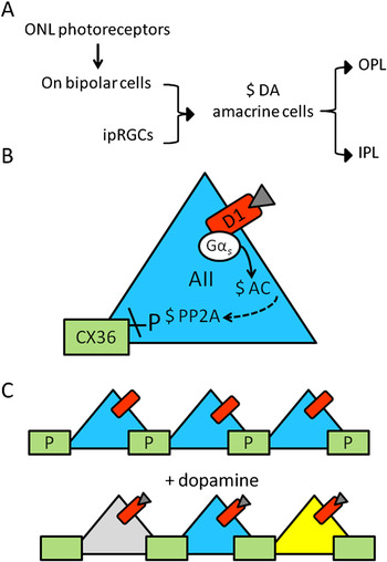
Retinal dopamine is generated by dopaminergic amacrine cells that project into both the inner plexiform layer (IPL) as well as the outer plexiform layer (OPL). The activity-dependent release of dopamine from these cells is regulated by input from intrinsically photosensitive retinal ganglion cells (Zhang et al., Reference Zhang, Wong, Sollars, Berson, Pickard and McMahon2008) as well as a subset of ON bipolar cells (Zhang et al., Reference Zhang, Zhou and McMahon2007; Dumitrescu et al., Reference Dumitrescu, Pucci, Wong and Berson2009). Light-dependent signals from photoreceptors in both the outer and inner retina converge on the dopaminergic amacrine cell and increase the spiking activity of these cells and presumably, the release of dopamine (Fig. 1A). One of the major effects of dopamine across the retina is to uncouple electrically coupled networks of cells to adjust retinal signaling for photopic conditions. Effects on amacrine cell coupling have been especially well studied in the rabbit retina.
Dopamine uncouples AII amacrine cells. (A) A scheme showing the pathway from photoreceptor stimulation to release of dopamine in the plexiform layers. (B) Activation of the D1 receptor on AII amacrine cells leads to activation of AC, which somehow leads to activation of PP2A and dephosphorylation of Cx36. (C) Under scotopic conditions, dopamine is absent and phosphorylated connexins support electrical coupling in the AII amacrine cell network. In the presence of dopamine, the pathway in (B) is activated and the cells become uncoupled.

Hampson et al. (1992) provided an early demonstration that AII amacrine cell dye coupling was reduced in response to dopamine, and a similar uncoupling response was demonstrated for photopic light stimuli (Bloomfield et al., Reference Bloomfield, Xin and Osborne1997; Bloomfield & Volgyi, Reference Bloomfield and Volgyi2004) and since then numerous groups have contributed to the scenario depicted in Fig. 1. AII amacrine cells form an electrically coupled network via expression of connexin 36 (Cx36)-containing (Feigenspan et al., Reference Feigenspan, Teubner, Willecke and Weiler2001; Mills et al., Reference Mills, O’Brien, Li, O’Brien and Massey2001) gap junctions. This Cx36-dependent coupling is reduced by dopamine (Xia & Mills, Reference Xia and Mills2004) working specifically at D1 receptors (Kothmann et al., Reference Kothmann, Massey and O’Brien2009). Using phospho-specific antibodies raised against Cx36, it was shown that D1 receptor activation leads to PKA-dependent dephosphorylation of the protein (Kothmann et al., Reference Kothmann, Massey and O’Brien2009) that is also dependent upon protein phosphatase 2A (PP2A, Fig. 1B and 1C). AII amacrine cells channel the rod photoreceptor signal into the ON and OFF cone bipolar pathway under low light conditions. The uncoupling of AII amacrines under photopic conditions will limit the spread of saturated rod responses into the ON and OFF pathways.
Gap junction permeability has also been studied for AC1 and AC2 amacrine cells in the rabbit retina. Here, D1 agonists also lead to uncoupling between AC1 cells, between AC1 and AC2 cells, and between AC1 and OFF α ganglion cells (Mills et al., Reference Mills, Xia, Hoshi, Firth, Rice, Frishman and Marshak2007). This uncoupling is also PKA dependent but further details of the pathway and the molecular nature of the connexins have not yet been established. Uncoupling of the AC1-AC1 network would also limit the lateral spread of signals and the spatial extent of signals shared with α OFF ganglion cells. Overall, the action of dopamine acting via D1 receptors on amacrine cell gap junctions likely enhances the relative strength of signaling in the vertical retinal pathway, thus preserving spatial resolution at the expense of sensitivity (Fig. 1C).
Aside from these network effects achieved by altering junctional conductance, dopamine-dependent regulation of chemical synapses has also been demonstrated. Both biochemical (Hensler & Dubocovich, Reference Hensler and Dubocovich1986) and physiological (Yeh et al., Reference Yeh, Battelle and Puro1984) studies provide evidence that dopamine enhances ACh release, presumably from amacrine cells. In amacrine cells recorded in an organotypic slice preparation from the rat, D1 receptor activation increased the amplitude of GABA-gated currents by increasing the sensitivity of GABAA receptors to GABA via PKA activity (Feigenspan & Bormann, Reference Feigenspan and Bormann1994). They also demonstrated that activating other G protein-coupled receptors linked to Gs including histamine (H1), adenosine (A2), and vasoactive intestinal peptide receptors increases GABAA receptor currents. This convergence of effects suggests that GABAA receptors are modulated by metabotropic signaling under a range of conditions.
Metabotropic glutamate receptors
Metabotropic glutamate receptors (mGluRs) comprise eight subtypes divided into three groups based on sequence similarity, agonist selectivity, and coupling to signaling pathways. Group I (mGluRs 1 and 5) is typically coupled to Gαq and the inositol 1,4,5 trisphosphate (IP3) pathway, although coupling to Gαs has also been demonstrated (Aramori & Nakanishi, Reference Aramori and Nakanishi1992; Joly et al., Reference Joly, Gomeza, Brabet, Curry, Bockaert and Pin1995) and the sequence responsible for this capability identified (Francesconi & Duvoisin, Reference Francesconi and Duvoisin1998). Group II receptors (mGluRs 2 and 3) and Group III receptors (mGluRs 4, 6, 7, and 8) couple to Gαi and inhibit the activity of AC. Most famously in the retina, mGluR6 inverts the sign of the cone to bipolar synapse establishing the ON signaling pathway in the visual system (Masu et al., Reference Masu, Iwakabe, Tagawa, Miyoshi, Yamashita, Fukuda, Sasaki, Hiroi, Nakamura, Shigemoto, Takada, Nakamura, Nakao, Katsoki and Nakanishi1995; Vardi & Morigiwa, Reference Vardi and Morigiwa1997). Amacrine cells also express mGluRs but the impact of their activation on visual signaling is not nearly as well understood.
Immunocytochemistry studies have demonstrated the expression of Group 1 receptors by amacrine cells in mammalian (Koulen et al., Reference Koulen, Kuhn, Wassle and Brandstatter1997) and avian retinas (Kreimborg et al., Reference Kreimborg, Lester, Medler and Gleason2001). mGluR1 has also been found on wide-field amacrine cells dissociated from the white bass retina (Vigh & Lasater, Reference Vigh and Lasater2003). Group II receptors have been localized to amacrine cells with an antibody that does not differentiate between mGluRs 2 and 3 in the cat retina (Cai & Pourcho, Reference Cai and Pourcho1999). With more selective antibodies, mGluR2, but not mGluR3, was localized to amacrine cells in the rat retina (Koulen et al., Reference Koulen, Malitschek, Kuhn, Wassle and Brandstatter1996; Quraishi et al., Reference Quraishi, Rush and Brown1996). Group III receptors mGluRs 4, 7, and 8 (mGluR6 is only found in the OPL) are found in the IPL (Quraishi et al., Reference Quraishi, Gayet, Morgans and Duvoisin2007) and have all been localized to amacrine cell processes (Brandstatter et al., Reference Brandstatter, Koulen, Kuhn, van der Putten and Wassle1996; Koulen et al., Reference Koulen, Malitschek, Kuhn, Wassle and Brandstatter1996; Koulen & Brandstatter, Reference Koulen and Brandstatter2002). Importantly, each of the expressed mGluRs was found postsynaptic to bipolar cells (Brandstatter et al., Reference Brandstatter, Koulen, Kuhn, van der Putten and Wassle1996; Koulen et al., Reference Koulen, Malitschek, Kuhn, Wassle and Brandstatter1996, Reference Koulen, Kuhn, Wassle and Brandstatter1997; Cai & Pourcho, Reference Cai and Pourcho1999; Koulen & Brandstatter, Reference Koulen and Brandstatter2002; Sen & Gleason, Reference Sen and Gleason2006), indicating they can respond to the release of synaptic glutamate.
In a study on wide-field amacrine cells from the white bass retina, Vigh and Lasater (Reference Vigh and Lasater2003) examined the mGluR1-dependent modulation of GABAA receptors. They demonstrated that activation of mGluR1-initiated IP3-dependent signaling that led to release of Ca2+ from internal stores and subsequent activation of the Ca2+/calmodulin-dependent phosphatase calcineurin. The outcome of this activity was a reduction in the GABA-gated current amplitude due either to dephosphorylation of these channels or an intermediate regulator. Interestingly, prolonged receptor activation was required to activate the pathway, suggesting that this inhibition is regulated by activity levels rather than being a constitutive process suppressing GABAA receptor function.
A role for mGluR1 has been demonstrated at the reciprocal synapse between the goldfish Mb (ON type) bipolar cell and GABAergic amacrine cells in a slice preparation (Vigh et al., Reference Vigh, Li, Hull and von Gersdorff2005). The inhibitory feedback from amacrine cells was enhanced when Mb bipolar cells were strongly depolarized (e.g., to 0 mV) or more physiologically relevant, with longer depolarization to more moderate voltages (e.g., 3 s, −20 mV). This effect was mimicked by a Group I agonist (LY367385) and blocked by a mGluR1-specific antagonist [(S)-3, 5-dihydroxyphenylglycine]. The implication from this work is that prolonged activity at the glutamatergic bipolar-to-amacrine synapse leads to enhancement of feedback that would be effective in reducing the synaptic output from the bipolar cell.
Multiple physiological effects of mGluR5 activation have been identified utilizing a culture system of identified amacrine cells derived from the chick embryo retina (Gleason et al., Reference Gleason, Borges and Wilson1993). These cells have been shown to express mGluR5 (and mGluR1, see below) based on immunocytochemical evidence (Kreimborg et al., Reference Kreimborg, Lester, Medler and Gleason2001). Activation of this receptor with an mGluR5-specific agonist leads to Ca2+ elevations initiated by release of Ca2+ from internal stores and followed by a highly variable phase due to flux of Ca2+ across the plasma membrane, possibly via transient receptor potential canonical (TRPC) channels (Sosa et al., Reference Sosa, Hoffpauir, Rankin, Bruch and Gleason2002). The Ca2+ elevations were dependent upon phospholipase C (PLC) activity indicating coupling of the receptor to Gαq and activation of the IP3 pathway. Interestingly, sequence analysis of the RNA encoding this receptor amplified from chicken retina also shows the potential for interaction with Gαs as previously demonstrated for mGluR5 from the rat retina (Francesconi & Duvoisin, Reference Francesconi and Duvoisin1998), but this interaction has yet to be tested experimentally in chick amacrine cells.
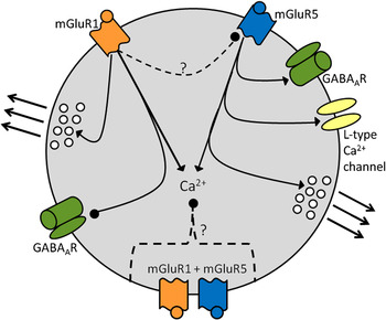
mGluR5 operating via this same PLC-dependent pathway also modulates GABAA receptor function as well as L-type Ca2+ channel function. Whole-cell GABA-gated currents were enhanced about 10% upon activation of mGluR5 and the activation was dependent upon Ca2+ and PKC activity. Measurements of the effects of mGluR5 activation on GABAergic quantal events revealed a 20% increase in amplitude, suggesting that synaptic GABAA receptors are preferentially modulated (Hoffpauir & Gleason, Reference Hoffpauir and Gleason2002). L-type Ca2+ currents are also enhanced (by ~40%) by mGluR5 activity, and this is a potential presynaptic effect because these voltage-gated Ca2+ channels can control neurotransmitter release from amacrine cells (Gleason et al., Reference Gleason, Borges and Wilson1994; Habermann et al., Reference Habermann, O’Brien, Wassle and Protti2003; Bieda & Copenhagen, Reference Bieda and Copenhagen2004; Vigh & Lasater, Reference Vigh and Lasater2004). The enhancement of these Ca2+ currents was dependent upon an elevation in cytosolic Ca2+ but was not PKC dependent. Instead, the effect could be mimicked by PKA agonists (Sosa & Gleason, Reference Sosa and Gleason2004). The link between mGluR5 and PKA could be due to an IP3-dependent increase in Ca2+ followed by the activation of a Ca2+-dependent AC. Alternatively, the enhancement of the Ca2+ current might be an outcome of coupling between mGluR5 and Gαs (Sosa et al., Reference Sosa, Hoffpauir, Rankin, Bruch and Gleason2002). Activation of mGluR5 has also been demonstrated to be a direct route to depolarization-independent neurotransmitter release from cultured GABAergic amacrine cells via the IP3 receptor dependent release of Ca2+ from internal stores (Warrier et al., Reference Warrier, Borges, Dalcino, Walters and Wilson2005). During depolarization-dependent release, mGluR5 activation also enhances evoked neurotransmitter release possibly due both to store contribution to cytosolic Ca2+ concentration and PKA-dependent enhancement of the Ca2+ current. Fig. 2 summarizes the outcomes of mGluR1 and mGluR5 activation on amacrine cells as described above.
Signaling through group 1 mGluRs can alter synaptic function in amacrine cells. Activation of mGluR1 generates Ca2+ elevations that stimulate calcineurin, which somehow leads to inhibition of GABAA receptors. Activation of these same receptors promotes the synaptic release of GABA. Activating mGluR5 also produces Ca2+ elevations, which result in GABAA receptor current enhancement, enhancement of L-type Ca2+, and stimulation of GABA release. Coactivation of mGluR1 and mGluR5 reduces the amplitude of Ca2+ elevations. This could be due to inhibition of Ca2+ flux mechanisms or to inhibition of mGluR5 signaling.

The prevalence of mGluR1 and mGluR5 on amacrine cell processes postsynaptic to bipolar terminals implies that the two receptors can be coexpressed (Koulen et al., Reference Koulen, Kuhn, Wassle and Brandstatter1997; Sen & Gleason, Reference Sen and Gleason2006). In cultured chick amacrine cells where immunocytochemistry indicates the two receptors are indeed coexpressed, the endogenous agonist glutamate produces both a phase of Ca2+ current enhancement and a phase of Ca2+ current inhibition (Sosa & Gleason, Reference Sosa and Gleason2004). There is pharmacological evidence that coactivation of the receptors depresses the magnitude of cytosolic Ca2+ elevations (Sosa et al., Reference Sosa, Hoffpauir, Rankin, Bruch and Gleason2002). The nature of this interaction has not yet been explored but might be due to inhibition of Ca2+ release or influx mechanisms or inhibition at the level of the receptor (Fig. 2).
A Group II (mGluRs 2 and 3) -selective agonist has been used in chick amacrine cells and whole-mount rabbit retina to examine the role of this class of mGluR in regulating amacrine cell function. In culture, (2S,1’R,2’R,3’R)-2-(2,3-dicarboxycyclopropyl) glycine (DCG-IV) suppressed AMPA receptor-dependent elevations in cytosolic Ca2+ (Gomes et al., Reference Gomes, Cunha, Nuriya, Faro, Huganir, Pires, Carvalho and Duarte2004). Interestingly, they also showed that D1 dopamine receptors on the same cells increased this AMPA receptor activity. In the rabbit retina, recordings from directionally selective ganglion cells in the presence of DCG-IV, revealed a robust response to stimuli in the null direction indicating that directional selectivity had been disrupted (Jensen, Reference Jensen2006). Because starburst amacrine cells (SACs) are known to express mGluR2 (Koulen et al., Reference Koulen, Malitschek, Kuhn, Wassle and Brandstatter1996; Cai & Pourcho, Reference Cai and Pourcho1999), and an acetylcholinesterase inhibitor reduced the effects of DCG-IV, it was suggested that activation of mGluR2 leads to inhibition of ACh release. The author suggests that GABA release is probably also reduced because directional selectivity is known to be dependent on GABAergic rather than cholinergic transmission and because the Group II receptors are known to inhibit presynaptic Ca2+ currents, which could affect release of both neurotransmitters. This scenario is yet to be tested experimentally. However, inhibition of ACh release due to inhibition of voltage-gated Ca2+ channels has been measured in response to activation of Group III receptors in cultured avian amacrine cells (Caramelo et al., Reference Caramelo, Santos, Carvalho and Duarte1999).
Most of what we know about Group III mGluRs in amacrine cells is confined to their localization rather than their function. In addition to the study on ACh release in culture mentioned above, the effects of the mGluR8-selective agonist (S)-3,4-dicarboxy-phenylglycine (DCPG) has been examined in the mouse retina. DCPG reduced the OFF response in OFF and ON-OFF ganglion cells, and part of this effect was due to a reduction in inhibition from presynaptic, presumably amacrine, cells. From these studies, it is clear that activation of mGluRs can have multiple effects on the function of amacrine cells. It is worth considering that most studies examining the action of particular mGluRs employ agonists specific to particular mGluRs in the retina; however, glutamate from bipolar cells is the endogeneous agonist for all of them. So although using agonists can elucidate receptor-specific changes in amacrine cell function, they cannot tell us when (or under what circumstances) each receptor will be activated. What is required are more studies like the elegant work of Vigh et al. (Reference Vigh, Li, Hull and von Gersdorff2005) at the Mb bipolar–amacrine cell reciprocal synapse, in which the investigators explore the conditions under which glutamate released from bipolar cells can engender mGluR1 receptor activation.
GABAB receptors
GABAB receptors function as obligate heterodimers of GABAB1 and GABAB2. These receptors signal via Gαi/o. It is well established in multiple preparations that the GABAB-dependent liberation of the beta gamma subunits of this G protein can lead to direct inhibition of most voltage-gated Ca2+ channels as well as activation of K+ channels including Kir3 and small conductance Ca2+-activated K+ channels (Filip & Frankowska, Reference Filip and Frankowska2008). Both in situ hybridization and immunocytochemical studies in rodent retina have indicated that amacrine cells express GABAB receptors (Koulen et al., Reference Koulen, Malitschek, Kuhn, Bettler, Wassle and Brandstatter1998; Zhang et al., Reference Zhang, Bettler and Duvoisin1998). At the electron microscopy level, GABAB receptors are found on both presynaptic and postsynaptic amacrine cell process, suggesting that these receptors could serve as autoreceptors (for GABAergic amacrine cells) as well as regulate postsynaptic responses. It was further established with colocalization studies that in the rabbit retina, GABAB receptors are expressed by SACs and other GABAergic amacrine cells but not glycinergic amacrine cells (see below, Zucker et al., Reference Zucker, Nilson, Ehinger and Grzywacz2005).
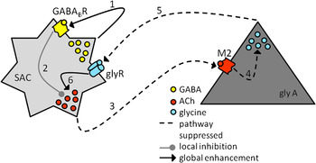
The GABAB receptor agonist baclofen has been shown in mudpuppy retina to increase amacrine cell transient light responses and also to suppress normally sustained light responses revealing an underlying transient component (Slaughter & Bai, Reference Slaughter and Bai1989). Neal and Cunningham (Reference Neal and Cunningham1995) looked at the effects of GABAB receptor activation on the light-stimulated release of ACh by the cholinergic/GABAergic SACs in the rabbit retina. They found that baclofen increased light-evoked ACh release and suppressed glycine release. They also found that activation of muscarinic receptors increased glycine release. A simple model was proposed in which GABA activates GABAB receptors on gycinergic amacrine cells, suppressing glycine release, disinhibiting SACs, and thereby increasing their cholinergic output. Ten years later, the localization study by Zucker et al. (Reference Zucker, Nilson, Ehinger and Grzywacz2005) required reform of this model because they found GABAB receptors on SACs but not on glycinergic amacrine cells. However, they also identified a costratifying population of glycinergic amacrine cells (4′-6′-diamino-2-phenylindole-3 cells) that express M2 muscarinic receptors (see below). The revised model (Fig. 3) proposes that GABAB receptors act as autoreceptors on a relatively small fraction of SAC varicosities where they mediate presynaptic inhibition and suppress cholinergic output locally. Reduction of cholinergic output then has a more global effect of reducing the activation of the more broadly expressed M2. Because M2 activation normally enhances glycine release, limiting M2 activation removes a major inhibitory input onto SACs (Neal & Cunningham, Reference Neal and Cunningham1995). Ultimately, this model explains the effect of GABAB activation in enhancing global light-stimulated ACh release but also important, sustains the notion that the dual neurotransmitter output from SACs results in dual activation of metabotropic receptors.
GABA and ACh released from SACs leads to activation of GABAB and muscarinic receptors. Release of GABA from SACs leads to activation of GABAB autoreceptors on a small subset of SAC dendrites (1). Local signaling through GABAB receptors inhibits (2) the release of ACh onto M2 muscarinic receptors on a glycinergic amacrine cell (3). Normally, M2 activity promotes the release of glycine (4) onto glycinergic receptors on the SAC (5), which ultimately removes inhibition of the SAC and enhances ACh output (After Zucker et al., Reference Zucker, Nilson, Ehinger and Grzywacz2005.)

Muscarinic receptors
Five muscarinic subtypes are currently recognized (M1–M5). Muscarinic receptors M1, M3, and M5 couple to Gαq and M2 and M4 couple to Gαi. Like the GABAB receptor, activation of M2 or M4 can result in inhibition of Ca2+ channels and activation of K+ channels with the overall effect being inhibitory. Through immunocytochemistry in the mammalian retina, M2 has been localized to the inner nuclear layer (INL) and IPL (Zucker & Ehinger, Reference Zucker and Ehinger2001; Yamada et al., Reference Yamada, Dmitrieva, Keyser, Lindstrom, Hersh and Marshak2003) and more specifically to glycinergic amacrine cells (Zucker et al., Reference Zucker, Nilson, Ehinger and Grzywacz2005). Muscarinic receptors M2, M3, and M4 have been localized to amacrine cells in the chicken retina (Fischer et al., Reference Fischer, McKinnon, Nathanson and Stell1998) and M2 has been found in amacrine cells in the salamander retina (but not in cholinergic amacrine cells, Cimini et al., Reference Cimini, Strang, Wotring, Keyser and Eldred2008). More recently, the expression of all five receptors has been examined in the rabbit retina (Strang et al., Reference Strang, Renna, Amthor and Keyser2010). Using receptor subtype-specific antibodies, each of the receptors was found in a population of amacrine cells and some receptor expression was overlapping. The expression of M2 was observed in SACs (see above), but the other four subtypes were also expressed by subsets of SACs. This group found limited expression in glycinergic amacrine cells, suggesting that it is GABAergic amacrine cells that dominate in muscarinic receptor expression. Their observation of M2 in SACs is at odds with Zucker et al. (Reference Zucker, Nilson, Ehinger and Grzywacz2005) as well as Yamada et al. (Reference Yamada, Dmitrieva, Keyser, Lindstrom, Hersh and Marshak2003, in primate retina). Also at odds with Zucker et al. (Reference Zucker, Nilson, Ehinger and Grzywacz2005) is the small fraction of glycinergic amacrine cells found to express M2. Strang et al. (Reference Strang, Renna, Amthor and Keyser2010) comment that the same M2 monoclonal antibody was used in each of the studies, but that the differences in apparent expression pattern might be due to differing fixation methods. These discrepancies are presently unresolved.
Much of what is known about the physiological effects of activating muscarinic receptors on glycinergic amacrine cells is described in the previous section. However, an additional role for M2 has been described in the salamander retina (Cimini et al., Reference Cimini, Strang, Wotring, Keyser and Eldred2008). Here, cyclic guanosine monophosphate-like immunoreactivity (cGMP-LI) was used as a proxy for nitric oxide (NO) production on the reasonable premise that a common effect of NO is to stimulate guanylate cyclase and increase cGMP levels. Stimulation of M2 produced enhanced levels of cGMP-LI in amacrine cells (by an unknown mechanism) that were blocked by inhibitors of the synthetic enzyme for NO, NO synthase. Evidence was also provided that the M2- and NO-dependent increase in cGMP-LI was occurring in GABAergic amacrine cells, and the cGMP-LI signal aligned with but did not overlap with choline acetyltransferase immunolocalization, suggesting that cholinergic amacrine cells were not stimulated to produce NO, but that they could provide the signal for its generation in GABAergic amacrine cells. It might be relevant to note that in cultured GABAergic amacrine cells, NO has been shown to increase cytosolic Cl− such that the inhibitory effects of incoming GABAergic or glycinergic signals would be transiently reduced or reversed (Hoffpauir et al., Reference Hoffpauir, McMains and Gleason2006). In the retina, it is possible that such a mechanism operates in the GABAergic amacrine cells that costratify with the cholinergic (and GABAergic) amacrine cell processes.
Cannabinoid receptors
The endogenous ligands of cannabinoid receptors are the endocannabinoids (ECs), which comprise a large group of arachidonic acid derivatives. The best studies of these are arachidonoyl ethanolamide (or anandamide) and sn-2 arachidonoyl glycerol (Yazulla, Reference Yazulla2008). The membrane permeable nature of these compounds and the absence of any known tethering mechanism support the concept that ECs are synthesized from membrane phospholipids on demand. The trigger for synthesis is elevation of cytosolic Ca2+ and activation of Ca2+-dependent phospholipases. The ECs signal through two metabotropic cannabinoid receptors (CB1R and CB2R), although knockout experiments indicate that there are other receptors (Begg et al., Reference Begg, Pacher, Batkai, Osei-Hyiaman, Offertaler, Mo, Liu and Kunos2005). CB1R and CB2R both signal through Gαi/o and, as such, are linked to decreased AC activity and reduction in cAMP levels. CB1Rs have been localized to amacrine cells (Straiker et al., Reference Straiker, Stella, Piomelli, Mackie, Karten and Maguire1999; Yazulla et al., Reference Yazulla, Studholme, McIntosh and Deutsch1999) in both nonmammalian and mammalian retinas. The endogenous agonist 2-arachidonoylglycerol has been isolated from rat retina (Straiker et al., Reference Straiker, Stella, Piomelli, Mackie, Karten and Maguire1999), and the expression of synthetic enzymes has been demonstrated in the mouse retina (Hu et al., Reference Hu, Arnold, Hutchens, Radicke, Cravatt, Wager-Miller, Mackie and Straiker2010). The CB2R receptor messenger RNA pattern is also consistent with amacrine cell expression (Lu et al., Reference Lu, Straiker and Maguire2000).
In the nervous system, ECs often function as a retrograde synaptic signaling system that is known to shape transmission at both excitatory and inhibitory synapses and can involve suppression of presynaptic calcium elevations (Kreitzer & Regehr, Reference Kreitzer and Regehr2001; Wilson et al., Reference Wilson, Kunos and Nicoll2001; Alger, Reference Alger2002). Warrier and Wilson (Reference Warrier and Wilson2007) have explored the consequences of CB1R activation on spontaneous synaptic transmission between cultured GABAergic amacrine cells. They found that in amacrine cells receiving low rates of spontaneous GABAergic minis, the CB1R agonist WIN 55212 (WIN) produced an increase in spontaneous mini frequency. Interestingly, there was an inverse relationship between the intensity of the agonist effect and the control rate of spontaneous mini frequency, suggesting that initially, high mini rate cells were already participating in EC signaling. The ability of a CB1R antagonist (SR 141716A) to suppress the mini rate in high mini rate cells supported this conclusion. This presynaptic effect of CB1R activation was not linked to changes in presynaptic Ca2+ but was dependent upon a reduction in cAMP levels. Another interesting twist to this story is that the asynchronous phase of evoked synaptic transmission seemed to be regulated by a distinct and inhibitory mechanism involving Ca2+ current suppression and possibly an undescribed WIN-sensitive receptor. The authors recognize that the novelty of their findings might be due to developmental factors, but they also make the intriguing suggestion that spontaneous release might have a special function and hence, separate regulatory mechanisms, in the extensive amacrine-to-amacrine network in the IPL.
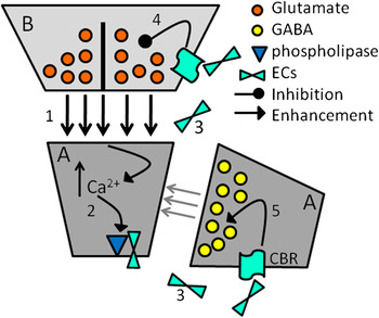
Looking one step back in retinal circuitry is useful in constructing a hypothesis for the activity of ECs in the inner retina. CB1R agonists cause suppression of bipolar cell L-type Ca2+ channels in salamander retinal slices (Straiker et al., Reference Straiker, Stella, Piomelli, Mackie, Karten and Maguire1999), which the authors proposed would inhibit glutamate release from bipolar terminals. In goldfish retina, CB1R agonists inhibited IK(V) and the predicted effect of this was to change the kinetics of the ON bipolar response making it more tonic (Yazulla et al., Reference Yazulla, Studholme, McIntosh and Fan2000; Yazulla, Reference Yazulla2008). For simplicity, the scenario depicted in Fig. 4 only considers the suppressive effect of CB1R activation on bipolar cell L-type Ca2+ channels. Because there is physiological evidence that amacrine cells can produce ECs (Warrier & Wilson, Reference Warrier and Wilson2007), an amacrine cell process is shown as the source of the ECs. Strong activation of glutamate receptors activates the Ca2+-sensitive phospholipases, generating ECs and diffusion of these molecules to receptors on neighboring amacrine cell processes as well as the bipolar cell terminal. Receptor activation on the neighboring amacarine cell increases the spontaneous release of GABA, and receptor activation on the bipolar terminal suppresses glutamate release. The net effect of EC signaling would be to reduce the activity of the EC-producing amacrine cell.
A potential scenario for the activation and actions of cannabinoid receptors on bipolar cells and amacrine cells. When excitatory input from bipolar cells (1) generates a calcium elevation in a postsynaptic amacrine cell dendrite (2), Ca2+-dependent phospholipases are activated and generate EC products. The ECs diffuse from the source cell (3), bind and activate receptors on bipolar cell terminals and a nearby amacrine cell process. The decrease in glutamate release from the bipolar cell (4) and the increase in spontaneous GABA release from the amacrine cell (5) suppress the activity of the EC-producing amacrine cell.

S1P receptors
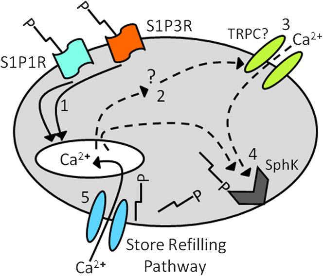
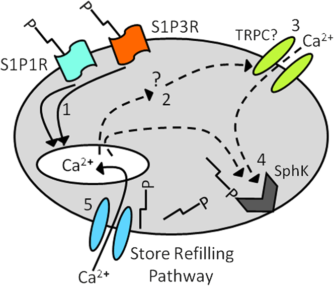
S1P has been implicated in development and in a number of disease states but also in normal physiological function in the central nervous system (Zhang et al., Reference Zhang, Fehrenbacher, Vasko and Nicol2006; Kajimoto et al., Reference Kajimoto, Okada, Yu, Goparaju, Jahangeer and Nakamura2007; Kanno et al., Reference Kanno, Nishizaki, Proia, Kajimoto, Jahangeer, Okada and Nakamura2010). S1P is synthesized when it is phosphorylated by sphingosine kinase (SphK, Ghosh et al., Reference Ghosh, Bian and Gill1994; Olivera et al., Reference Olivera, Rosenthal and Spiegel1996). SphK itself is thought to be regulated by phosphorylation, and the enzyme can be activated by Ca2+/calmodulin (Hla et al., Reference Hla, Lee, Ancellin, Liu, Thangada, Thompson and Kluk1999) possibly via translocation to the plasma membrane (Sutherland et al., Reference Sutherland, Moretti, Hewitt, Bagley, Vadas and Pitson2006). Once synthesized, S1P, which is poorly membrane permeant, is thought to be released via an ATP-binding cassette transporter as it is synthesized (Kim et al., Reference Kim, Takabe, Milstien and Spiegel2009). Extracellular S1P activates a family of five receptors (Sanchez & Hla, Reference Sanchez and Hla2004), S1P1R–S1P5R (S1P4R with low affinity so this receptor may have an alternative endogenous ligand). S1P1R is known to couple to Gαi and is linked to suppression of AC and also activation of PLC (Okamoto et al., Reference Okamoto, Takuwa, Gonda, Okazaki, Chang, Yatomi, Shigematsu and Takuwa1998). S1P2R and S1P3R couple to Gαi, Gαq, and Gα13 and S1P5R has been reported to signal via Gαi and Gα12. Immunocytochemical and physiological evidence suggests that S1P1R and S1P3R (Crousillac et al., Reference Crousillac, Colonna, McMains and Gleason2009) as well as SphK (Borges et al., Reference Borges, Lindstrom, Walters, Warrier and Wilson2008; Crousillac et al., Reference Crousillac, Colonna, McMains and Gleason2009) are expressed in cultured avian GABAergic amacrine cells. S1P is known to function both intracellularly by a poorly understood mechanism and extracellularly via its receptors. In store-depleted cultured GABAergic amacrine cells, Borges et al. (Reference Borges, Lindstrom, Walters, Warrier and Wilson2008) demonstrated that one receptor-independent action of S1P was to activate a store-refilling Ca2+ influx pathway based on Ca2+ imaging of dendrites. Inhibition of the synthetic enzyme for S1P (SphK) suppressed basal activation of this pathway, suggesting that SphK is expressed and active in amacrine cells. Receptor-mediated signaling of S1P via S1P1R and S1P3R has also been described in similar cultures of GABAergic amacrine cells (that were not store depleted). S1P was demonstrated to elicit cytosolic Ca2+ elevations consisting of release from internal stores followed by an often prolonged phase dependent upon Ca2+ influx, possibly via TRPC channels. An S1P-dependent current and the influx phase had a similar time course, were both sensitive to La3+, and had similar pharmacology, suggesting that they represent the same process. Interestingly, the prolonged Ca2+ elevations often engendered by receptor activation were made more transient when SphK activity was inhibited. These results suggest a scenario where extracellular S1P stimulates Ca2+ signaling, which in turn can increase SphK activity and boost cytosolic S1P in order to refill the Ca2+ store (Fig. 5). The Ca2+-imaging data from Crousillac et al. (Reference Crousillac, Colonna, McMains and Gleason2009) were collected from cell bodies only so the distribution of these receptor-mediated events is not yet known. Because the store-refilling event described above (Borges et al., Reference Borges, Lindstrom, Walters, Warrier and Wilson2008) occurred in dendrites, the machinery involved likely resides throughout the cell raising the possibility that this form of Ca2+ signaling has the potential to contribute to neurotransmitter release at amacrine cell synapses as has been shown for synapses elsewhere in the brain (Kajimoto et al., Reference Kajimoto, Okada, Yu, Goparaju, Jahangeer and Nakamura2007; Kanno et al., Reference Kanno, Nishizaki, Proia, Kajimoto, Jahangeer, Okada and Nakamura2010). The endoplasmic reticulum protein stromal interaction molecule 1 (STIM1) and the plasma membrane protein Orai1 are known to mediate store depletion-dependent Ca2+ influx in nonexcitable cells and some excitable cells (Liou et al., Reference Liou, Kim, Heo, Jones, Myers, Ferrell and Meyer2005; Prakriya et al., Reference Prakriya, Feske, Gwack, Srikanth, Rao and Hogan2006), and they may be involved in the S1P response in amacrine cells. However, the expression and function of these proteins have not yet been demonstrated in the inner retina. Intriguingly, a recent report demonstrates that the STIM1- and Orai1-dependent store-refilling mechanism can involve recruitment of TRPC1 channels to the plasma membrane via vesicular trafficking (Cheng et al., Reference Cheng, Liu, Ong, Swaim and Ambudkar2011).
Activation of sphingosine-1-phosphate receptors (S1PRs) lead to calcium signaling in amacrine cells. Cellular signaling is initiated by activation of S1P1R and S1P3R in cultured GABAergic amacrine cells are shown. Receptor activation leads to release of Ca2+ from internal stores. The elevation in cytosolic Ca2+ either directly or indirectly (2) activates a nonspecific cation channel on the plasma membrane (3, possibly a TRPC channel). Either Ca2+ from stores or Ca2+ crossing the plasma membrane (or both) activates SphK to generate internal S1P (4). S1P interacts with a transport mechanism that mediates Ca2+ store refilling (5). The initial Ca2+ influx pathway (3) and the store-refilling pathway may be the same mechanisms or they might be separate, as depicted.
Conclusion
There is good evidence that both classical retinal neurotransmitters and some less well-established signaling molecules modify amacrine cell function via interactions with metabotropic receptors. In the case of dopamine, an extensive change in amacrine cell electrical connectivity is initiated, which has a global effect on information flow in the retina. Immunocytochemical and physiological evidence indicates that mGluRs 1, 2, 4, 5, 7, and 8 can be expressed on amacrine cells. The role of endogenous glutamate acting on these receptors will be challenging to elucidate, but as specific pieces of IPL circuitry become defined, it should become possible to determine the circumstances under which specific mGluRs become activated and how their activity shapes the processing of signals in the inner retina. The work of Vigh et al. (Reference Vigh, Li, Hull and von Gersdorff2005) serves as a model for the usefulness of such an approach. GABAB receptors are predominately expressed on GABAergic amacrine cells indicating that they often function as autoreceptors and suppress the release of GABA and in the case of SACs, ACh as well. There is immunocytochemical evidence that all five muscarinic receptor subtypes can be expressed by amacrine cells. Most is known about the function of M2 and a function for these receptors had been determined in both glycinergic amacrine cells (promotion of glycine release) and GABAergic amacrine cells (generation of NO). Although investigations of the physiological consequences of lipid signaling in the inner retina is in its early stages, the evidence thus far indicates that both ECs and S1P interact with their metabotropic receptors to alter amacrine cell function.
It would be gratifying to assemble these pieces of the puzzle so that a clear picture emerged regarding the role of these receptors in facilitating amacrine cell processing of visual signals. Until we understand more about the specific contributions of different amacrine cell subtypes, and the stimulus conditions under which different metabotropic receptors become activated, much of that picture will be missing.
Acknowledgements
Special thanks to Emily McMains for her careful reading of the manuscript. The author's work was supported by NIH grant EY012204.