Introduction
Metastatic lesions of the temporal bone are very rare. The most common presenting otological symptom is hearing loss, which may be either conductive or sensorineural. Presentation as facial nerve palsy has also been reported. The usual mechanism of otological symptoms is bony invasion causing eustachian tube dysfunction, compression of the cochlear nerve fibres at the internal acoustic meatus, or direct compression of the cochlea itself.
Temporal bone metastases commonly arise from breast malignancy (29 per cent) followed by lung malignancy (11 per cent).Reference Cumberworth, Friedmann and Glover1
We present a rare case of a 57-year-old man who presented only with post-aural ache, secondary to metastatic primary adenocarcinoma of the lung.
Case report
A 57-year-old man was referred to our ENT out-patient clinic suffering from persistent left post-aural pain of four months' duration. The pain had gradually worsened and had begun to affect his sleep, despite routine analgesics. There were no other ear, nose or throat symptoms. He was an ex-smoker, having given up 30 years ago, and was otherwise fit and healthy, with no significant co-morbidities.
Upon examination, the patient showed extreme tenderness in the post-aural region. The ENT examination was otherwise normal, with no other abnormalities.
Flexible fibre-optic nasal endoscopy and audiometric testing were also normal.
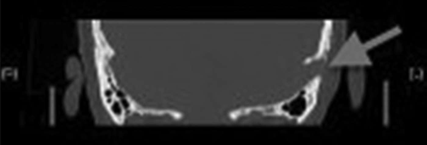
Diagnostic imaging was undertaken, utilising computed tomography (CT) of the temporal bone. This showed a bone-eroding lesion in the petrous temporal bone (Figure 1). A diagnosis of glomus or arteriovenous malformation could not be excluded at this time.
Coronal computed tomography scan of the temporal bone and internal auditory meatus, showing an irregular soft tissue mass with bony destruction over the left petrous temporal bone (arrow).

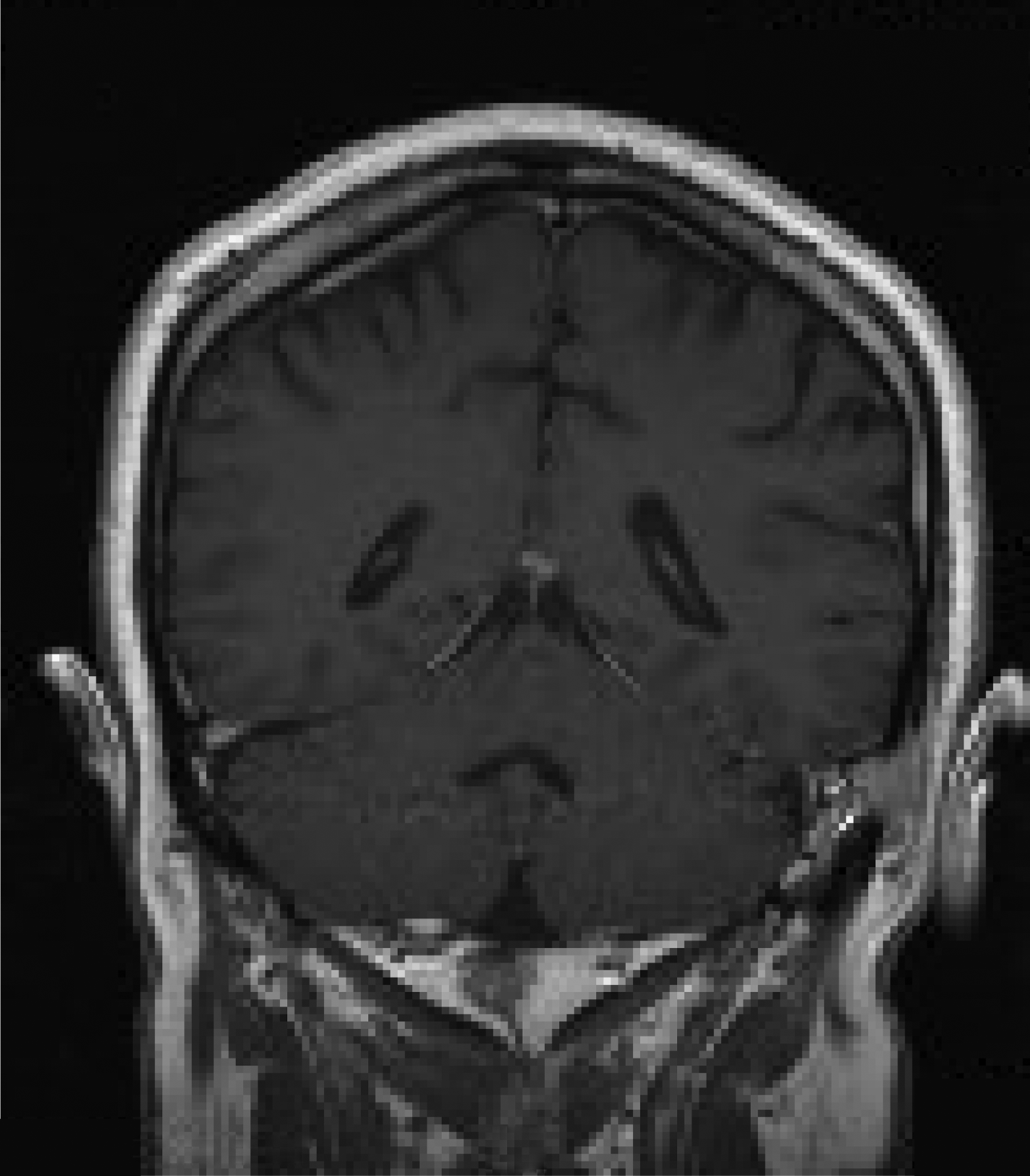
Further imaging with contrast-enhanced magnetic resonance imaging (MRI) demonstrated that the lesion involved the left petrous temporal bone and adjacent occipital bone, with sparing of the sigmoid sinus (Figure 2). The lesion was reported as being highly suggestive of infiltrative neoplasm. Following MRI, the differential diagnosis also included unusual infection or a granulomatous process.
Coronal, contrast-enhanced magnetic resonance imaging scan of the head, showing a poorly defined, spiculated mass involving the posterior and outer aspect of the left petrous temporal bone.
Due to the radiological findings and inconclusive diagnosis, an incisional biopsy was carried out. Histopathological examination of the biopsy specimen showed fibro-adipose tissue bearing an infiltrative tumour composed of irregular glandular structures lined by atypical epithelial cells with large, pleomorphic, clear nuclei and moderate amounts of cytoplasm (Figure 3). These findings were suggestive of invasive, moderately differentiated adenocarcinoma.
Photomicrograph showing adenocarcinoma organised in glandular structures. (H&E; ×20)

Additional immunohistochemical analysis showed the tumour cells to be strongly positive for pancytokeratin, cytokeratin 7, BerEP4 antigen and thyroid transcription factor 1. They did not stain for cytokeratin 20, carcinoembryonic antigen, prostate-specific antigen, BRST2 antigen, cluster of differentiation 56 glycoprotein, chromogranin-A and thyroglobulin (Figure 4).
Photomicrograph showing negative thyroglobulin immunohistochemical staining. (×20)
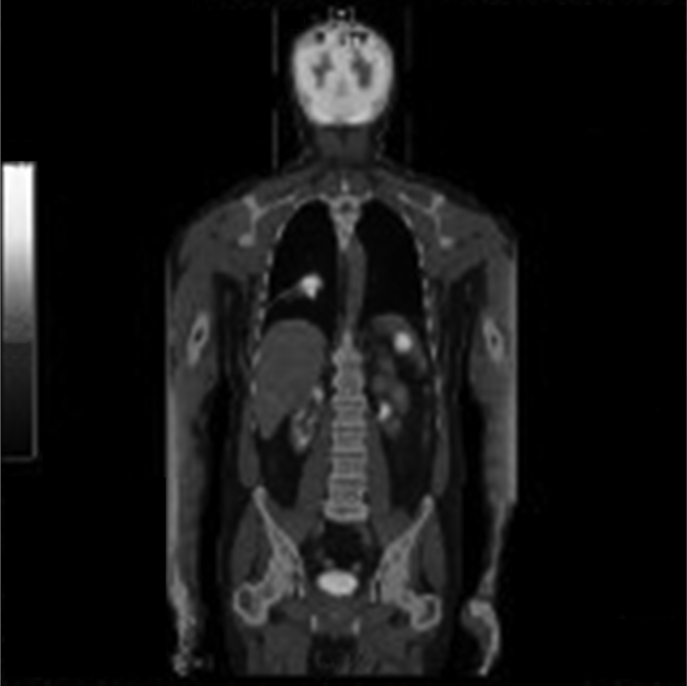
The overall features suggested a metastatic adenocarcinoma, with the lung as the most likely primary source. After discussion in the multidisciplinary team meeting, a whole body positron emission tomography (PET) scan was arranged, which confirmed marked uptake within the lobulated apical right lower lobe of the lung (suggesting a primary lesion), with a low density lesion in the spleen (Figure 5). The tumour-node-metastasis (TNM) staging of the malignancy was T2b N3 M1b.
Whole-body fluoro-deoxy-glucose positron emission tomography scan showing marked uptake (maximum standardised uptake value, 14.6) within a lobulated, 5.6 cm, apical right lower lobe mass which appears to have an endobronchial component. There is also marked uptake in an approximately 2.9 cm, low density lesion in the spleen.
These findings were discussed in the lung multidisciplinary team meeting, and an opinion from a regional skull base surgeon was also obtained.
Following these discussions, the patient was treated with a combination of chemotherapy and radiotherapy.
Discussion
Metastasis of lung adenocarcinoma to the temporal bone is an uncommon clinical entity. In 1976, Hill and Kohut presented a case of temporal bone metastasis from colonic carcinoma. They went on to review the 102 published case reports of temporal bone metastasis, and found that 12 per cent of these were metastases from a lung primary, which was the second commonest primary site.Reference Hill and Kohut2
In 1985, Belal reported that metastasis to the temporal bone was probably more common that originally thought, and presented a histopathological report of 13 patients with known temporal bone metastasis. The most common primary site was breast, with other identified primary sites being brain, prostate, lung, thyroid and blood.Reference Belal3 More recent reviews have agreed that the three commonest primary sites are kidney, breast and lung.Reference Nelson and Hinojosa4
There are now a plethora of case reports of temporal bone metastases, including distant spread from lung adenocarcinoma, and it is widely recognised that there are distinctive patterns of involvement and presentation of metastatic tumours to the temporal bone. In 1980, Berlinger et al. suggested five discrete types of metastatic temporal bone involvement: (1) direct invasion from a local primary; (2) isolated haematogenous metastasis from a distant primary; (3) meningeal carcinomatosis; (4) leptomeningeal extension from an intracranial primary; and (5) leukaemic or lymphomatous infiltration.Reference Berlinger, Koutroupas, Adams and Maisel5
Haematogenous spread is the commonest of these types, with most metastases occurring within the petrous region of the bone; this is due not only to the ability of the petrous marrow to remove circulating tumour cells, but also to the sluggish blood flow within the marrow, which favours tumour cell deposition.Reference Proctor and Lindsay6 Metastatic involvement of the aerated parts of the temporal bone is less common, and is often due to vascular permeation from the petrous part of the bone. Whilst invasion of the mastoid air cells and middle ear normally occurs secondary to petrous bone metastasis, the otic capsule and inner ear appear to be quite resistant to invasion.Reference Schuknecht, Allam and Murakami7
• Metastatic lesions to the temporal bone are increasingly encountered
• Cases with persistent symptoms and no physical signs should receive early imaging
• Lack of an audiological deficit does not exclude a temporal bone lesion
Diagnosis of haematogenous metastasis to the temporal bone can be a challenge, as patients presenting with symptoms will often have very advanced disease. Temporal bone metastases often stay silent until they extend from the petrous bone into other areas, causing otological symptoms. A 2004 review by Cureoglu et al. reported that the commonest symptom is hearing loss.Reference Cureoglu, Tulunay, Ferlito, Schachern, Paprella and Rinaldo8 Conductive hearing loss occurs due to disruption of the eustachian tube (causing secondary otitis media), middle-ear invasion (causing ossicle destruction) or tympanic membrane dysfunction (causing various otological manifestations). Sensorineural hearing loss can also occur due to cochlear nerve invasion, compression of the internal auditory meatus, or inner ear invasion via the otic capsule.Reference Schuknecht, Allam and Murakami7 Direct invasion of the internal auditory meatus causes earlier otological symptoms. Facial nerve paralysis, hearing loss and tinnitus can all occur; onset can be insidious, or sudden in one-third of patients.
Conclusion
Most patients presenting with hearing impairment, tinnitus or balance disturbance will have a non-neoplastic cause for these symptoms; however, it is important to consider the diagnosis of primary or metastatic carcinoma.Reference Devaney, Boschman, Willard, Ferlito and Rinaldo9, Reference Imamura and Murakami10 In the case reported here, the patient had no symptoms other than post-auricular pain and tenderness. Following review of the existing literature, it appears very unusual for a patient with temporal bone metastasis to present with such non-specific symptoms, especially in the absence of classical otological symptoms such as hearing loss.