1. Introduction
The Phyllolepida are a peculiar group of the Arthrodira (Placodermi), widespread in the Givetian–Famennian of Gondwana (Australia, Antarctica, Turkey, South America: e.g. Janvier, Reference Janvier1983; Long, Reference Long1984, Reference Long2003; Ritchie, Reference Ritchie1984, Reference Ritchie2005; Young, Reference Young1984, Reference Young1988, Reference Young and Tingey1991, Reference Young2005a, Reference Youngb, Reference Youngc; Young & Moody, Reference Young and Moody2002; Young & Long, Reference Young and Long2005; Young, Long & Turner, 1993; Young, Moody & Casas, 2000) and in the Famennian of Euramerica (North America, Greenland, West Europe, Russia: e.g. Agassiz, Reference Agassiz1844; Denison, Reference Denison and Schultze1978; Heintz, Reference Heintz1930; Lane, Cuffey & Daeschler, 2001; Leriche, Reference Leriche1931; Lohest, Reference Lohest1888; Newberry, Reference Newberry1889; Rohon, Reference Rohon1900; Stensiö, Reference Stensiö1934, Reference Stensiö and Piveteau1969). They are characterized by a dorsoventrally flattened dermal armour, the lack of rostral and pineal plates, and the presence of a large unpaired centronuchal plate, for which no consensus exists as to its origin (either due to the fusion of nuchal and central plates, or to the loss of central plates). The centronuchal plate is surrounded by a ‘ring’ of perinuchal plates. There is no external foramen for the endolymphatic duct on the paranuchal plates. Post-marginal plates are lacking, and anterior and/or posterior median ventral plates are reduced. The ornamentation mainly consists of ridges lacking semi-dentine.
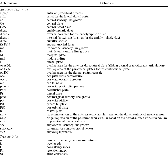
Abbreviations used in text and figures

First considered as belonging to the ‘coelacanths’ (Agassiz, Reference Agassiz1844), the ‘crossopterygians’ (Zittel, Reference Zittel1887–90), the Placodermi (Newberry, Reference Newberry1889) and the Heterostraci (Woodward, Reference Woodward1915), the Phyllolepida were finally assigned with certainty to the Placodermi (Stensiö, Reference Stensiö1934). Subsequently, they were considered as either sharing an immediate common ancestor with the Arthrodira (Denison, Reference Denison and Schultze1978), belonging to the Actinolepidoidei (Long, Reference Long1984), or being of indetermined position within the Arthrodira (Goujet & Young, Reference Goujet and Young1995). They were considered as close to Wuttagoonaspis (Long, Reference Long1984; Young, Reference Young1980; Young & Goujet, Reference Young and Goujet2003) and/or to Antarctaspis (Denison, Reference Denison and Schultze1978; Long, Reference Long1984). More recently, a sister-group relationship between the Phyllolepida (as a crownward member of the top of an ‘actinolepidoid’ paraphyletic ensemble) and the Phlyctaenioidei (‘Phlyctaenii’ plus Brachythoraci) was proposed, and Antarctaspis and Wuttagoonaspis are the most inclusive taxa among the Arthrodira (Dupret, Reference Dupret2004). However, the latter hypothesis was not followed by Young (Reference Young2005a, Reference Youngb, Reference Youngc), who argued that the ridged ornamentation of the Phyllolepida and Wuttagoonaspis is homologous and therefore is an important synapomorphy of this ensemble.
Because the phyllolepid remains were abundantly encountered in the eastern margin of Gondwana before the Famennian, and because the earliest known and most primitive forms were found in Australia, it was commonly accepted that this group originated from Australia. However, the most plesiomorphic phyllolepids so far known (Cobandrahlepis Young, Reference Young2005c, and Yurammia Young, Reference Young2005c, both dated as Givetian) already possess many phyllolepid diagnostic features (e.g. the presence of the centronuchal plate). As a consequence, the phylogenetic relationships between the Phyllolepida and the other groups of Placodermi have long been debated, without leading to a consensus.
It is also noteworthy that despite the close connection between South China and Australia during the Devonian Period (after the Pragian: Zhu, Reference Zhu, Blieck and Turner2000; Zhu & Zhao, Reference Zhu, Zhao and Rong2006; Zhu, Wang & Wang, 2000), no phyllolepid remains (or closely related forms) have been found in China. The new form Gavinaspis convergens described here throws some new light onto the systematic and palaeogeographic origin of the Phyllolepida.
2. Material and methods
All of the referred specimens are housed in the IVPP (Institute of Vertebrate Palaeontology and Palaeoanthropology, Beijing, People's Republic of China). They were collected by the Early Vertebrate Research Group of IVPP during recent field trips (1998–2006) to Qujing, Yunnan, and have been prepared mechanically.
Abbreviations for placoderm dermal bones and other structures, and phylogenetic abbreviations as used in the text and figures, are listed in Table 1.
3. Geological setting
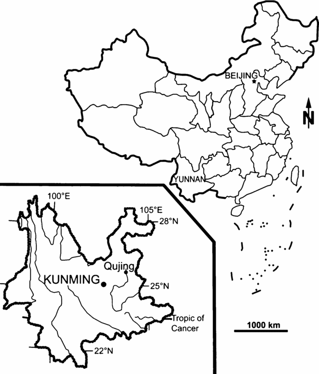
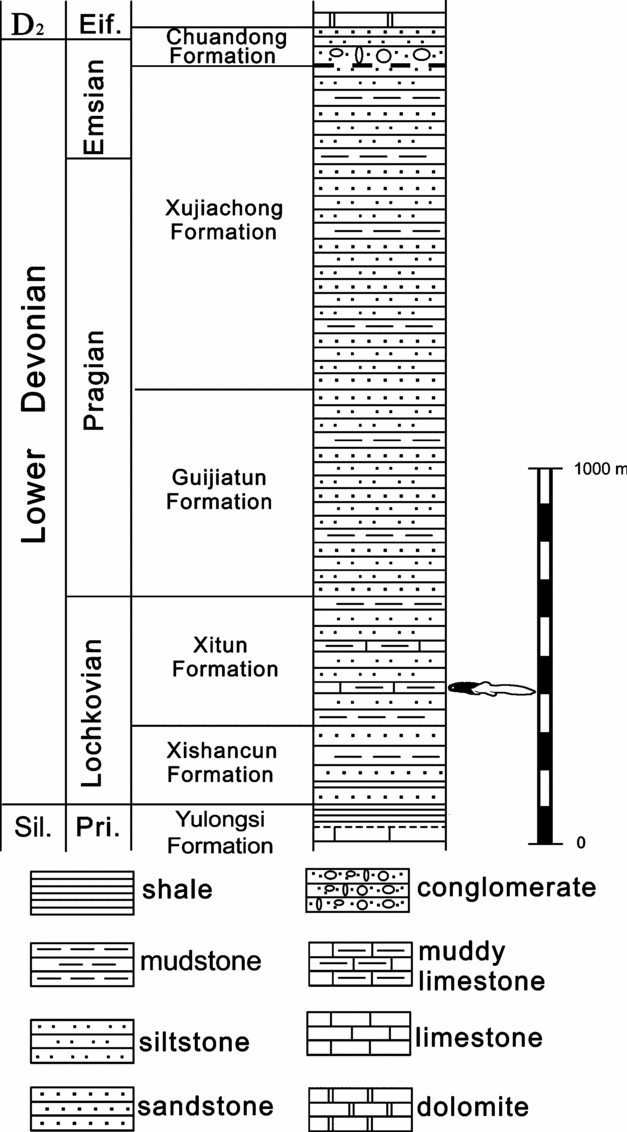
The specimens described herein come from the middle part of the Xitun Formation at a locality close to Xitun village in the suburb of Qujing (Yunnan, South China, Figs 1, 2; Diabolepis–Nanpanaspis macrovertebrate assemblage II of Zhu, Wang & Wang, 2000; Late Lochkovian; only one locality). The matrix of the specimens is a light-grey clayey limestone. The Xitun Formation of Qujing yields remains of the Galeaspida Eugaleaspis changi, Nanpanaspis microculus, Laxaspis qujingensis, Cyclodiscaspis ctenus (Liu, Reference Liu1965, Reference Liu1975), Microholonaspis microthyris, Hyperaspis acclivi (Pan, Reference Pan1992), the Thelodonti Parathelodus scitulus, P. asiatica, P. catalatus, P. trilobatus, P. cornuformis (Wang, Reference Wang1997), the Chondrichthyes Gualepis elegans, Changolepis tricuspidus, Peilepis solida, Ohiolepis? xitunensis, the Acanthodii Nostolepis sp., Youngacanthus gracili (Wang, Reference Wang1984), the Sarcopterygii Youngolepis praecursor (Chang & Yu, Reference Chang and Yu1981), Diabolepis speratus (Chang & Yu, Reference Chang and Yu1984), Psarolepis romeri (Yu, Reference Yu1998; Zhu, Yu & Janvier, 1999), Achoania jarviki (Zhu, Yu & Ahlberg, 2001), Styloichthys changae (Zhu & Yu, Reference Zhu and Yu2002), and Meemannia eos (Zhu et al. Reference Zhu, Yu, Wang, Zhao and Jia2006), the Antiarcha Yunnanolepis chii (Liu, Reference Liu1963), Y. parvus, Y. porifera, Phymolepis cuifengshanensis, P. guoruii, Zhanjilepis aspratilis, Chuchinolepis gracilis, C. qujingensis, C. sulcata, C. robusta (Chang, Reference Chang1978; Zhang, Reference Zhang1978, Reference Zhang1984; Zhu, Reference Zhu1996), the Arthrodira Szelepis yunnanensis (Liu, Reference Liu1979, Reference Liu1981) and the Petalichthyida (Zhu, Reference Zhu, Blieck and Turner2000; pers. obs.).
Position of Qujing, Yunnan, South China.

Stratigraphic column of the Lower Devonian deposits in Yunnan, South China. The fish icon represents the stratigraphic position of Gavinaspis convergens gen. et sp. nov. Modified after Zhu & Zhao, Reference Zhu, Zhao and Rong2006.

4. Systematic palaeontology
PLACODERMI McCoy, Reference McCoy1848
ARTHRODIRA Woodward, Reference Woodward1891
PHYLLOLEPIDA Stensiö, Reference Stensiö1934
Diagnosis. ‘Actinolepidoid’ Arthrodira in which an unpaired centronuchal plate is present and no paired central plates are identified. Centronuchal plate is surrounded by four or five paired bones including preorbital, postorbital, marginal and paranuchal plates. Post-marginal plates are absent.
Remarks. The diagnosis above and systematic rank are modified after Young (Reference Young2005b, p. 175) to incorporate new data on the Chinese phyllolepid. Despite the presence of an unambiguous centronuchal plate, we consider that the skull roof pattern and the outline of the associated neurocranium of the new form described herein do not fit the recently emended diagnosis of the family Phyllolepididae Woodward, Reference Woodward1891 (non rank-order Phyllolepida Stensiö, Reference Stensiö1934) proposed by Young (Reference Young2005b, Reference Youngc) or by Ritchie (Reference Ritchie2005). Moreover, the phylogenetic analysis attempted herein clearly indicates a sister-group relationship between Gavinaspis gen. nov. and the Phyllolepididae. Other characters mentioned in Young's (2005b) diagnosis are still applicable for the family Phyllolepididae Woodward, Reference Woodward1891 (all Phyllolepida except Gavinaspididae fam. nov.): centronuchal plate as broad as or broader than long, and surrounded by five smaller paired bones; rostral and pineal plates absent from skull roof; posterolateral plate absent from trunk armour; median dorsal plate without an inner keel, and anterior dorsolateral plate with a narrow elongate area; posterior ventrolateral plate of triangular shape, lacking a lateral lamina; dermal ornament mainly consisting of smooth concentric ridges, with some tubercles and tubercle rows.
Family gavinaspididae fam. nov
Diagnosis. Same as for the type genus and its type species, by monotypy.
Type genus. Gavinaspis gen. nov. Genus
Gavinaspis gen. nov
Diagnosis. Same as for the type species, by monotypy.
Etymology. To acknowledge the major contribution to the study of phyllolepid placoderms by Dr Gavin C. Young, Australian National University, Canberra.
Type species. Gavinaspis convergens gen. et sp. nov.
Gavinaspis convergens gen. et sp. nov.
Figures 3–7
Holotype. Associated postethmoid part of a skull roof in ventral and dorsal views (IVPP V 15085-1, 2), with its neurocranium attached (in dorsal view, IVPP V 15085-2).
Other material. Three left paranuchal plates (in internal and external views; IVPP V 15086.1-3); thoracic material referred to this taxon is unknown.
Etymology. Suggesting the convergence of middle and posterior pitlines in the midline, as well as the fusion of paired central plates into a single unpaired unit.
Locality and horizon. From a locality close to the Xitun village in the suburb of Qujing, Yunnan, southeastern China, Xitun Formation, Late Lochkovian, Early Devonian.
Diagnosis. Phyllolepid with the occipital portion of neurocranium very elongated and slender; dermal rostral capsule not fused to the postethmoid part of skull roof; postorbital plate contacting the paranuchal plate mesially to infraorbital and main lateral sensory line grooves (at least on the internal side of skull roof); marginal plate not extending mesially to infraorbital and main sensory line grooves in internal view; ornamentation consisting of elongated tubercles, sometimes merging into ridges.
Description. The specimen is tectonically deformed by a dextral shear (the right side is posteriorly displaced relative to the left side).
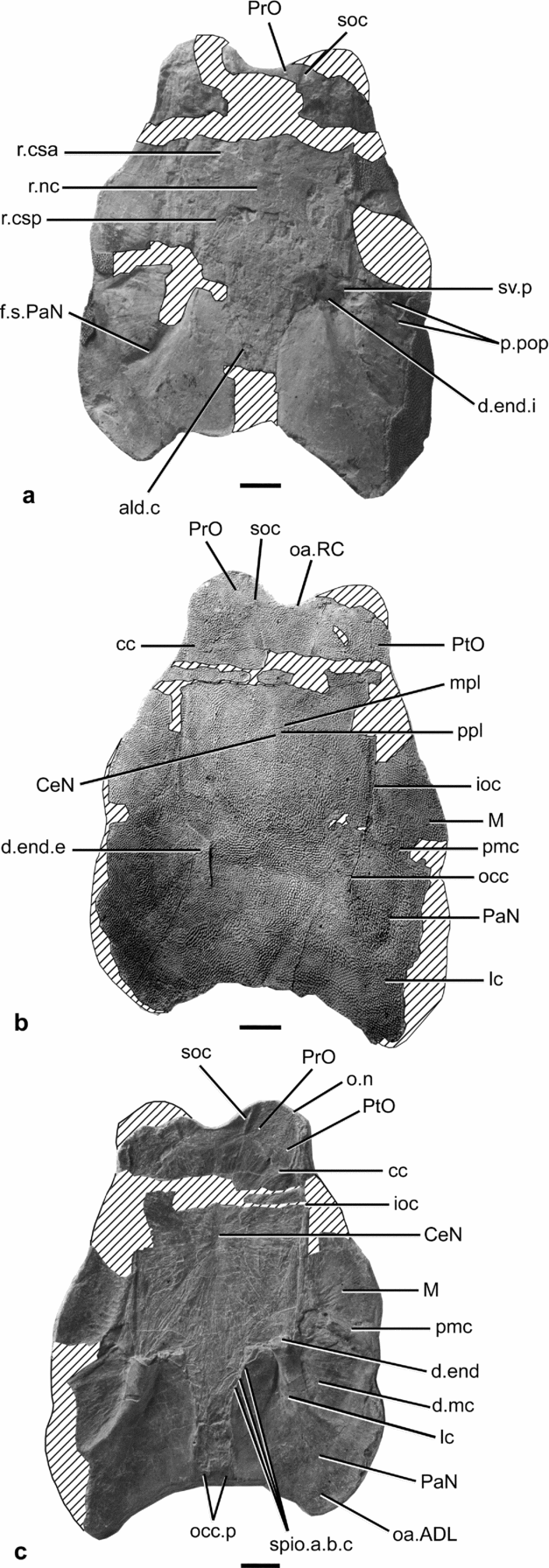
Neurocranium. This is only visible in dorsal view, but is poorly preserved in its pre-vagal portion, consisting of a natural mould of the overlying radiating fibres of the dermal plates and sensory line grooves. No trace of an antorbital or a supraorbital process is visible. The anterior postorbital process (a.po.p, Fig. 4a) is barely visible and situated around a crack in the specimen; nevertheless, it seems to be as large as that in non-phyllolepid ‘actinolepidoids’. The posterior postorbital process (p.po.p, Figs 4a, 5a), though its outline is hardly distinguishable, is well developed and bifid, as in all ‘Dolichothoraci’. The supravagal process (sv.p, Figs 4a, 5a) is poorly developed, and its tip is not preserved. In the otic region, two shallow ridges corresponding to the underlying anterior and posterior semicircular canals are visible (r.csa, r.csp, Figs 4a, 5a). In the mid-plane of the otic region, a quite large longitudinal ridge may be interpreted as the impression of the underlying neural canal surrounding the medulla oblongata (r.nc, Figs 4a, 5a). The most surprising feature of the neurocranium is the postvagal (occipital) region, which is very long and narrow, and thus resembles more that of some Petalichthyida (e.g. Macropetalichthys in Stensiö, Reference Stensiö and Piveteau1969, Fig. 21) than that of the Arthrodira or the Placodermi in general. A vascular plexus is visible at the limit between the neurocranium and the overlying dermal bones. The subparanuchal fossa (f.s.PaN, Figs 4a, 5a) is well marked. In the occipital part, the lateral walls of the perichondral bone of neurocranium are poorly preserved on the ventral side of the skull roof but show at least three foramina for the spino-occipital nerves (spio.a-c, Figs 4c, 5c); since the occipital region is very elongated, it is possible that there were more than three pairs of spino-occipital nerves. The posterior wall of the neurocranium, though poorly preserved, shows the two posterior occipital processes (occ.p, Figs 4c, 5c), separated by a shallow embayment. A poorly preserved oblique tube, along the lateral side of the occipital part of the neurocranium, is interpreted as the canal for the lateral dorsal aorta (ald.c, Figs 4a, 5a).
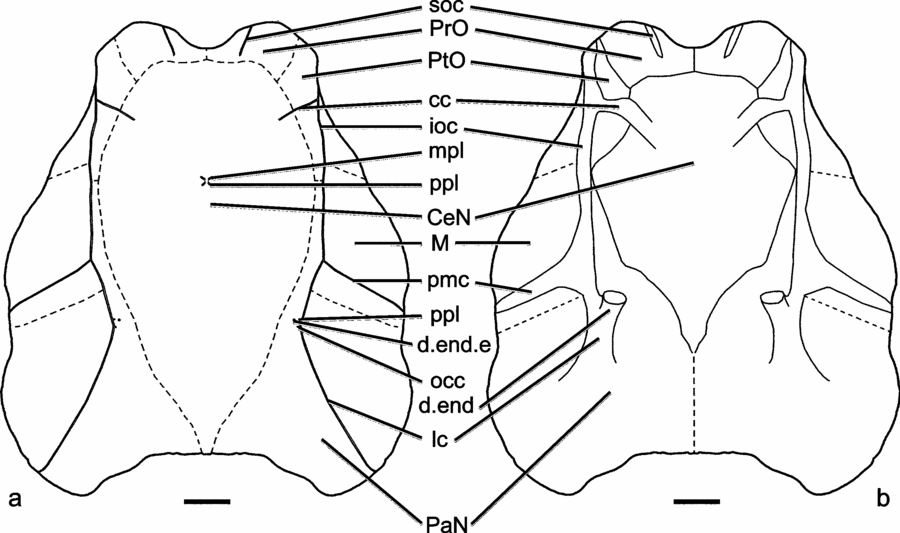
Schematic reconstruction of the skull roof of Gavinaspis convergens gen. et sp. nov. Tectonic dextral shear distortion has been corrected by means of computerized assistance (torsion 5°). (a), dorsal view; (b) ventral view. Scale bar 1 cm. For key to abbreviations, see Table 1.

Head of Gavinaspis convergens gen. et sp. nov. (a–c, holotype). (a) Dorsal view of the neurocranium and impression of skull roof, no. IVPP V 15085-2; (b) dorsal (external) view of the skull roof, no. IVPP V 15085-1; (c) ventral (internal) view of the skull roof, no. IVPP V 15085-1. All scale bars 1 cm. For key to abbreviations, see Table 1.
Head of Gavinaspis convergens gen. et sp. nov. (a–c, holotype). (a) Dorsal view of the neurocranium and impression of skull roof, holotype no. IVPP V 15085-2; (b) dorsal (external) view of the skull roof, no. IVPP V 15085-1; (c) ventral (internal) view of the skull roof, no. IVPP V 15085-1. All scale bars 1 cm. For key to abbreviations, see Table 1.

Skull roof. The dermal rostral capsule (usually composed of the pineal, rostral and postnasal plates) is missing, as is the case in most ‘actinolepidoids’, but its position is indicated by an anterior embayment of the preorbital plates, and a slight overlap area on its anterior margin (oa.RC, Figs 4b, 5b).
The postethmoid region of the skull roof is well preserved and is longer than wide (Figs 3, 4b–c, 5b–c). The sensory line system is more conspicuous on the internal side (broad thickenings) than on the external side (very narrow grooves, when visible). The very tiny and dense tuberculated ornamentation on the external side of the skull roof obscures bone sutures.
The preorbital plates (PrO, Figs 3, 4a–c, 5a–c) are broad and very short paired plates. Their anterior edge shows a shallow embayment for the insertion of the dermal rostral capsule. The supraorbital sensory line groove (soc, Figs 3–5) extends back only to the radiation centre of the preorbital plate. The contact between the preorbital and postorbital plates seems to be a butting contact, with no overlap. A low thickening suggests that the centronuchal plate slightly overlaps the preorbital plates. The orbital notch (o.n, Figs 4c, 5c) is shared by the edges of the preorbital and postorbital plates.
The radiation centre of the postorbital plates (PtO, Figs 3, 4a–c, 5b–c) is situated at the junction of the infraorbital (ioc, Figs 3, 4a–c, 5c–c, 6) and central (cc, Figs 3, 4b–c, 5b–c) sensory line grooves. The central sensory line groove does not reach the radiation centre of the centronuchal plate. Mesially to the infraorbital sensory line groove, the postorbital plate shows a long and very slender posterior process contacting the similarly slender anterior process of the paranuchal plate (PaN, Figs 3, 4c–c, 5b–c). The junction of these two processes (along the infraorbital sensory line groove) prevents the centronuchal (CeN, Figs 3, 4b–c, 5b–c, 6) from contacting the marginal plates (M, Figs 3, 4a–c, 5b–c, 6), at least on the internal side of the skull roof. This character is a derived feature shared by the superfamily Kujdanowiaspidoidea (sensu V. Dupret, unpub. Ph.D. thesis, Muséum National d'Histoire Naturelle, 2003; Dupret, Goujet & Mark-Kurik, 2007) and by the derived Phyllolepida (Austrophyllolepis and Phyllolepis; Cowralepis displays a fenestra rather than a real contact between the postorbital and paranuchal plates; see Ritchie, Reference Ritchie2005, Fig. 20). The condition in Placolepis (Ritchie, Reference Ritchie1984) and in Cobandrahlepis (Young, Reference Young2005c) resembles that in the family Actinolepididae and in the Phlyctaenioidei, where the marginal plate separates the postorbital from the paranuchal plates. It is noteworthy that the postorbital plates contact the paranuchal plate in Wuttagoonaspis, but via neither a blade nor a process (Ritchie, Reference Ritchie1973).
The marginal plates (M, Figs 3, 4a–c, 5b–c, 6) are large elements, gently convex laterally, and forming the lateral edge of the skull roof, although their anterior and posterior boundaries are rather unclear. The radiation fibres on the internal side of the skull roof clearly show that their radiation centre is situated at the junction between the infraorbital, postmarginal (pmc, Figs 3, 4b–c, 5b–c, 6) and main (lc, Figs 3, 4a–c, 5b–c, 6) sensory line grooves, and that the plate does not extend mesially to the infraorbital and main sensory line grooves. This is a derived character displayed by the family Kujdanowiaspididae (sensu V. Dupret, unpub. Ph.D. thesis, Muséum National d'Histoire Naturelle, 2003 and Dupret, Goujet & Mark-Kurik, 2007, non Berg, Reference Berg1955, Reference Berg1958), but unknown in the Phyllolepida. It is noteworthy that the outline of the marginal plate is poorly known in the early Arthrodira Wuttagoonaspis (Ritchie, Reference Ritchie1973) and Antarctaspis (White, Reference White1968, non Denison, Reference Denison and Schultze1978). The postmarginal sensory line (pmc, Figs 3, 4b–c, 5b–c, 6) groove is very large on the internal side of the plate. The postmarginal plates are clearly absent, as there is no overlap area (nor bone suture) for them on the marginal or on the paranuchal plates.
Since the median unpaired element is huge and the central plates are not individualized, we propose to term this median dermal component of the skull roof the centronuchal plate (CeN, Figs 3, 4b–c, 5b–c), by homology with the pattern displayed by the Phyllolepididae. This plate is much longer than wide and its radiation centre is anteriorly placed (at the level of the first third of the plate length). It contacts the preorbital plates anteriorly, without separating them. A low thickened area around this contact suggests a possible overlap of the preorbital plates by the centronuchal plate, as is the case in the known phyllolepids.
The external side exposes particularly well the posterior end of the middle pitline and the anterior end of the posterior pitline (mpl, ppl, Figs 3a, 4b, 5b); both are located at the radiation centre of the plate. The anterior and posterior ends of the posterior pitlines are not connected (that is, they do not form a continuous groove on the dermal bones), thereby implying a superficial course. The central sensory line groove does not reach onto the radiation centre. A thin, posteromesially directed groove that runs from almost the posterior end of the supraorbital groove toward the radiation centre of the centronuchal plate is interpreted here as an ornamentation artefact, and not as the continuity of the supraorbital sensory line groove.
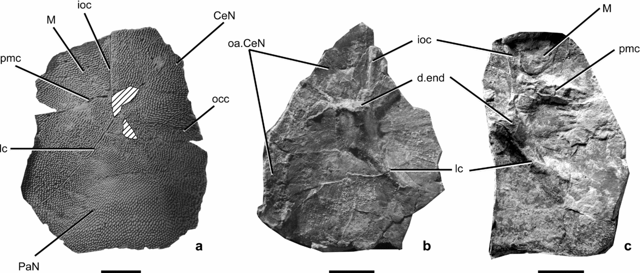
The paranuchal plates (PaN, Figs 3, 4b–c, 5b–c, 6) are also large elements of the skull roof. Their radiation centre is anteriorly situated and close to the external foramen for the endolymphatic duct (d.end.e, Figs 3a, 4a, 5b), as is the case in the non-phyllolepid Actinolepidoidei; in the Phyllolepididae, the radiation centre of the paranuchal plate is close to the posterior edge of the plate. Nevertheless, it is noteworthy that, as in the Phyllolepididae, the radiation centre is at the level of the junction between the curved main lateral line groove and the posterior pitline, which roughly corresponds to the concave anteromesial margin of the paranuchal plate and to the level of the proximal end of the median oblique process of Young (Reference Young2005c, Fig. 4A) and Ritchie (Reference Ritchie2005, Fig. 7F–G) in the Phyllolepididae. The paranuchal plates are widely overlapped mesially by the centronuchal plate as in the Phyllolepididae, although in the latter the posterior part of the paranuchal plate is much shorter. Posteriorly to the radiation centre on the internal side, there is a thick, long and posterolaterally tapering ridge that slightly bends laterally from the endolymphatic duct (d.end, Figs 3b, 4c, 5c, 6) toward the posterior part of the main sensory line groove. This divides the plate into two almost equal parts. At mid-length of the paranuchal plate, the oblique ridge divides in two parts: one straight and tapering posterolaterally bears the main sensory line canal; the other one is more laterally directed and does not seem to have any particular function, unless perhaps an attachment area for the levator muscles of the head, as it is situated along the cucullaris fossa (d.mc, Figs 4c, 5c). The posterolateral edge of the paranuchal plate is smooth, corresponding to a sliding neck-joint type of dermal craniothoracic articulation (oa.ADL, Figs 4c, 5c), as is the case in the ‘Actinolepidoidei’ (and in the Phyllolepida).
Three left paranuchal plates of Gavinaspis convergens gen. et sp. nov. (a) dorsal (external) view of left paranuchal plate, specimen no. IVPP V 15086.1; (b, c) ventral (internal) view of left paranuchal plate, specimen no. IVPP V 15086.2 (b) and specimen no. IVPP V 15086.3 (c). All scale bars 1 cm. For key to abbreviations, see Table 1.

Externally, the occipital cross commissure (occ, Figs 3, 4b, 5b, 6) and the posterior end of the posterior pitline (ppl, Figs 3a, 4b, 5b) are in the normal location, at the level of the plate radiation centre. The occipital cross commissure is far in front of the posterior skull roof margin, so it could not have run posteriorly to the skull roof (in a ‘nuchal gap’ or on an extrascapular element as in some other placoderms), but rather had a transverse course.
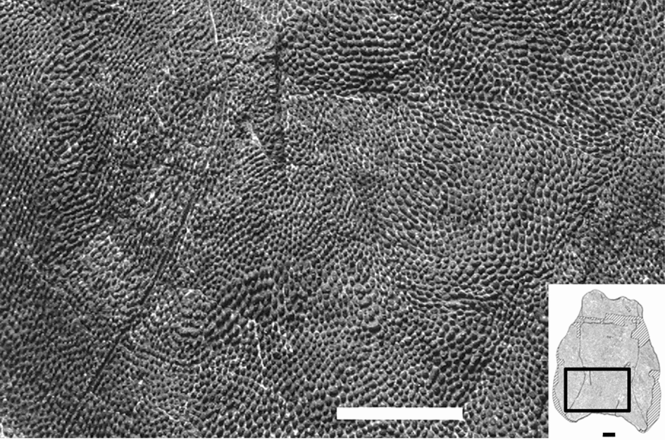
Ornamentation. On the skull this mainly consists of minute elongated tubercles, some of which merge into short ridges reaching a few millimetres in length. The tubercles do not resemble those of the Phyllolepididae (e.g. Austrophyllolepis in Long, Reference Long1984, Figs 5, 19), since in the Gondwanan forms the tubercles are clearly rounded. Nevertheless, the ridge formation seems to be same in Gavinaspis and the Phyllolepididae, except for the fact that, in the latter, ridges are osseous and do not bear dentine-like tissues.
Restoration. Computerized assistance, using a distortion algorithm of Adobe® Photoshop®, was used for the restoration of Gavinaspis convergens gen. et sp. nov. diplayed in Figure 3. The midline axis is defined by the position of the supraorbital sensory line grooves on the preorbital plates, the radiation centre of the centronuchal plate and the endolymphatic ducts on the paranuchal plates. A transverse axis is indicated by the endolymphatic ducts (ventral view). These axes intersect at an angle of 85°, digitally restored to 90° to remove the oblique distortion. An inverse 5° distortion was made for the ventral view.
5. Discussion
5.a. The endolymphatic duct in the Phyllolepida
The endolymphatic duct is a tubular structure that connects the labyrinth to the external environment. It opens externally by a foramen, and internally into the saccula of the inner ear by a proximal foramen. The endolymphatic duct is long and oblique in the Arthrodira (except for the Phyllolepididae), as in Gavinaspis. An endolymphatic duct is unknown in the Phyllolepididae (except possibly for Cowralepis; see below), where an external foramen on the paranuchal plate has never been found. Consequently, some characters referring to these structures in the previous phylogenetic analyses were coded as not applicable (Dupret, Reference Dupret2004; Dupret, Goujet & Mark-Kurik, 2007). Optimization of these ‘missing data’ by the software supported a close relationship between the Phyllolepida and the Phlyctaenioidei (Dupret, Reference Dupret2004; Dupret, Goujet & Mark-Kurik, 2007), but Young (Reference Young2005b) considered this evidence of relationship as artificial (along with other character discussions mentioned below). Nevertheless, the discovery in Cobandrahlepis (Young, Reference Young2005c, Figs 3A–C, 4A–B, m.pr) and in Cowralepis (Ritchie, Reference Ritchie2005, Figs 7A–G, mp.PNu) of an anterior oblique internal mesial process on the paranuchal plate could be interpreted as a vestigial endolymphatic duct in the Phyllolepida. A very short ‘craniospinal process ridge’ on the internal side of a paranuchal plate of Austrophyllolepis youngi was also described (Long, Reference Long1984, Fig. 19A). Ritchie (Reference Ritchie2005) pointed out the lack of an external foramen for the endolymphatic duct on the paranuchal plate in Cowralepis, and suggested that the opening might have been situated in the gap between the paranuchal and centronuchal plate. However, the actual absence of an endolymphatic external pore in all other phyllolepidids suggests that its function was progressively lost, with a vestigial stage of a blind endolymphatic duct possibly present in Cobandrahlepis and Austrophyllolepis.
5.b. Which definition for the central plates?
The central plates are conspicuous in most placoderms except for the Antiarcha. As for the Phyllolepida, there is no consensus about the absence of these plates, whether they fused to the nuchal plate, or are completely lost and replaced by the nuchal plate. A third hypothesis, suggested here by Gavinaspis, has never before been proposed: the central plates are fused in a single element and the nuchal plate is lost. In order to attempt an explanation, it is necessary to consider the definition and homologies of the central plate. The central plates may be identified on the basis of three major criteria: the position of the plate, the presence of a central sensory line groove/canal, and the presence of the middle and posterior pitlines. (1) The topographic position of the plate on the skull roof may be used but though convenient, it is not accurate enough. (2) The presence of a central sensory line groove/canal is applicable in most cases but some taxa show a more or less long central sensory line groove that does not reach the central plate (e.g. the ‘Phlyctaenii’ Dicksonosteus arcticus with a very short groove, see Goujet, Reference Goujet1984; in the Antarctaspidae with a rather elongated groove as in Toombalepis tuberculata Young & Goujet, Reference Young and Goujet2003, or Yujiangolepis liujingensis, Wang, Pan & Wang, 1998 (see also Young & Goujet, Reference Young and Goujet2003), Antarctaspis mcmurdoensis White, Reference White1968 (non Denison, Reference Denison and Schultze1978); this condition has led Denison to believe in the absence of central plates in Antarctaspis mcmurdoensis). In the Macropetalichthyidae Lunaspis broilii Gross, Reference Gross1937 and the Quasipetalichthyidae Eurycaraspis incilis Liu, Reference Liu1991, this groove/canal is simply absent. (3) The presence of the middle and posterior pitlines (when visible) is the most accurate character that can be used to identify the central plates, at least in the Arthrodira. Indeed, in closely related groups like the Macropetalichthyidae, the posterior pitline can be located on the anterior paranuchal plate (e.g. Lunaspis broilii in Gross, Reference Gross1937). It is also noteworthy that some Quasipetalichthyidae display a second posterior pitline on the central plate (e.g. Eurycaraspis incilis Liu, Reference Liu1991). Nevertheless, in the known Antarctaspididae (basal Arthrodira), the anterior ends of the posterior pitlines are located around the radiation centre of the nuchal plate (see Young & Goujet, Reference Young and Goujet2003). In the latter case, only the topographic position of the central plate is used as a criterion. In other words, we face a circular argument situation, even though we consider that the most accurate definition refers to the position of the middle and posterior pitlines.
Tuberculated ornamentation of Gavinaspis convergens gen. et sp. nov. (specimen no. IVPP V 15085-1). Scale bar 1 cm.

The hypothesis that the central plates have fused into a single median element in the new taxon may be compared with the family Actinolepididae (genera Bollandaspis and Actinolepis; see Schmidt, Reference Schmidt1976; Mark-Kurik, Reference Mark-Kurik1973, Reference Mark-Kurik1985), in which the preorbital plates are fused in a single unit. This may be the case in the Phyllolepida, if we refer to the position of the posterior pitlines, but it is also challenged (yet not refuted) by the antarctaspidid skull roof pattern. Nevertheless, Graham-Smith (Reference Graham-Smith1978), based on the numerous abnormal specimens of the antiarch Bothriolepis, concluded that the sensory lines could have become anchored to different combinations of the bone rudiments (‘primordium’ of Stensiö, Reference Stensiö1947) at an early stage of the skeletogenesis, and that during later growth they were consequently drawn along different courses. Such a variable feature may then become fixed following a speciation event, and this scheme is proposed for Antarctaspididae herein: the pitline terminations would have been anchored to the nuchal plate primordium, therefore the adult pattern does not show any association between the pitlines and the central plates.
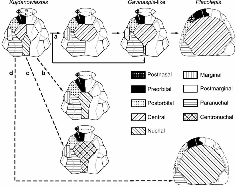
5.c. Hypotheses about processes leading to an unpaired centronuchal plate pattern (Fig. 8)
Here we consider the hypothetical transformation of the skull roof pattern from an actinolepid ancestor (Kujdanowiaspis podolica Brotzen, Reference Brotzen1934) to a phyllolepid pattern (Placolepis budawangensis Ritchie, Reference Ritchie1984).
Four hypothetical morphological transformations from a classical actinolepid condition (Kujdanowiaspis podolica) to a basal phyllolepid condition (Placolepis budawangensis), depending on the initial homology assumption about the centronuchal plate (external view). (a) The most likely hypothesis (plain line), where the process involves a fusion of the paired central plates and the loss of the nuchal plate. The other three hypotheses (dashed lines) are: (b) the process involves the loss of the central plates; (c) the process involves the fusion of central and nuchal plates; (d) the so-called postnasal plate of phyllolepids would be homologous with the central plate of other arthrodires (Young, Reference Young2005c). Not to scale. For key to abbreviations, see Table 1.

In the hypothesis favoured here (fig. 8a), the central plates fuse altogether and the nuchal plate is lost. Ideally, the posterior part of the centronuchal plate would become rather narrow or perhaps not form the posterior edge of the skull roof, the paranuchal plates would become the most important components of the posterior part of the skull roof, and the centre of radiation of the unique central plate would be anteriorly placed as are the ends of the middle and posterior pitlines. This almost perfectly fits the pattern displayed by Gavinaspis.
If we consider a loss of the central plates (fig. 8b), all the peri-central plates would become larger (not only the nuchal plate), the centre of radiation of the nuchal plate should be at the geometric centre of the plate, the posterior part of the nuchal plate is not narrowed, and the middle and posterior pitlines may be lost (lacking anchoring points, if not attached to the nuchal primordium). This does not fit the pattern of Gavinaspis convergens gen. et sp. nov.
If we consider the fusion of the central and nuchal plates (fig. 8c), the posterior part of the centronuchal plate is not narrowed, and the radiation centre coincides with the geometric centre of the plate. This again does not fit the pattern displayed by Gavinaspis convergens gen. et sp. nov.
Recently, Young proposed a possible fourth homology, according to which the phyllolepid so-called ‘postnasal plate’ could be homologous to the arthrodiran central plate (fig. 8d), ‘because interpretation as the central plate of other placoderms seems equally likely using the two criteria of relationship to adjacent bones and possession of sensory grooves’ (Young, Reference Young2005c, p. 262). This hypothesis, though alluring, is not confirmed by the pattern in Gavinaspis convergens gen. et sp. nov.
5.d. Remarks concerning the ornamentation
There are different hypotheses of homology about this character, all leading to interesting discussions. Before the description of Wuttagoonaspis (Ritchie, Reference Ritchie1973), its ridged plates were attributed to phyllolepid remains (e.g. Rade, Reference Rade1964, on the authority of Prof. Tør Orvig). Despite the presence of ridges in Wuttagoonaspis, Ritchie considered this genus only distantly related to the Phyllolepida. Dupret (Reference Dupret2004) regarded the ridged ornament as a non-homologous character; the alternative hypothesis (e.g. Long, Reference Long1984; Miles, Reference Miles1971; Young, Reference Young1980; Young & Goujet, Reference Young and Goujet2003) is that the ridged ornamentation is one of the synapomorphies shared by Wuttagoonaspis and the Phyllolepida. However, the Phyllolepida and Wuttagoonaspis are not the only placoderms that show a ridged pattern. In many groups of Arthrodira or related forms, at least one genus displays this kind of ornamentation (possibly together with tubercles): Actinolepis (Mark-Kurik, Reference Mark-Kurik1973, Reference Mark-Kurik1985) and Baringaspis (Miles, Reference Miles1973) for the non-phyllolepid ‘Actinolepidoidei’, Diadsomaspis (Gross, Reference Gross1937) for the phlyctaeniids, Holonema (e.g. Newberry, Reference Newberry1889) for the Brachythoraci, and Lunaspis for the Petalichthyida (Gross, Reference Gross1961). It is also noteworthy that the tissular structures are different in the selected groups; in some Phyllolepida, the ridges are osseous and lack dentinous tissue, whereas, as in the forms cited above, the ridges are composed by bone and semi-dentine like any tubercle. Nevertheless, the histological structure of the tubercles of Austrophyllolepis and Placolepis remains unknown (see Long, Reference Long1984, Fig. 19B; Ritchie, Reference Ritchie1984, Figs 6B, 7A, C, E–G, K, M). Moreover, the dermal ridges in Wuttagoonaspis display a different shape than those of phyllolepids: in the former the ridges are thicker and display small nipple-like structures all along.
5.e. Phylogenetic analysis
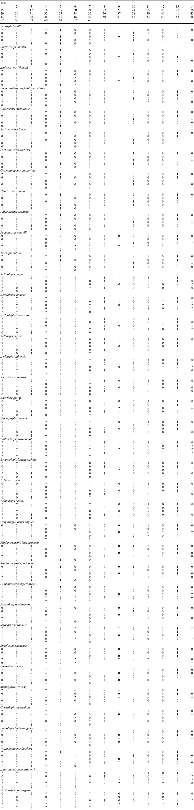
The ingroup is composed of 38 taxa, among which are the best known actinolepids (nineteen species), some phlyctaeniids (seven species), brachythoracids (three species), basal arthrodires (Wuttagoonaspis fletcheri and Antarctaspis mcmurdoensis) and phyllolepids (five species, including Gavinaspis convergens gen. et sp. nov. and Cowralepis mclachlani Ritchie, Reference Ritchie2005). The outgroup is composed of the petalichthyids Lunaspis broilii Gross, Reference Gross1937 and Eurycaraspis incilis Liu, Reference Liu1991. The complete list of the 63 characters is given in Appendix 1 (character state codings do not indicate any a priori primitive or derived condition); these are scored for the 38 taxa in the data matrix in Appendix 2.
In order better to accommodate the various hypotheses about the centronuchal plate homologies, three different coding strategies have been attempted. (1) The centronuchal plate consists of the fused central plates and the nuchal plate is lost; in this coding, every character referring to the nuchal plate is coded as not applicable for phyllolepids and Gavinaspis; there are thus 63 characters in the matrix. (2) The centronuchal plate consists of the nuchal plate and the central plates are lost; in this coding, every character referring to the central plates is coded as not applicable for phyllolepids and Gavinaspis; there are thus 62 characters in the matrix. (3) The centronuchal plate consists of the fused central and nuchal plates; there are thus 62 characters in the matrix.
Each data matrix was treated with Nexus Data Editor 0.5.0 (Page, Reference Page2001), and the analysis performed by P.A.U.P. 4.0.b10 (Swofford, 1989–1997). The heuristic search logarithm was used, because of the large number of taxa. All characters were unordered and unpolarized a priori, and the trees were rooted with the two petalichthyid taxa as outgroup. Wagner optimization was used because it accepts both convergences and reversions. The optimization of missing data was carried out using ACCTRAN (favouring reversions).
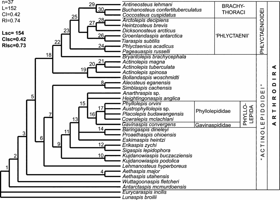
The resulting trees are different depending on the chosen coding for the centronuchal plate (coding 1: n=37, L=152, CI=0.4211, RI=0.7381; coding 2: n=38, L=149, CI=0.4228, RI=0.7346; coding 3: n=37, L=152, CI=0.4145, RI=0.7351). It is nevertheless noteworthy that the three strict consensus trees obtained have the same topology. In this respect, the results discussed below only refer to the strict consensus tree obtained with the first coding (Fig. 9; L=154, CI=0.4156, RI=0.723).
Phylogenetic relationships among Arthrodira, and classification of the Phyllolepida.

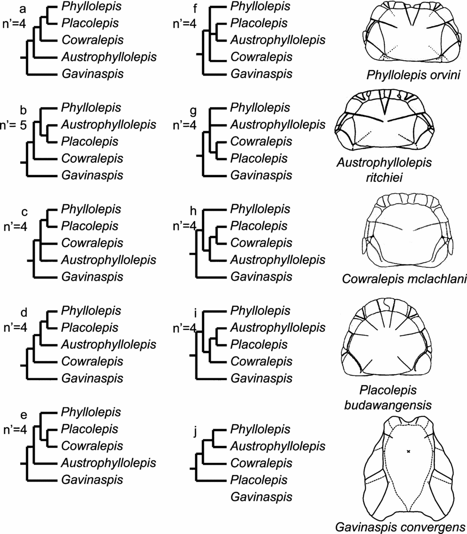
(a–i) The nine possible intra-Phyllolepida topologies encountered among the 37 equally parsimonious trees obtained from the entire data matrix used here. (j) Ritchie's (2005) phylogeny.

In the strict consensus tree, the ‘Actinolepidoidei’ are paraphyletic (nodes 1 to 22) but their internal relationships are different from those published earlier (Dupret, Reference Dupret2004; Dupret, Goujet & Mark-Kurik, 2007). The Arthrodira are monophyletic (node 1). Antarctaspis and Wuttagoonaspis are still the successive most inclusive arthrodire taxa (nodes 1 to 2). The genus Aethaspis appears here as monophyletic (node 4). The family Actinolepididae (node 19) is still phylogenetically independent of other actinolepids (closer to Bryantolepis and the Phlyctaenioidei than to other actinolepids), and Aleosteus is the sister group of Simblaspis in this new scheme (node 8). The Phlyctaenioidei are a monophyletic group (node 23), but the ‘Phlyctaenii’ appear paraphyletic, because the family Phlyctaeniidae (Phlyctaenius acadicus and Pageauaspis russelli) shares many symplesiomorphies with the more basal actinolepids (nodes 23 to 25).
The Phyllolepida are monophyletic (node 16), and in this new consensus tree they are not closely related to the Phlyctaenioidei. It is noteworthy that the deletion of both Gavinaspis and the family Phlyctaeniidae yields a close relationship between the Phyllolepida and the Phlyctaenioidei, similar to that proposed by Dupret (Reference Dupret2004). Except for the sister-group relationship between Gavinaspis and the family Phyllolepididae, the internal relationships of the Phyllolepididae are not resolved (polytomy node 17); this might be due to the large number of characters involved in this analysis. Among the 37 equiparsimonious trees, nine intra-Phyllolepida topologies are encountered (fig. 10a–i), but none of them corresponds to Ritchie's phylogeny hypothesis for the group (Ritchie, Reference Ritchie2005, p. 225, Fig. 20B; fig. 10j). Ritchie considered that there was a gradual cline between the basal Placolepis and the more derived Phyllolepis, and that the intermediate forms of this cline (Cowralepis and Austrophyllolepis) illustrate a progressive modification of the paranuchal plate (and subsequently the centronuchal plate) shape and/or of the main lateral sensory line groove, inducing a progressive loss of contact between the centronuchal and marginal plates (primitive feature displayed by Placolepis) that is replaced by a contact between the postorbital and paranuchal plates (Austrophyllolepis and Phyllolepis). Even though our topologies do not mirror this cline, we agree with Ritchie's hypothesis, and we suggest that a phyllolepid data matrix at the species level should be attempted (thus avoiding homoplasic interferences from other taxa). Nonetheless, the clear sister-group relationship between Gavinaspis and the Phyllolepididae, as well as the peculiar skull roof pattern of the former, support the erection of a new distinct, but monogeneric, family for Gavinaspis: the Gavinaspididae fam. nov.
5.f. Palaeobiogeographic and palaeogeographic implications
It is commonly considered that the Devonian palaeoglobe was divided into two major landmasses by the Rheic and Palaeo-Tethys oceans, with a northern Laurussian and a southern Gondwanan landmass group. One of the exceptions consists of the main body of the China palaeocontinent, belonging to neither Gondwana, nor Laurussia, and hence being termed the Pan-Cathaysian landmass group (Zhu & Zhao, Reference Zhu, Zhao and Rong2006). As a consequence, a high level of vertebrate endemism is observed in South China during Early Devonian times. In the Early Devonian global reconstructions of Scotese (Reference Scotese1997), the Gondwana and Pan-Cathaysian landmass groups are placed well apart from each other.
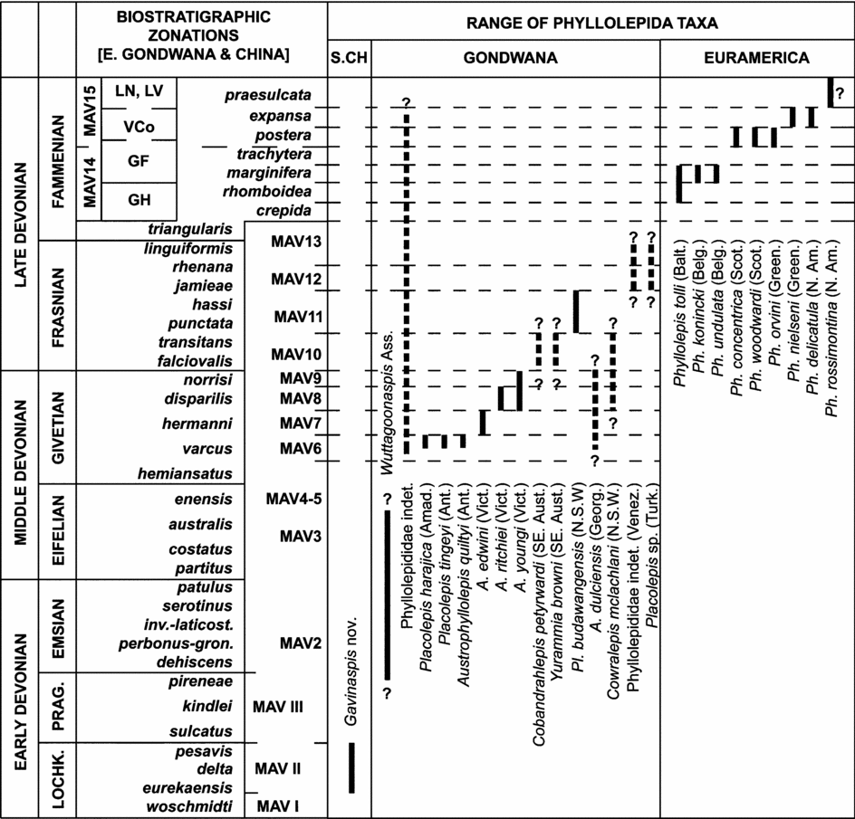
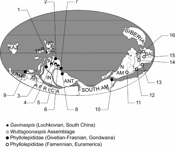
Since the hitherto most ancient known Phyllolepida have been encountered in eastern Gondwana (Placolepis harajica Young, Reference Young2005b from the Early Givetian of Amadeus Basin, central Australia; Placolepis tingeyi Young & Long, Reference Young and Long2005 and Austrophyllolepis quiltyi Young & Long, Reference Young and Long2005 from the Early Givetian of the Aztec siltstones, southern Victoria, Antarctica; see Figs 11, 12), a Gondwanan origin for the Phyllolepida was proposed (Young, Reference Young2005a). Since the new genus Gavinaspis is the oldest non-Gondwanan representative of the Phyllolepida (see phylogenetic analysis), and the Pan-Cathaysian landmass is isolated from the Gondwanan one (before the Emsian), this hypothesis seems untenable. This also challenges the hypothesis of a more or less large ocean between the South China block and the northern Gondwanan margin proposed in most palaeogeographic reconstructions (e.g. Cocks & Torsvik, Reference Cocks and Torsvik2002; Torsvik & Cocks, Reference Torsvik and Cocks2004), at any rate from the Givetian (Middle Devonian), as far as Phyllolepida are concerned. When studying other early vertebrate groups (e.g. Sarcopterygii), a shallow marine connection between South China and northeastern Gondwana may have occurred as late as the Pragian–Emsian boundary (E'Em bioevent). Indeed, before the E'Em bioevent, the Placodermi and other early vertebrate taxa (e.g. Galeaspida) show a well-marked endemism in South China. Most of them became extinct during and after this episode, most probably because of the arrival of, and the subsequent competition with, eastern Gondwanan forms (Zhu, Reference Zhu, Blieck and Turner2000). This event is proposed for dating the southward migration of Phyllolepida into eastern Gondwanan margins.
Summary of known and supposed stratigraphic ranges (Lochkovian–Famennian) for the Wuttagoonaspida and Phyllolepida (Arthrodira) of South China (S.CH), Gondwana and Euramerica (for Gondwana: Amad. – Amadeus Basin; Ant. – Antarctica; Georg. – Georgina Basin; N.S.W. – New South Wales; SE. Aust. – southeastern Australia; Turk. – Turkey; Venez. – Venezuela; Vict. – Victoria. For Euramerica: Balt. – Baltic States; Belg. – Belgium; Green. – Greenland; N. Am. – Northern America (Pennsylvania, USA); Scot. – Scotland (UK). Supposed stratigraphic ranges indicated by dashed lines with a question mark. Australian stratigraphic range after Young, Reference Young2005a, Reference Youngb; Young & Long, Reference Young and Long2005) (updated after Young, Reference Young and Long1993, Reference Young1999). Conodont zonation from Zhu, Wang & Wang, 2000 (Lochkovian) and Talent et al. Reference Talent, Mawson, Aitchison, Becker, Bell, Bradshaw, Burrow, Cook, Dargan, Douglas, Edgecombe, Feist, Jones, Long, Phillips-Ross, Pickett, Playford, Rickards, Webby, Winchester-Seeto, Wright, Young, Zhen, Wright, Young and Talent2000 (Pragian–Famennian). Lochkovian macrovertebrate assemblages (MAV I–III, left sided of the column) after Zhu, Wang & Wang, 2000; Pragian–Famennian macrovertebrate assemblages (MAV2–15, right side of the column), miospore (GH, GF, VCo, LN, LV) and conodont zone is approximate (modified after Young, Reference Young, Young and Laurie1996; Young & Turner, Reference Young, Turner, Blieck and Turner2000; after Young, Reference Young2005a, Reference Youngb; Young & Long, Reference Young and Long2005).

Global distribution pattern for the Phyllolepida and Wuttagoonaspida, plotted on a Devonian palaeogeographic reconstruction (after Zhu & Zhao, Reference Zhu, Zhao and Rong2006). Gavinaspis is indicated by a black star; the Wuttagoonaspis assemblages by white stars; Givetian–Frasnian Phyllolepida by closed circles; Famennian Phyllolepida (genus Phyllolepis) by open circles. Chinese Lochkovian locality: 1 – Qujing (Yunnan, South China); Gondwanan Givetian–Frasnian localities: 2 – Townsville area (Queensland, Australia: Young, Reference Young2005b); 3 – Carnarvon Basin (Western Australia: J. A. Long, unpub. data, in Young, Reference Young2005b); 4 – Amadeus Basin and 5 – Georgina Basin (central Australia: Young, Reference Young2005a, Reference Youngb; Young & Goujet, Reference Young and Goujet2003); 6 – West-central New South Wales (Australia: Hills, Reference Hills1931, Reference Hills1936; Ritchie, Reference Ritchie1973; Young, Reference Young and Long1993, Reference Young1999); 7 – Braidwood–Pambula–Mount Howitt (south east Australia: Long, Reference Long1984; Ritchie, Reference Ritchie1984; Young, Reference Young1983); 8 – Transantarctic Mountains, southern Victoria land (Antarctica: Young & Long, Reference Young and Long2005); 9 – Upper Antalya Nappe (Western Lycian Taurus, Turkey); 10 – Sierra de Perija (Venezuela: Young & Moody, Reference Young and Moody2002; Young, Moody & Casas, 2000); Euramerican Famennian localities: 11 – Virginia–Pennsylvania (USA: Daeschler, Frumes & Mullison, 2003; Lane & Cuffey, Reference Lane and Cuffey2005); 12 – Scotland (UK: Agassiz, Reference Agassiz1844; Woodward, Reference Woodward1915); 13 – Wallonia (Belgium: Leriche, Reference Leriche1931; Lohest, Reference Lohest1888); 14 – East Greenland (Heintz, Reference Heintz1930; Stensiö, Reference Stensiö1934, Reference Stensiö1939); 15 – Baltic States (Vasiliauskas, Reference Vasiliauskas, Grigialis and Karatajute-Talimaa1963); 16 – Timan (Russia: Esin et al. Reference Esin, Ginter, Ivanov, Lebedev, Luksevics, Avkhimovich, Golubtsov and Petukhova2000). ANT – Antarctica; AR – Arabia; ARM – Armorica; AU – Australia; BAL – Baltica; G – Greenland; IN – India; KAZ – Kazakhstan; N AM – North America; NC – North China Block; SC – South China Block; SOUTH AM – South America; TAR – Tarim.

Although the Wuttagoonaspida and Phyllolepida are not closely phylogenetically related, it is remarkable that the most ancient ‘Wuttagoonaspis assemblage’ occurrence in Australia is dated as end-Pragian or Early Emsian (Fig. 11; Young, Reference Young2005b, Fig. 4; Young, Reference Young2005a, Fig. 5). Nevertheless, a recent fieldtrip in Zhaotong (Pragian, Yunnan, South China) yielded an incomplete skull roof that could belong to the Wuttagoonaspida; this specimen is presently under study. In other words, both the Phyllolepida and Wuttagoonaspida could have originated from South China, and would have invaded Gondwana during the E'Em bioevent.
If one can consider that the Chinese origin for the Phyllolepida is settled, the dispersal process of this group is more problematic. It can nevertheless be divided into four steps.
(1) Invasion into Gondwana took place during the E'Em event (without evidence of a later return into China since this group is still unknown in younger Chinese strata; the latter remark can be explained by biological considerations, Gavinaspis being a rather bigger and non-flattened organism than Gondwanan phyllolepids and hence being probably more nektonic than sub-benthic, a ‘come-back’ to China was perhaps impossible for the latter).
(2) Eastward (to Venezuela, Fig. 12) and westward (to Turkey, Fig. 12) dispersal occurred in Gondwana until the Late Frasnian. It is also possible, though without fossil evidence, that the Turkish forms dispersed more westwardly to South America (Fig. 12).
(3) An invasion northward into Laurussia/Euramerica occurred during the Frasnian–Famennian boundary. The only hitherto known Euramerican Phyllolepida are dated as Famennian, and all belong to the genus Phyllolepis Agassiz, Reference Agassiz1844. It is noteworthy that not only the Phyllolepida invaded Euramerica at the end of the Frasnian or the beginning of the Famennian. A northward invasion by the Groenlandaspididae (Placodermi, ‘Phlyctaenii’), the Megalichthyidae and the Rhizodontida (Sarcopterygii) is coeval with a southward dispersal of the genera Asterolepis (Placodermi, Antiarcha) and Holoptychius (Sarcopterygii) (Dupret, Clément & Janvier, 2005). The discovery of similar Frasnian vertebrate faunas in Turkey (Western Lycian Taurus: Janvier, Reference Janvier1983; Janvier, Clément & Cloutier, 2007), in Colombia (Cuche Formation, Department of Boyacá: Janvier & Villarroel, Reference Janvier and Villarroel2000) and in Venezuela (Sierra de Perijá: Young & Moody, Reference Young and Moody2002; Young, Moody & Casas, 2000) sheds light on two possible dispersal routes. The main question is then which dispersal route may have been followed for this invasion, that is, whether it is through a Middle Eastern or a South American route. Comparisons between the phylogenies of the different vertebrate groups involved should be considered in order to supply an answer, although it is noteworthy that only the Colombian fauna includes some Euramerican forms (Asterolepis and Holoptychius); moreover, the Turkish groenlandaspidids may be endemics and would not have led to any descent outside this area (Dupret, Clément & Janvier, 2005). Consequently, a South American route is preferred here. A last, but unlikely, possibility would be a circum-Rheic Ocean migration between Turkey and South America slightly before and during the Frasnian–Famennian faunal interchange.
(4) The Famennian phyllolepid invasion into Euramerica is confusing (Figs 11, 12). Indeed, it is noteworthy that the earliest Euramerican phyllolepids are encountered in the Baltic States (close to the palaeoequator), whereas the latest are encountered in Pennsylvania (close to the southern palaeotropic). Paradoxically, the supposedly most primitive forms (morphologically close to Placolepis and Austrophyllolepis) are encountered in Belgium (Young, Reference Young2005a, p. 207) and North America (Lane & Cuffey, Reference Lane and Cuffey2005).
6. Conclusions and summary
The new form Gavinaspis convergens gen. et sp. nov. demonstrates the systematic and palaeogeographic origins of the suborder Phyllolepida. It is dated from the Late Lochkovian of Yunnan (South China) and provides some interesting anatomical characters, intermediate between a classical actinolepid arthrodire and a more derived phyllolepid. Its peculiar centronuchal plate permits the suggestion of a new hypothesis concerning the formation of this dermal element, that is, the fusion of the central plates into a single element along with with the loss of the nuchal plate. Its phylogenetic relationship with other phyllolepids leads to the erection of the new family Gavinaspididae, the sister family to the Gondwanan and Euramerican Phyllolepididae. Its early age suggests a possible South Chinese origin for the Phyllolepida, rather than the Gondwanan origin previously proposed. The invasion of Gondwana by the Phyllolepida is likely to have occurred at the end of the Pragian, during the E'Em bio-event, together with other early vertebrate migrations.
Further investigations of the Late Frasnian vertebrate faunas and the Euramerican Phyllolepis species records are needed, in order to elaborate more precise dispersal processes.
Acknowledgements
We thank Zhao Wenjin and Jia Liantao (IVPP, P.R. China) for the help in the field and with photography, and Xiong Cuihua for the preparation. Edouard Poty (Université de Liège, Belgium) permitted V. D. to access the Famennian phyllolepid collections. Wang Junqing (IVPP) added interesting remarks concerning the Early Devonian Arthrodires of Yunnan. John Long (Museum Victoria, Australia) and Gavin Young (Australian National University, Canberra ACT, Australia) contributed motivating discussions concerning Phyllolepida and basal Arthrodira during the IPC 2006 in Beijing. Two anonymous reviewers greatly enhanced the quality of the manuscript. This work was supported by the Major Basic Research Projects (2006CB806400) of MST of China, the Chinese Foundation of Natural Sciences (40332017 and 40602005), and UNESCO-IGCP 491. The authors thank the two anonymous reviewers for their constructive remarks and corrections.
Appendix 1. List of characters
1. Link between the two neurocranial components (ethmoid and postethmoid parts):
0. no link (‘loose nose’ fishes)
1. fusion (by means of either osseous trabecles, or complete fusion)
2. Position of the foramen for the hyomandibular branch of the facial nerve (fVIIHm) in relation to the anterior postorbital process:
0. foramen in the distal part of the anterior postorbital process
1. foramen in a proximal and posterior position to the process
3. Neurocranial supraorbital process:
0. absent
1. present
4. Neurocranial basal process:
0. absent
1. present
5. Rostral, pineal or rostropineal plates:
0. absent
1. present
6. Pineal or rostropineal plate separates the preorbital plates:
0. no
1. yes
7. Rostral and pineal plates fused into a single rostropineal component:
0. no
1. yes
8. Preorbital plates show an embayment for the insertion of the pineal or the rostropineal plate:
0. no, or very shallow
1. yes, very deep
9. Pineal or rostropineal plate fused to the skull roof:
0. no
1. yes
10. Postnasal plates fused to the preorbital plates:
0. yes
1. no
11. Position of the orbits in the skull roof:
0. dorsal
1. lateral
12. Preorbital plates:
0. separate
1. fused
13. External morphology of the sensory line system:
0. canals with external pores
1. grooves
14. Supraorbital sensory lines:
0. separate
1. meet posteriorly
15. Infraorbital and main sensory lines grooves run along the mesial margin of the marginal plate:
0. no
1. yes
16. Central plates:
0. fused into a centronuchal plate
1. individualized paired elements
17. Shape of suture between central plates:
0. straight
1. sinuous
18. Pineal (or rostropineal) plate contacts the central plates:
0. no
1. yes
19. Posterior edge of the preorbital plates indents the anterior edge of the central plates:
0. no
1. yes
20. Contact between central and preorbital plates:
0. yes
1. no
21. Contact between central and marginal plates:
0. yes
1. no
22. Contact between postorbital and paranuchal plates:
0. yes
1. no
23. Nuchal plate:
0. absent
1. present
24. Nuchal plate separates the central plates:
0. no
1. yes
25. Contact between orbits and central plates:
0. no
1. yes
26. Preorbital plates are part of the orbital margin:
0. yes
1. no
27. Central sensory line groove extends from the postorbital plate to the radiation centre of the central plates:
0. no
1. yes
28 Posterior pitline present on both central and paranuchal plates:
0. anterior and posterior ends clearly connected
1. anterior and posterior ends not connected, implying a superficial course
29. Central sensory line groove:
0. absent
1. present
30. Postmarginal plate:
0. absent
1. present
31. Morphology of the anterior external nuchal – central plates contact suture:
0. nuchal plate indents the central plates
1. straight suture
32. Contact between the pineal and nuchal plates:
0. no
1. yes
33. Number of paranuchal plates pairs:
0. one pair
1. two pairs
34. Occipital cross commissure:
0. on both nuchal and paranuchal plates
1. only on paranuchal plates
35. Posterolateral edge of the paranuchal plates:
0. convex
1. concave
36. Posterior process of the paranuchal plate behind the nuchal plate (external side):
0. absent
1. present
37. Position of the external foramen for the endolymphatic duct:
0. well anteriorly to the posterior edge of the paranuchal plate, or on the anterior paranuchal plate for the Petalichthyida
1. near to the posterior edge of the paranuchal plate
38. Type of exoskeletal dermal craniothoracic articulation:
0. actinolepid ‘sliding neck joint’
1. ginglymoid phlyctaenioid type
2. ‘spoon-like’ petalichthyid type
39. Dermal articular condyle of anterior dorsolateral plates:
0. close together
1. well apart
40. Ventral keel on the internal side of the median dorsal plate:
0. absent
1. present
41. Unornamented (overlapped) area on the anterior margin of the median dorsal plate:
0. absent
1. present
42. The unornamented zone on the anterior margin of the median dorsal plate is:
0. simple
1. double
43. Extrascapular plate:
0. absent
1. present
44. Dorsolateral groove (for an accessory sensory line) on the anterior dorsolateral plate:
0. absent
1. present
45. Posterolateral plate:
0. absent
1. present
46. Pectoral notch of the anterior ventrolateral plate:
0. shallow
1. deep
47. Prepectoral process of the anterior ventrolateral plate:
0. short
1. long
48. Anteroventral plates:
0. absent
1. present
49. Anterolateral and anterior ventrolateral plates connected behind the pectoral fenestra:
0. no
1. yes
50. Anterior median ventral plate:
0. absent
1. present
51 Posterior median ventral plate:
0. absent
1. present
52. Mutual overlap of posterior ventrolateral plates:
0. simple overlapping
1. sinusoidal/double overlapping
53. Spinelets on the mesial side of spinal plate:
0. absent
1. present
54. Postmedian dorsal plates:
0. absent
1. present
55. Width/length ratio of the preorbital plates:
0. (W/L) > 0.5
1. (W/L) ≤ 0.5
56. Central plates length ratio to the skull roof length (from the anterior edge of preorbital plates to posteriormost edge of the skull roof):
0. (LC/LSR) < 45%
1. (LC/LSR) ≥ 45%
57. Length/width ratio of the nuchal plate:
0. (L/W) ≤ 1.5
1. (L/W) > 1.5
58. Length/width ratio of the median dorsal plate:
0. (L/W) < 1.5
1. (L/W) ≥ 1.5
59. Length/height ratio of the anterior dorsolateral plate:
0. (L/H) < 1
1. (L/H) ≥ 1
60. Length/height ratio of the posterior dorsolateral plate:
0. (L/H) < 2
1. (L/H) ≥ 2
61. Angle between interolateral and spinal plates:
0. angle < 110°
1. angle ≥ 110°
62. (LSp beard by AVL/LSp) ratio = RSp:
0. RSp < 60%
1. RSp ≥ 60%
63. Length/width ratio of posterior ventrolateral plates:
0. (L/W) < 1.5
1. (L/W) ≥ 1.5
Appendix 2. Data matrix