Introduction
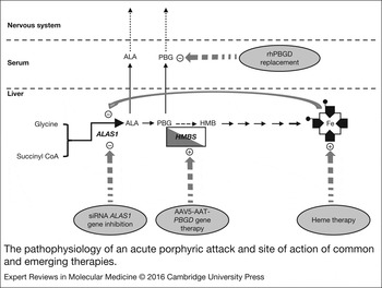
Acute intermittent porphyria (AIP, MIM 176000) is an autosomal dominant metabolic disease caused by a genetic deficiency of porphobilinogen deaminase (EC 4.3.1.8; PBGD) or hydroxymethylbilane synthase (HMBS, HGNC: 4982), the third enzyme of the heme synthesis pathway (Refs Reference Puy, Gouya and Deybach1, Reference Harper and Sardh2, Reference Kauppinen3). AIP is characterised by acute attacks triggered by metabolic, hormonal and environmental factors. Such factors have in common their ability to reduce the regulatory heme pool in the liver resulting in the strong up-regulation of the first and rate-limiting enzyme of the hepatic heme synthesis pathway, 5′-aminolevulinate synthase 1 (ALAS1, EC 2.3.1.37, HGNC: 396) (Fig. 1). As a consequence, the production of porphyrin precursors aminolevulinic acid (ALA) and porphobilinogen (PBG) is highly induced in porphyric livers, and the plasma concentrations of both molecules are markedly increased (Refs Reference Meyer, Schuurmans and Lindberg4, Reference Kauppinen and Mustajoki5). Clinical symptoms are associated with this high porphyrin precursor accumulation and include severe abdominal pain, hypertension, nausea, vomiting and constipation (Ref. Reference Harper and Sardh2). Some patients display severe neurological complications including peripheral motor neuropathy and central nervous system involvement such as acute encephalopathy, convulsions, anxiety, insomnia, depression, disorientation, hallucinations or confusional states (Refs Reference Puy, Gouya and Deybach1, Reference Kauppinen3).
The pathophysiology of an acute porphyric attack and site of action of common and emerging therapies. An acute attack is triggered in HMBS/PBGD mutation carriers under conditions leading to increased hepatic heme demand. The biochemical feature is a high induction of hepatic ALAS1 activity associated with high production of potentially neurotoxic porphyrin precursors (ALA and PBG). Current Standard of Care in AIP is intravenous heme administration, which provides exogenous heme for the negative feedback inhibition of ALAS1, decreasing further ALA and PBG production. rhHMBS replacement therapy induces the rapid metabolisation of the PBG metabolite accumulated in the plasma, but its administration results in little clinical efficiency because it does not prevent the production of new precursors. PBGD-gene therapy induces a very long-term restoration of the heme synthesis pathway. However, in order to exert tangible clinical effects, it would be necessary to increase the vector dose and/or to develop more efficient vectors. The siRNA ALAS1 generates a durable blockade of hepatic ALAS1 protein expression (more than 40 days). Further studies are needed to understand whether reduced heme supply could modify the excretion of hydrophobic and toxic compounds, change drug pharmacokinetic properties and increase the sensitivity to other triggering factors associated with acute attacks. ALA, delta-aminolevulinic acid; ALAS1, delta-aminolevulinic acid synthase; siRNA ALAS1, interfering RNA-mediated silencing of hepatic ALAS1; HMB, hydroxymethylbilane; PBG, porphobilinogen; PBGD, porphobilinogen deaminase or HMBS, hydroxymethylbilane synthase; rhPBGD, recombinant human PBGD, AAV5-AAT-PBGD, adeno-associated virus vector serotype 5 leading to liver specific expression of the human HMBS protein.

AIP is the most common of the acute porphyrias (Ref. Reference Elder6). The prevalence of an HMBS mutation can be high (1/1675) (Ref. Reference Nordmann7) but the clinical penetrance in Europe is approximately 5.4/ million (1/185 000) (Ref. Reference Elder6). Retrospective studies (Refs Reference Kauppinen and Mustajoki5, Reference von und zu Fraunberg8) revealed a decreasing incidence of new acute attacks over recent decades, probably due to a better understanding of the natural history of the disease that helped to identify triggering factors, family screening and preventive counselling, rapid and specific diagnostic tests, which avoid delays in diagnosis and availability of specific treatments (Refs Reference Marsden9, Reference Bonkovsky10).
Treatment and prevention of porphyric attacks
Current Standard of Care for acute attacks is based on heme therapy and carbohydrate loading. Intravenous administration of heme (Normosang® heme arginate in Europe and South Africa and Panhematin® hemin in USA, Recordati Rare Diseases) replenishes the hepatocyte heme pool and down-regulates ALAS1, leading to decreased production of porphyrin precursors and improvement of the clinical condition (Ref. Reference Aarsand, Petersen and Sandberg11). Heme controls hepatic ALAS1 by down-regulating ALAS1 transcription but also by inducing mRNA destabilization or by blocking the mitochondrial import of the mature enzyme (Ref. Reference Ajioka, Phillips and Kushner12).
One limitation of heme therapy is that the effect on reducing plasma ALA and PBG levels is delayed 2–3 days. Indeed, in a double-blind, placebo-controlled trial performed with 12 patients, in which heme treatment was delayed 2 days, no statistical benefit was shown (Ref. Reference Anderson13). For this reason, heme therapy should be given without delay in severe acute attacks and maintained during a period of 4 days (3–4 mg/kg heme/day). This is the current procedure in known HMBS mutation carriers. However, there is usually a delay in AIP diagnosis in unknown HMBS mutation carriers, holding back the introduction of the heme therapy.
A high carbohydrate diet or intravenous glucose administration also inhibits hepatic ALAS1 transcription (Ref. Reference Handschin14), but heme therapy is more effective than glucose in reducing ALA and PBG accumulation (Refs Reference Puy, Gouya and Deybach1, Reference Bonkovsky10). There is a general consensus that treatment with glucose infusion may be sufficient in milder attacks although it does not lead to a significant decrease in porphyrin precursor overproduction. This is probably because intracellular regulatory heme pool levels are not replenished and thus ALAS1 expression is reinduced soon after.
Most symptomatic patients have only one attack, but around 5% of women and 3% of men experience recurrent hyper-activation of the hepatic heme synthesis pathway (Ref. Reference Elder6). Endocrine factors and steroid hormones, in particular, are the most important precipitating factors in women. Some women develop premenstrual attacks, which can be treated by the administration of gonadotropin-releasing hormone (GnRH) analogues (Refs Reference Harper and Sardh2, Reference Anderson15). However, in a recent observational survey, the recurrence of acute attacks persisted in 11 of 15 women treated with GnRH agonists (Ref. Reference Marsden9). In these cases and those women in whom the relationship between recurrent acute attacks and menstrual cycle phase is unpredictable, prophylactic heme therapy should be considered the alternative (Ref. Reference Stein16). Among the methods for preventing acute attacks, heme therapy was reported by patients to be the most effective in reducing episodes of abdominal pain and improving other clinical manifestations (Refs Reference Marsden9, Reference Bonkovsky10). Many centers offered an individually tailored regimen of prophylactic heme therapy for patients with recurrent porphyric crises.
While intravenous heme therapy is generally well tolerated (only a few patients complain of headaches or pyrexia), recurrent treatment requires replacement of venous access due to thromboembolic disease and increases the risk of liver fibrosis (Ref. Reference Willandt17) and iron overload in the liver (a heme arginate dose of 250 mg contains 22.7 mg of iron) (Refs Reference Marsden9, Reference Goetsch and Bissell18).
Although prophylactic heme is increasingly used, it is uncertain whether such a therapy can prevent accumulation of porphyrin precursors and porphyrins in subsequent events. Heme infusion has been linked to the induction of hepatic heme oxygenase 1 expression (HMOX1, EC 1.14.99.3, HGNC: 5013) (Refs Reference Doberer19, Reference Yoshida20), the key enzyme of heme catabolism, resulting in the reduction of regulatory heme pool levels in the hepatocyte and consequently in the up-regulation of ALAS1 expression. This effect on hepatic heme catabolism reduces the therapeutic efficacy of heme therapy administration and generates the tolerance reported in some patients (Refs Reference Dover21, Reference Herrero22).
To date, the only established curative treatment for AIP is orthotopic liver transplantation (Refs Reference Dowman23, Reference Soonawalla24, Reference Yasuda25). This procedure has an excellent long-term outcome for liver-based metabolic and genetic diseases (Ref. Reference Junge26). In a report from the UK Transplant Registry based on data from 10 patients who received transplants due to acute porphyria, the survival rate was 80% (Ref. Reference Dowman23). However, the authors also reported a high percentage of hepatic artery thrombosis (40%) concluding that this procedure should be reserved for patients with severe recurrent acute attacks and greatly impaired quality of life. Indeed, advanced neuropathies may persist after liver transplantation suggesting that less invasive alternatives must be implemented before neuronal damage becomes irreversible.
Long-term complications in acute porphyrias
Among chronic medical conditions, systemic arterial hypertension and chronic kidney disease show a high prevalence in patients with AIP (Refs Reference Bonkovsky10, Reference Pallet27). In an observational study including 90 AIP subjects attended between 2010 and 2012 in US centres for porphyria, chronic kidney disease occurred in up to 29% of subjects (vs 1% in the control population group) (Ref. Reference Bonkovsky10). Indeed, the incidence of chronic renal failure (defined as eGFR < 60 ml/min/1.73 m2) was higher among symptomatic patients compared with asymptomatic HMBS mutation carriers (59% versus 11% respectively) as reported by the French Porphyria center in a recent population-based study including 74 AIP patients and 62 asymptomatic HMBS mutation carriers (Ref. Reference Pallet27). Experimental studies showed that high porphyrin precursor excretion enhances endoplasmic reticulum stress, apoptosis and epithelial phenotypic changes in proximal tubular cells (Ref. Reference Pallet27).
The development of end-stage renal disease is a devastating and life-threatening complication in AIP patients with chronic active disease (Refs Reference Unzu28, Reference Sardh29). Indeed, progressive renal insufficiency aggravates porphyria attacks (Ref. Reference Unzu28) and worsens preexisting neuropathies (Ref. Reference Wahlin30). For these patients, combined liver-kidney transplantation should be considered (Ref. Reference Wahlin30). Therefore, these data suggest that effective therapies controlling high porphyrin precursor production and excretion may significantly reduce the progression to end-stage renal disease.
Long-term complications of AIP also include hepatocellular carcinoma (HCC). This deadly tumour is the most frequent primary liver cancer and affects twice as many men as women. In the majority of cases, chronic hepatic injury is triggered by hepatitis B or C virus infection, heavy alcohol consumption and metabolic syndrome-associated non-alcoholic steatohepatitis and diabetes (Ref. Reference Bruix, Gores and Mazzaferro31). However, both retrospective (Ref. Reference Sardh32) and prospective (Refs Reference Elder6, Reference Andant33, Reference Innala and Andersson34) studies performed over the past 30 years have clearly shown a remarkably increased incidence of HCC in AIP patients compared with the general population (Ref. Reference Stewart35). Interestingly, these studies have also shown that most AIP patients with HCC did not have any of the previously mentioned aetiologic factors associated with HCC development (Refs Reference Sardh32, Reference Andant33, Reference Innala and Andersson34, Reference Bjersing, Andersson and Lithner36, Reference Andersson, Bjersing and Lithner37). Moreover, histological features such as inflammatory infiltrate, fibrosis or necrotic tissue damage, characteristic of long-standing parenchymal injury, were not commonly observed in the liver of AIP subjects with HCC (Ref. Reference Deybach and Puy38). These observations, together with the fact that HCC cases in AIP patients were predominantly found in females (as opposed to the male predominance of HCC in the general population), further support the existence of a porphyria-specific risk factor for this neoplasia (Refs Reference Innala and Andersson34, Reference Stewart35). Therefore, HCC is now fully recognised as a frequent serious late complication of AIP, and annual screening of patients with AIP for HCC has been recently recommended, in line with the surveillance implemented for other groups of liver patients at risk of HCC (Refs Reference Sardh32, Reference Innala and Andersson34).
The mechanisms of hepatocarcinogenesis in AIP are not fully understood, and there is no apparent correlation between HCC development and the presence of specific somatic mutations in the HMBS gene (Refs Reference Elder6, Reference Sardh32). Increased oxidative stress in the liver parenchyma of AIP patients has been suggested as an important pro-carcinogenic factor. Certainly, oxidative stress induced by persistent inflammation has been involved in the progression of chronic liver disease and in the development of HCC by different aetiologic agents (Ref. Reference Takaki and Yamamoto39). Mechanistically, reactive oxygen species (ROS) may cause DNA damage and mutagenesis by the direct oxidation of deoxyguanosine residues, or through the formation of DNA adducts with lipid peroxidation products (Ref. Reference Urtasun, Berasain, Avila, Albano and Parola40). In the case of AIP-related HCC, two non-mutually exclusive mechanistic hypotheses have been proposed to explain tumourigenesis, both involving the generation of oxidative stress. A number of studies have shown that ALA is a pro-oxidant molecule under physiological conditions (Ref. Reference Monteiro41). Through its auto-oxidation, ALA can produce ROS that damage DNA, as has been shown in vitro and in vivo (Refs Reference Onuki42, Reference Onuki43). Moreover, ALA can also cause oxidative damage in liver mitochondria, inducing structural and functional alterations in this organelle (Refs Reference Hermes-Lima44, Reference Hermes-Lima45, Reference Laafi46, Reference Homedan47, Reference Heilmann48). Further evidence on the role of excessive ALA accumulation and the risk of HCC development over time comes from the higher proportion of patients with repeated acute attacks, and thus higher ALA levels that are found among those developing the malignancy (Refs Reference Sardh32, Reference Innala and Andersson34, Reference Lithner and Wetterberg49). Although additional studies are needed to fully demonstrate the hepatocarcinogenic capacity of ALA, the observations summarised above suggest that therapies effective at controlling the production of this metabolite may significantly reduce the risk of HCC development over the AIP patient's lifetime. These therapies could also be applied to asymptomatic HMBS mutation carriers that still display high levels of ALA excretion, as these patients are also at risk of developing HCC although at later stages than symptomatic AIP individuals (Ref. Reference Andant33).
The second mechanistic hypothesis on the development of HCC in AIP relates to the impaired synthesis of heme. The defective enzymatic activity of mutant HMBS leads to a reduced availability of heme, which is an essential cofactor for many enzymes involved in antioxidant defence, as well as in xenobiotic metabolism and mitochondrial respiration (Ref. Reference Karim50). Moreover, it has recently been shown that hemin, the oxidative form of heme, may have anti-tumourigenic effects (Ref. Reference Lee51), while older reports had already shown the impairment of heme synthesis in cancer models and predicted the pro-tumourigenic consequences of this defect (Ref. Reference Batlle52). Clearly, further experimental studies are required to cogently demonstrate a direct implication of defective heme synthesis in AIP-associated HCC. However, a recent study identified somatic mutations in the HMBS and protoporphyrinogen oxidase (PPOX, EC 1.3.3.4, HGNC: 9280) genes in the HCC tissues of two patients with variegate porphyria and AIP respectively that were located in trans to the respective germline mutations (Ref. Reference Schneider-Yin53). These somatic mutations were not found in HCC tissues from ten non-porphyria associated HCCs. The authors hypothesised that PPOX and HMBS genes might act as tumour suppressors in liver cancer. Thus, somatic second-hit mutations could result in nearly complete inactivation of these enzymes and an active development of HCC (Ref. Reference Schneider-Yin53). However, analysis of two independent sets of gene expression microarray data in liver samples from patients with HCC involving a total of 500 subjects (Refs Reference Lamb54, Reference Roessler55) showed that the mRNA levels of these two genes were induced about two-fold in the tumour tissue when compared with the adjacent non-tumour area. These data would be at variance with the hypothesis of a tumour suppressive role for PPOX and HMBS genes in hepatocarcinogenesis. Nevertheless, the observations of Schneider-Yin et al. (Ref. Reference Schneider-Yin53) are indeed thought-provoking findings. However, HCCs are heterogeneous tumours from a molecular perspective, accumulating numerous passenger mutations during their development along with profound changes in gene expression (Ref. Reference Schulze56). It would be interesting to explore whether these or functionally equivalent somatic mutations, could be detected in preneoplastic or early neoplastic lesions in the liver of these porphyric patients.
Emerging therapies for acute intermittent porphyria
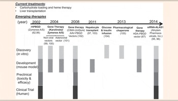
Despite the excellent long-term outcome of organ transplantation (Refs Reference Dowman23, Reference Wahlin30) research efforts must be directed toward the development of safer, non-invasive and more efficient therapies able to reverse the neurologic damage before it becomes irreversible and to reduce long-term complications associated with AIP. A summary of the emerging therapies for AIP and their clinical development status is shown in Table 1. Of these potential therapies tested in vitro and/or in experimental mouse models of AIP, three targeting precise points along the heme synthesis pathway have been or are being assayed in the corresponding clinical trials (Fig. 1). The present review provides a summary of the main results obtained in clinical trials of HMBS replacement therapy and recent gene therapy trials aiming to over-express HMBS in hepatocytes or to inhibit hepatic ALAS1 over-expression.
Chronological summary of emerging therapies for acute intermittent porphyria and current level of development.

Enzyme replacement therapy (ERT) for the treatment of acute intermittent porphyria
The supplementation with the protein that is deficient in the specific disease is a potential strategy for the treatment of monogenic genetic diseases. This concept was first demonstrated for the treatment of Gaucher disease type I, a type of lysosomal storage disease (Ref. Reference Brady57). In these diseases, the genetic defect affects an enzyme located in the lysosomes. The early success of ERT was due to a natural targeting mechanism of the lysosomal enzymes to accumulate in these organelles. This targeting mechanism is based on carbohydrates such us mannose-6-phosphate, which is recognised by ubiquitous specific receptors (Ref. Reference Kornfeld58). ERT has been refined and currently several enzymes are commercially available for the treatment of different lysosomal storage diseases such as mucopolysaccharidosis I and VI, Fabry disease, Pompe disease, Gaucher disease and Hunter syndrome. The concept has also expanded to other dysfunctions. Pancreatic enzyme replacement is indicated for the treatment of exocrine pancreatic insufficiency due to cystic fibrosis or other conditions (Ref. Reference Somaraju and Solis-Moya59). Finally, ERT is also approved for the treatment of adenosine deaminase deficiency and some urea cycle disorders (Ref. Reference Shemesh60).
The clinical experience accumulated in these three decades of development has demonstrated that a successful ERT strategy must fulfill two requirements:
-
(i) A production and purification method should be available to produce high quantities of a highly active and purified enzyme. Posttranslational modifications are important for the targeting properties and therefore the selection of the type of cells or microorganisms for the production is a critical issue.
-
(ii) The enzyme must reach the subcellular compartments in the specific organ or organs affected by the disease where the toxic metabolites accumulate. If the natural proteins do not have such a tropism, the protein must be engineered to provide the specific targeting. Bone and brain represent a challenge for the development of ERT as the enzymes needed do not have easy access to these tissues.
All these clinical successes fostered the development of an ERT for the treatment of AIP (Refs Reference Johansson61, Reference Sardh62). The intravenous and subcutaneous administration of high doses of recombinant human HMBS/PBGD (rhPBGD, Porphozym®, Zymenex) protein reduced plasma PBG accumulation during an acute attack induced after phenobarbital challenge in a mouse model of AIP (Ref. Reference Johansson61). In June 2002, the European Medicines Agency (EMA) granted Porphozym® an orphan designation (EU/3/02/103) and clinical trials were performed in healthy subjects, asymptomatic HMBS-deficient subjects who have increased porphyrin precursor excretion (Ref. Reference Sardh62) and patients with repeated attacks (Ref. Reference Andersson63). The clinical trials showed that the enzyme was able to detoxify PBG metabolites in the plasma (Fig. 1) but its short half-life in circulation and the lack of liver targeting impaired any clinical benefit (Table 2). Thus, from these early clinical trials we can identify the critical criteria that must be fulfilled for effective ERT for AIP:
-
(i) The protein must be targeted to the cytosol of the hepatocytes to restore the heme synthesis pathway and reduce PBG and ALA accumulation.
-
(ii) The half-life in circulation must be prolonged to detoxify the toxic metabolites from the bloodstream.
-
(iii) Ideally, the protein must cross the blood-brain barrier to detoxify the metabolites accumulated in the central nervous system.
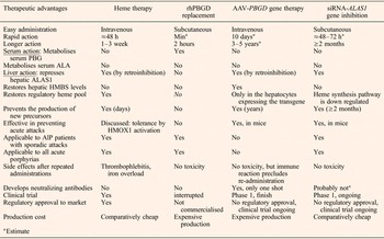
Comparison of advantages and disadvantages of clinically tested therapies for acute porphyrias.

AAV5, adeno-associated viral vector serotype 5; ALA, δ-aminolevulinic acid; ALAS1, ALA synthase; siRNA-ALAS1, interfering RNA-mediated silencing of hepatic ALAS1; HMOX1, heme oxigenase 1; rhPBGD, recombinant human porphobilinogen deaminase.
Potential of liver gene therapy for acute intermittent porphyria
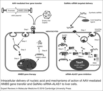
Gene therapy is a versatile procedure consisting of the introduction of genetic information into host cells to achieve sustained clinical correction (Fig. 2, Left). Given that the liver parenchyma remains strictly normal throughout the course of the disease and accessible to transfer vectors, gene therapy represents a potential alternative to liver transplantation for AIP. Gene therapy can be executed by delivering a transgene expression cassette (gene addition, Fig. 2, Left), by blocking/disrupting gene expression (gene silencing, Fig. 2, Right) or by delivering a fragment of nucleic acid to repair a mutated gene (gene repair). Given the great variability in the mutations responsible for AIP (>386 mutations) (Ref. 64) and the risk of manipulating the cell genome, gene repair is the least attractive option. Clinical trials using the first two strategies, HMBS-gene therapy and interference RNA for ALAS1 gene inhibition, were recently conducted in patients with AIP. Advantages and disadvantages of these two gene therapy strategies are compared with other therapeutic options available for AIP in Table 2.
Intracellular delivery of nucleic acid and mechanisms of action of AAV-mediated HMBS gene transfer and GalNAc-siRNA-ALAS1 to liver cells. (Left) AAV-mediated gene transfer. Recombinant AAV vectors are produced in packaging cell lines and subsequently purified. AAV cell entry is mediated by specific receptors that internalise viral particles (Step 1). Then, the AAV particles traffic to the endosome/lysosomes (Step 2). AAVs are released from the late endosome and transported towards the perinuclear region (Step 3). Finally, AAVs are transported through nuclear pores by a VP-1 dependent mechanism (Step 4). Capsids are degraded and viral genomes are released (Step 5). Second DNA strands are synthesised (Step 6). Viral DNAs are either episomal-maintained (≥97%) or integrated into the genome (Step 7). Recombinant HMBS protein is expressed (Step 8). (Right) Liver-targeted delivery of a siRNA-ALAS1 for posttranscriptional gene silencing strategy. A double-stranded (ds) RNA is conjugated to a trivalent GalNAc carbohydrate cluster to enhance liver targeting through the asialoglycoprotein receptor (ASGPR). dsRNA is internalised into an endosome vesicle and released into the cytoplasm (Step 2). The RISC complex processes dsRNA (Step 3). The passenger strand is removed and the guide strand remains to target mRNA for silencing. The resulting mature RISC complex may silence gene expression triggering AGO2-mediated cleavage of the ALAS1 mRNA target (Step 4). ALAS1 mRNA is cleaved and mRNA degraded (Step 5). AAV, adenoassociated vector; ASGPR, asialoglycoprotein receptor; GalNAc, N-Acetylgalactosamine; PBGD, porphobilinogen deaminase or HMBS, hydroxymethylbilane synthase; rHMBS, recombinant HMBS protein; RISC, RNA-induced silencing complex; siRNA, small interfering RNA.

HMBS gene addition therapy: vector choice and preclinical and clinical studies
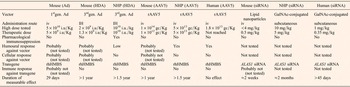
The addition of a functional HMBS gene to parenchymal liver cells is a potential alternative to restore normal function in the liver of patients with AIP. Regulation of transgene expression is not required because HMBS is not the enzyme that regulates the hepatic heme synthesis pathway and long-term HMBS over-expression was safe in mice (Refs Reference Yasuda65, Reference Unzu66) and non-human primates (Refs Reference Unzu67, Reference Paneda68). Indeed, experiments performed in a mouse model of the disease confirmed that HMBS over-expression in a low proportion of hepatocytes (about 5–10%) fully protected against phenobarbital-induced porphyrin precursor accumulation (Refs Reference Unzu66, Reference Unzu69). Different viral vectors have been developed (Table 3) and demonstrated safe and stable transfer of the HMBS gene into hepatocytes able to prevent biochemical attacks induced by phenobarbital challenge in a mouse model of AIP. However, to date, the most promising results in the clinical arena of liver gene transfer have been achieved with an adeno-associated virus (AAV) vector for the treatment of hemophilia B (Refs Reference Nathwani70, Reference Nathwani71). Indeed, the first AAV vector-based gene therapy product, Glybera, for the treatment of lipoprotein lipase deficiency received market approval by the EMA in 2012 (Ref. Reference Bryant72). Clinical compatibility with the vector used in these studies, and experimental evidence of the therapeutic efficacy of an AAV vector serotype 5 leading to liver-specific expression of human HMBS protein (AAV5-AAT-PBGD vector) (Ref. Reference Unzu66) in the AIP mouse, strongly support its translational potential towards the patient. In May 2009, the EMA granted this gene therapy vector an Orphan Drug Designation for the treatment of AIP (EMA/COMP/147330/2009) and a European consortium (AIPgene) (Ref. 73) was created to perform a first-in-human phase 1 clinical trial of liver-directed gene therapy (Refs Reference D'Avola74, Reference D'Avola75).
Comparison of experimental design between preclinical and clinical studies in gene therapy for AIP.

AAV5, adeno-associated viral vector serotype 5; Ad, first generation adenoviral vectors; ALAS1, delta-aminolevulinic acid synthase; GalNAc, N-Acetylgalactosamine; HDA, helper dependent adenoviral vector; IH, intrahepatic; iv, intravenous; rhHMBS, recombinant human hydroxymethylbilane synthase; siRNA, small interfering RNA; siRNA-ALAS1, interfering RNA-mediated silencing of hepatic ALAS1.
The trial was designated as an open-label, single dose and dose-escalating trial. Eight AAV5-seronegative patients with severe AIP were included by pairs in four cohorts and received a single intravenous administration of the AAV5-AAT-PBGD vector ranging from 5 × 10¹¹ to 1.8 × 10¹³ gc/kg. Treatment was well tolerated in all cases. No treatment-related serious adverse events were observed following administration. Moreover, no changes in blood cell count, liver and renal tests, inflammatory cytokine production were observed during the 12-month follow-up (Refs Reference D'Avola75, Reference D'Avola76).
For any clinical application of gene therapy, a set of potential safety concerns applies. Even if the presence of AAV5-AAT-PBGD genome has been detected in body fluids for several weeks, experience in preclinical and different clinical studies ruled out horizontal and vertical transmission of the AAV vector (Refs Reference Arruda77, Reference Manno78, Reference Paneda79, Reference Couto, Parker and Gordon80). Genotoxicity is also a potential concern because the expression of rep gene in wildtype AAVs has been associated with the integration of AAV genome into known liver cancer driver genes leading to their overexpression (Ref. Reference Nault81). However, the rep gene was deleted in the recombinant AAV5-AAT-PBGD vector and the clinical batch used in the clinical trial for AIP did not contain detectable replication-competent particles. Finally, the integration events of the vector in the host genome were rare (<3%), random and outside potential oncogenic genes as demonstrated in non-human primate liver (Ref. Reference Paneda68) and in liver biopsies taken 1 year after AAV-AAT-PBGD administration in the six patients tested (Ref. Reference D'Avola76). Long-term follow-up of these AAV-treated patients will help to evaluate the real impact in humans.
The safety and efficacy considerations on AAV-mediated gene transfer in clinical trials showed immunogenicity against the vector and/or transgene product as the main concern. Immune response directed against the transgene product is an important issue in gene therapy trials in patients who lack endogenous production of the transgene. Given that patients with AIP are already synthesizing subnormal levels of the HMBS enzyme (or normal levels of HMBS with reduced catalytic activity), the potential immune response against the HMBS transgene protein is not expected. Lack of immune reaction against the transgene was confirmed in three preclinical assays performed in non-human primates (Refs Reference Unzu67, Reference Paneda68, Reference Unzu82), even in those animals in which human HMBS was over-expressed in the liver for more than 70 weeks (Ref. Reference Unzu67).
All subjects included in the trial were previously screened for preexisting immunity against the wild type AAV5 capsid protein. Low levels of neutralizing antibodies (>1:5) completely abrogate rAAV2-mediated liver transduction (Ref. Reference Manno78). Moreover, the generation of cytotoxic T lymphocytes CTLs specific for the rAAV2 capsid protein causing the extinction of transgene expression was observed in patients with pre-existing immunity against wild-type AAV2 capsid proteins in two clinical trials of hemophilia B gene therapy using an rAAV2 vector (Ref. Reference Mingozzi and High83). Given that the frequency of serum anti-AAV IgGs in the human population is estimated to be lower for serotypes different from AAV2 (70% versus around 40% for serotype 5 or 8 (Ref. Reference Boutin84), those serotypes 5 and 8 lacking cross-reaction with serotype 2 (Ref. Reference Davidoff85) are less efficient at developing T-cell immune responses in their clinical application. In line with this, the administration of a short course of oral corticosteroids around the time of detection of the immune response blocked the CD8+ T-cell responses against the vector capsid in an AAV8 liver gene transfer trial of hemophilia B (Ref. Reference Nathwani71). In the clinical trial for AIP, no T-cell response was detected against capsid protein or the recombinant HMBS protein in any of the patients (Refs Reference D'Avola75, Reference D'Avola76).
Intravenous infusion of the vector resulted in liver transduction, as confirmed by the presence of the recombinant vector genome in liver biopsies taken 12 months after the administration. In order to estimate therapeutic efficacy patients were followed for at least 6 months before (observational period) and 12 months after treatment (interventional phase) by analyzing the changes in urinary PBG and ALA levels and the impact on symptoms, use of heme therapy and pain medications, number and duration of hospitalisations, psychological alterations and quality of life. Even if some patients experienced an improvement in the health-related quality of life (Refs Reference D'Avola76, 86), porphyrin precursor over-excretion was not modified and more efficient gene transfer vectors or higher doses are needed to reduce ALA and PBG accumulation. Biochemical and clinical analysis of efficacy endpoints is detailed in a recent publication (Ref. Reference D'Avola76).
Strategies for optimizing HMBS gene delivery in AIP
In an attempt to improve the efficacy and performance of gene therapy vectors for AIP, we assayed the insertion into the vector promoter of the minimal functional sequence of the transcriptional enhancer of the human ALAS1 gene (Ref. Reference Podvinec87). The inclusion of this sequence in phase with the promoter of the vector strongly induced transgene expression (up to 10-fold) in the presence of different porphyrinogenic stimuli, such as estrogens, starvation and certain drugs (International application patent N° PCT/EP2015/070204, filing date 4 September 2015) (Ref. Reference Fontanellas and Unzu88). Thus, this element is able to enhance the expression of the therapeutic transgene when it is most needed, at the time when porphyrinogenic factors increase the demand for hepatic heme and precipitate the acute porphyria attacks.
We are also interested in understanding the complexities of the vector–host interactions in the specific context of patients with frequent acute attacks and regular prophylactic administration of heme. Experimental data suggested that AAV5-liver gene transfer was negatively affected after a chronic administration of heme but it was not modified by high porphyrin precursor concentration induced in AIP mice after phenobarbital challenge (unpublished data).
In human trials, evidence of stable transgene expression following AAV vector-mediated gene transfer to the liver comes from the hemophilia B studies, with a follow-up of approximately 3.5 years (Ref. Reference Nathwani71). However, the genome of the rAAV vector, and also that of adenoviral vectors, persists predominantly in extra-chromosomal forms. Although the physiological turnover of liver cells is normally very low in adult mammals (Ref. Reference Borzio89), sustained transgene expression throughout the life of the patient will require repeated administrations. However, first exposures to viral vector capsid preclude repeated administration of the same vector due to the formation of neutralizing antibodies. Sequential administration of rAAV pseudotyped with different capsid proteins (AAV5 and AAV8) (Ref. Reference Unzu66) or sequential injections of rAAV and Helper-dependent adenoviral (HDA) (Ref. Reference Unzu67) vectors carrying the same therapeutic cassette are conceivable strategies to circumvent anti-vector immunity and prolong transgene HMBS expression.
Given the fact that liver transplantation requiring lifelong immunosuppression is the only alternative for patients who develop repeated and severe porphyria attacks, the use of transient and intensive pharmacological immunosuppression can be considered to support translational approaches in gene therapy mediated by viral vectors. Thus, delivery of adenoviral viral vectors concomitant to a combination of immunosuppressive agents safe for AIP (Refs Reference Barone90, Reference Vicari-Christensen91, Reference Wavamunno and Chapman92, 93) was effective in curtailing the immune response towards this vector in macaques (Ref. Reference Unzu67). Unfortunately, this strategy was ineffective against AAV5 (Ref. Reference Unzu82) and doses and agents must be optimised for this vector.
siRNA ALAS1 gene inhibition
After several decades of technological development in the laboratory, RNA-based technologies are finally reaching the clinical trial arena. The stability and the delivery of the RNAs have been improved by advances in the design of cis-acting structural elements and in the RNA packaging and targeting technologies (Ref. Reference Vallazza94). There are three main RNA-based technologies under development: aptamers, modified messenger RNAs and small interfering RNA (siRNA). siRNAs targeting hepatic ALAS1 expression are being tested in clinical trials as a novel approach for acute porphyrias (Fig. 1) (Table 1) (Ref. Reference Yasuda95). This siRNA is a double-stranded RNA molecule of about 20 base pairs in length and conjugated with N-Acetylgalactosamine (GalNAc) to target the liver (Fig. 2, Right). This is an amino sugar binding the asialoglycoprotein receptor expressed on the outer surface of the hepatocyte plasma membrane. The GalNAc-siRNA conjugate shows robust transfer efficiency to the liver and does not trigger an immune response, which supports its potential for repeated administration without the development of neutralizing antibodies (Table 2 and 3). The siRNA technology has the advantage that it is activated in the cytoplasm and can generate repeated cycles of silencing (Fig. 2, Right).
In September 2015, Alnylam Pharmaceuticals, Inc. announced initial positive results from its ongoing Phase 1 clinical trial with a siRNA targeting hepatic ALAS1 performed in asymptomatic high excretors patients (Ref. 96). A total of 16 asymptomatic high excreting patients were enrolled in four single ascending dose cohorts (N = 4 per group), with subjects receiving 0.035, 0.1, 0.35, or 1.0 mg/kg doses of siRNA ALAS1. Exosomal mRNA preparations from serum and urine demonstrated that siRNA ALAS1 treatment resulted in potent, dose-dependent and durable silencing of ALAS1 mRNA in the liver (Source Alnylam) (Ref. 96). As a consequence of this inhibition, results showed that a single subcutaneous dose of siRNA ALAS1 resulted in an up to 82% lowering of ALA and an up to 93% lowering of PBG urinary excretion. Considering the dose of drug required to reduce the accumulation of ALA and PBG, the siRNA proved to be much more effective in humans than in experimental animals (Table 3).
The reduction of precursors reached the maximum value 3 days after siRNA ALAS1 administration. In this regard, the administration of the interference RNA provides no therapeutic advantage over heme therapy for patients with sporadic attacks (Table 2) but it could provide an effective therapeutic approach for prophylaxis in patients with recurrent acute attacks or asymptomatic high excretors.
Nevertheless, the main concern regarding this therapeutic approach would be that siRNA ALAS1 has a very long half-life and the ALAS1 inhibitory effect was still almost at maximum levels after 40–45 days after siRNA ALAS1 administration. Given that the half-life of mitochondrial ALAS1 is among the shortest of all mitochondrial proteins (≈70 min), extended downregulation of the hepatic heme synthesis pathway could markedly reduce heme production. Therefore, any demand of heme from respiratory cytochromes, bile acid synthesis enzymes or drug and xenobiotic-metabolising cytochrome P450 enzymes will be reduced. Under these circumstances, we could speculate that the process of drug metabolism will be slowed down, changing drug pharmacokinetic properties and increasing the patient's sensitivity to drugs and other triggering factors associated with acute attacks.
Further, siRNA ALAS1 was found to be generally well tolerated with no clinically significant drug-related adverse events in patients with elevated urinary levels of ALA and PBG but without a recent history of porphyria attacks or disease activity. Liver ALAS1 mRNA was elevated by over 3-fold compared with healthy subjects. Alnylam Pharmaceuticals expects to initiate a clinical trial in AIP patients suffering from recurrent attacks in early 2016 (Ref. 96). It will be interesting to know whether this technology works during an acute attack, where liver ALAS1 mRNA is significantly induced (between 10 and 200 times) and urinary ALA and PBG levels are increased above asymptomatic levels by 6.3- and 2.5-fold, respectively.
Conclusions
There is a pressing need for disease-modifying therapies for patients suffering recurrent porphyria attacks or showing a permanent over-excretion of porphyrin precursors. As discussed above, currently used treatments may entail significant adverse effects, and in some cases do not prevent the progression of irreversible neurologic damage, kidney disease, malignancy or the impairment in quality of life. From the economic point of view, the development of a more effective therapy will lower the cost of medical care of patients with AIP by reducing hospitalisations and pain medications. Also, an effective therapy will enable the patient to resume a productive life.
The present review provides an overview of active translational research aimed at developing more efficient treatments for AIP, improving patients’ quality of life and protecting against the long-term complications associated with high porphyrin precursor excretion. The most efficient therapy for sporadic attacks must be easy to administer and able to generate rapid action in the three compartments involved in the acute attack: (i) it must restore the heme synthesis pathway in the liver to prevent further accumulations of ALA and PBG; (ii) it must lead to a rapid and sustained reduction in serum PBG and ALA; (iii) ideally, it must be able to cross the blood-brain barrier and protect the brain from neurotoxic precursors accumulated in the plasma during the acute attack.
In patients suffering from recurrent attacks, a single dose of an AAV-AAT-PBGD gene therapy would avoid recurrence of acute attacks for years. The first-in-human Phase 1 clinical trial confirmed the safety and tolerability of AAV5-AAT-PBGD gene therapy but higher doses and/or more efficient vectors are needed to obtain therapeutic expression of the transgene. The siRNA ALAS1 represents an efficient treatment option to prevent high accumulation of porphyrin precursors. It remains to be determined whether the effect of the prolonged down-regulation of hepatic heme synthesis could modify excretion of hydrophobic and toxic compounds and increase the sensitivity to drugs and other triggering factors associated with acute attacks. In this way, the co-administration of siRNA ALAS1 with low doses of heme infusion should be further explored as a means to replenish the hepatic heme pool without inducing heme oxygenase 1 and reducing iron overload in the liver.
Acknowledgements
The authors acknowledge helpful discussions with Professor Jesús Prieto, Professor Ignacio Melero and Professor Rafael Enríquez de Salamanca, and they are grateful to the generosity of the patients and the Spanish Porphyria Foundation for their assistance and support (non-financial).
Conflict of interest
The authors declare no conflict of interest.
Financial support
This work was supported in part by grants from Fundación de Investigación Médica Aplicada (A.F., M.A.A, P.B., CIMA-University of Navarra); Spanish Fundación Mutua Madrileña to A.F. and P.B; Spanish Fundación Eugenio Rodríguez Pascual to A.F. and M.A.A., the European VII Framework program—Project AIPGENE (A.F., grant FP7-Health-2010-261506) and Spanish Institute of Health Carlos III (FIS) co-financed by European FEDER funds (P.B., grant number PI13/00207; A.F., grant numbers PI061475, PI09/02639, PI12/02785, PI15/01951).