Isolation of the left innominate artery from the right aortic arch connecting with the main pulmonary artery via the left ductus arteriosus is a rare anomaly of the aortic arch.Reference Paul, Hugh, David, Robert and Timothy 1 Herein, we report an extremely rare case of coarctation, or incomplete isolation, of the left innominate artery complicated with double-outlet right ventricle and severe pulmonary stenosis.
Case report
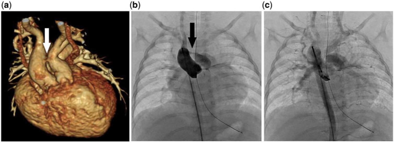
A 3-month-old female infant born at the gestational age of 31 weeks and weighing 891 g at birth was referred to our centre for the management of congenital heart disease. On physical examination, she had dysmorphic features, including a depressed nasal bridge and a smaller left arm compared with the right arm. The blood pressure values were 108/56, 56/32, and 104/58 mmHg in the right arm, left arm, and right leg, respectively. Oxygen saturation was 90% in both arms and legs. Transthoracic echocardiography demonstrated double-outlet right ventricle, subaortic ventricular septal defect, severe pulmonary stenosis, right aortic arch, left patent ductus arteriosus, and persistent left superior caval vein draining into the roof of the left atrium. Inverse blood flow from the brain to the left common carotid artery was observed. Contrast-enhanced computed tomography revealed severe stenosis at the junction between the proximal ascending aorta and the left innominate artery (Fig 1a). In the presence of worsening right ventricular outflow obstruction, her oxygen saturation gradually decreased to 70% in room air and 85% in 2 L nasal oxygen at 4 months of age when aortic angiogram revealed a narrow jet of blood flow from the ascending aorta to the main pulmonary artery (Fig 1b) and delayed inverse flow from the left common carotid artery draining into the main pulmonary artery via the arterial duct (Fig 1c). No chromosomal or genetic anomalies, including 22q11 microdeletion, were detected. Surgical repair was performed at the age of 6 months, comprising ventricular septal defect patch closure, right ventricular outflow tract reconstruction using an expanded polytetrafluoroethylene trivalved conduit, re-routing of the left superior caval vein to the right atrium, re-implantation of the left innominate artery to the ascending aorta, and ligation of the arterial duct. Blood flow in the left carotid artery normalised after surgery with no blood pressure difference between the arms. A small bilateral subdural hematoma was detected 27 days after surgery in brain MRI image. There were no ischemic changes in the cerebral parenchyma. At the 2-year follow-up, the patient was doing well except for mild pulmonary hypertension likely due to chronic lung disease associated with prematurity.
( a ) Anterior view of contrast-enhanced cardiac CT image showing a narrow connection between the ascending aorta and the left innominate artery (white arrow). ( b ) Aortic angiogram showing a very narrow jet from the ascending aorta to the main pulmonary artery via the left innominate artery and ductus arteriosus (black arrow). ( c ) Delayed inverse flow from the left common carotid artery draining into the main pulmonary artery via the arterial duct (see Supplementary Figure S1).

Discussion
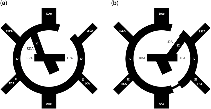
A review of the Edwards’ “double arch hypothesis” is essential to elucidate the embryological aetiology of the current case (Fig 2).Reference Edwards 2 We postulated that our patient had a right aortic arch with mirror-image branching with the exceptions of the incompletely regressed left branch of aortic sac and left patent ductus arteriosus. There are several case reports of isolated left innominate artery due to the interruption of the left branch of the aortic sacReference Kreeger, Schlosser and Sallee 3 , Reference Gowda, Bhat and Jayaranganath 4 , Reference Dubey, Gupta and Kothari 5 , Reference Reeves, Colen, Sheridan and Ward 6 , Reference Singh, Satyapal, Moodley and Rajaruthnam 7 ; however, to the best of our knowledge, there is only one case report on incomplete isolation of the left innominate artery.Reference Lee, Kim, Lee and Kim 8 In our patient, pulmonary stenosis worsened over time in the setting of aggravating right ventricular outflow obstruction; the pulmonary blood flow was solely delivered via the left carotid artery and the narrow connection between the ascending aorta and the left innominate artery when she underwent surgery.
Diagrams of the double aortic arch hypothesis illustrating the variations caused by the regression of segments. ( a ) Right aortic arch with mirror-image branching. ( b ) Right aortic arch with incomplete isolation of the left innominate artery and the left arterial duct. AAo = ascending aorta; DAo = descending aorta; LCA = left common carotid artery; LDA = left ductus arteriosus; LPA = left pulmonary artery; LSCA = left subclavian artery; RCA = right common carotid artery; RDA = right ductus arteriosus; RPA = right pulmonary artery; RSCA = right left subclavian artery; III, IV, VI refer to the corresponding embryonic arches.

The difference in blood pressure between the arms is a significant clinical sign of this anomaly.Reference Gowda, Bhat and Jayaranganath 4 The blood flow is reduced in the left arm because it is supplied either by backward flow in the left carotid artery starting from the right carotid artery and the right vertebral artery using the circle of Willis or by collateral arteries. Although there was a narrow connection between the left innominate artery and the ascending aorta, the left arm of our patient was already smaller compared with the right arm at 3 months of age. Moreover, cerebral ischemia should also be considered, particularly in the presence of a left ductus arteriosus with significant left-to-right shunting.Reference Reeves, Colen, Sheridan and Ward 6 , Reference Singh, Satyapal, Moodley and Rajaruthnam 7 Given its bilaterality and the absence of cerebral ischemia in the images, a subdural hematoma in the present patient was probably irrelevant with cerebral blood steal. To minimise all the risks, surgery to re-implant the left innominate artery to the ascending aorta should be performed as soon as feasible due to the promising outcomes.Reference Kreeger, Schlosser and Sallee 3 , Reference Reeves, Colen, Sheridan and Ward 6 , Reference Singh, Satyapal, Moodley and Rajaruthnam 7 , Reference Lee, Kim, Lee and Kim 8
Conclusion
We experienced a case of an extremely rare aortic arch anomaly with incomplete isolation of the left innominate artery associated with double-outlet right ventricle and the right aortic arch. The differential blood pressure between the arms with inverse flow in the left carotid artery was the key for this unusual diagnosis. Surgical repair is advised to avoid cerebral ischemia and growth delay in the left arm.
Acknowledgement
The authors thank the cardiovascular surgery team at Chukyo Children Heart Center, who performed the operation on this patient.
Supplementary Material
To view supplementary material for this article, please visit https://doi.org/10.1017/S1047951119000702
Financial Support
This research received no specific grant from any funding agency or from commercial or not-for-profit sectors.
Conflicts of Interest
None.
Ethical Standards
The authors assert that all work reported complies with the ethical standards of the Helsinki convention, and institutional and national research committee.