Introduction
Strongyloides stercoralis is a nematode of clinical importance that causes strongyloidiasis and affects people worldwide, especially in tropical and subtropical countries, remaining one of the most neglected and under-reported helminthic infections (Olsen et al Reference Olsen, van Lieshout, Marti, Polderman, Polman, Steinmann, Stothard, Thybo, Verweij and Magnussen2009). The global prevalence of the disease in 2017 was estimated to be 8.1%, corresponding to 613.9 million people infected (Buonfrate et al Reference Buonfrate, Bisanzio, Giorli, Odermatt, Fürst, Greenaway, French, Reithinger, Gobbi, Montresor and Bisoffi2020), but global prevalence is basically unknown as a result of limited surveillance (Gordon et al Reference Gordon, Utzinger, Muhi, Becker, Keiser, Khieu and Gray2024). In immunocompromised patients such as those with HTLV or HIV infections, those using corticosteroids, with organ transplants, or those with systemic lupus erythematosus, the infection can lead to hyperinfection or disseminated syndromes, which may result in 60%–85% mortality in these susceptible individuals (Merz Reference Merz2015; Nutmann Reference Nutman2017; Page and Spare Reference Page and Speare2016; Paula and Costa-Cruz Reference Paula and Costa-Cruz2011).
Corticosteroid treatment is the most frequent cause of hyperinfection in patients with previous undiagnosed chronic strongyloidiasis (Dominguez et al Reference Domínguez, Pérez-López, Sánchez, Contreras, Guerrero and Abenza Abildúa2022). During the COVID-19 pandemic, several case studies worldwide reported severe manifestations of strongyloidiasis (Strongyloides hyperinfection syndrome and disseminated strongyloidiasis) due to immunosuppression associated with COVID-19 treatment (Sharma et al Reference Sharma, Islam, Alam, Das, Islam and Ghose2024). Considering this, it is important to assess for Strongyloides when using immunosuppressive medications for patients who have risk factors (Hamze et al Reference Hamze, Tai and Harris2023).
Due to the ethical and practical limitations of studying hyperinfection and disseminated strongyloidiasis directly in humans, particularly in immunocompromised patients, animal models are essential for advancing our understanding of the disease. Among these, Strongyloides venezuelensis has been used in rodent models of Strongyloides infection and in biological and immunological studies. Wistar rats are used for laboratory maintenance because L3 migrates through the lungs before penetrating the duodenal mucosa, similar to S. stercoralis cycle (Viney and Kikuchi Reference Viney and Kikuchi2017).
Parasitic helminths release a variety of molecules into their host, generally referred to as excretory−secretory (E/S) products, which impact host-parasite interaction, e.g. modulate the host immune response (Maizels et al Reference Maizels, Smits and McSorley2018). In immunocompetent rats, Strongyloides venezuelensis infection is self-limiting, with elimination occurring around 28 days post-infection. However, corticosteroid administration alters this course. Dexamethasone prevents worm expulsion and increases parasitic load, creating an environment to study the onset of hyperinfection. This model enables the evaluation of E/S antigens during acute hyperinfection (8 days post-infection), a phase difficult to assess in clinical studies since patients typically present with chronic infections. Therefore, the primary objective of this study was to evaluate the diagnostic performance of E/S antigens from S. venezuelensis L3 larvae for the specific detection of IgG antibodies in this acute window, using serum samples from immunosuppressed and experimentally infected rats as a preclinical proof-of-concept.
Material and methods
Animals and experimental groups
Male Rattus norvegicus (Wistar) rats weighing 100–120 g, between 6 and 8 weeks were used in experiments. Animals were Specific Pathogen Free (SPF) and were kept in the facilities of Rede de Biotérios de Roedores of Universidade Federal de Uberlândia (REBIR-UFU), with provision of sterilized feed and water ad libitum and kept in cages with a maximum density of four rats. Colony room ambient temperature was 22 ± 2 °C, with artificial lighting 12h:12h light-dark cycle.
Rats were divided into four groups: immunocompetent and infected with S. venezuelensis (IC+) (n = 6) or non-infected (IC−) (n = 6), immunosuppressed and infected with S. venezuelensis (IS+) (n = 6) or non-infected (IS−) (n = 6). Prior to infection, immunosuppressed groups (IS+, IS−) received 5 μg/mL of dexamethasone disodium phosphate (Medley, Brazil) diluted in water for 5 days, as previously described by Yang and Healey (Reference Yang and Healey1993). Animals from IC+ and IS+ groups were inoculated subcutaneously in the abdominal region with 1500 S. venezuelensis larvae. All procedures involving animals were conducted in accordance with the ethical principles of animal experimentation and were approved by the Ethics Committee on Animal Use of the Federal University of Uberlândia (CEUA/UFU, protocol no. 019/15 and 075/08). A sample size of n = 6 per group was selected based on previous studies using this specific model (Gonçalves et al Reference Gonçalves, Silva, Ueta and Costa-Cruz2012) and in strict adherence to the 3Rs principle (Replacement, Reduction, and Refinement), aiming to minimize animal use while maintaining sufficient statistical power for the proposed analyses.
Biological samples and evaluation of parasitic burden
The estimated number of eggs per gram of faeces (EPG) was performed using the method of Cornell-McMaster (Gordon and Whitlock Reference Gordon and Whitlock1939). Rats of IC+ and IS+ groups were placed on clean, damp paper towels to defecate on 7 and 8 days post-infection (dpi), and faeces were collected.
To recover parthenogenetic females (PF) and serum samples, rats were anaesthetised with ketamine 60 mg/kg and xylazine 7 mg/kg (Syntec, Brazil) and euthanized on 8 dpi. Blood was collected by cardiac puncture and serum samples were obtained after centrifugation. After euthanasia, the small intestines were removed, sectioned longitudinally, and incubated in Petri dishes (phosphate buffered saline (PBS) 0.1 mol/L, pH 7.2, 37 °C, 2 h). The solution was collected and centrifuged at 1,000 rpm, and the count of parasitic females was performed (Sato and Toma Reference Sato and Toma1990).
Strongyloides venezuelensis L3 larvae
Faeces from animals infected with S. venezuelensis L3 were collected after 8 days of infection and cultured in charcoal. Cultures were maintained at 28 °C for 72 h, and L3 larvae were harvested and concentrated using the method described by Rugai et al (Reference Rugai, Mattos and Brisola1954). Larvae were transferred to a conical tube containing PBS 0.1 mol/L, pH 7.2 and 0.25% sodium hypochlorite, supplemented with 100 U/mL penicillin and 100 μg/mL streptomycin. The mixture was then centrifuged at 1280 × g for 5 min. After centrifugation, the supernatant was discarded, and the larvae were washed three times with PBS supplemented with 100 U/mL penicillin and 100 μg/mL streptomycin. Between each washing step, the tube was centrifuged at 1280 × g for 5 min. At the end of the washing cycles, the larvae were collected and used to produce antigenic extracts.
Larval cultures were maintained under sterile conditions and inspected daily for contamination. This inspection included macroscopic observation for turbidity or fungal growth, as well as microscopic examination of larvae to identify any fungal contamination or other microbial presence. Only cultures containing larvae with preserved motility and structural integrity, and free of visible fungal or bacterial contamination, were used for antigen collection.
Antigenic extracts from S. venezuelensis L3 larvae
Total saline extract
Total saline extract (TS) was produced according to Gonzaga et al. (Reference Gonzaga, Ribeiro, Cunha-Junior, Ueta and Costa-Cruz2011), with modifications. Approximately 300,000 S. venezuelensis L3 filarioid larvae were resuspended in PBS (0.1 mol/L, pH 7.2) containing protease inhibitors (ULTRA minicomplete, Roche, Germany) and ruptured by 10 cycles of freeze (1 min, −196 °C) and thaw cycles. The solution was then centrifuged at 12400 × g for 30 min at 4 °C, and the supernatant was collected (TS). The protein concentration was determined by the method of Lowry et al. (Reference Lowry, Rosebrough, Farr and Randall1951), and the TS of S. venezuelensis L3 larvae was then stored at −20 °C until use.
Excretion/secretion antigens (E/S) in RPMI or PBS
Excretion/secretion (E/S) antigens were produced in vitro according to Soblik et al (Reference Soblik, Younis, Mitreva, Renard, Kirchner, Geisinger, Steen and Bratting2011) and Cunha et al (Reference Cunha, de Carvalho, de Sousa and Costa-Cruz2017). L3 larvae were transferred to 12-well culture plates at a concentration of 30,000 larvae/mL/well. Supernatants obtained from each larval culture were pooled and concentrated on an Amicon® Ultra-15 10K Centrifuge Filter (Merck Millipore, Ireland) and centrifuged at 5000 × g for 20 min at 4 °C. The antigen products after concentration in both RPMI and PBS mediums were quantified according to the method of Lowry et al. (Reference Lowry, Rosebrough, Farr and Randall1951) and stored at −20 °C.
ELISA to detect anti-Strongyloides venezuelensis IgG antibodies in rat serum samples
Rat serum samples were analysed using an enzyme-linked immunosorbent assay (ELISA) for anti-S. venezuelensis IgG detection, according to Gonçalves et al (Reference Gonçalves, Silva, Ueta and Costa-Cruz2012). Low-affinity microtiter plates (Greiner Bio-one, Germany) were selected to minimize non-specific background binding commonly observed with complex antigenic mixtures. Plates were coated with TS, RPMI E/S or PBS E/S antigen preparations (10 μg/mL of each antigenic extract in 0.06 M carbonate–bicarbonate buffer (pH 9.6)). After washing, plates were incubated with serum samples diluted at 1:10. This dilution was established based on pilot checkerboard titrations to optimize the signal-to-noise ratio for detecting the early antibody response at 8 dpi. Samples were incubated for 45 min at 37 °C and subsequently with the secondary antibody consisting of peroxidase-labelled goat anti-rat IgG (Sigma) at the ideal dilution of 1:8000 for 45 min at 37 °C. The reaction was revealed by adding the enzyme substrate (0.03% H2O2 and o-phenylenediamine [OPD] in 0.1 M citrate–phosphate buffer, pH 5.0) and incubated for 15 min at room temperature. The reaction was stopped by adding 2 N H2SO4. The optical density (OD) value was determined for each well in a microplate reader (Epoch™ Microplate Spectrophotometer, BioTek Instruments, USA) using a 492 nm filter.
Statistical analysis
Normality was assessed using the Shapiro-Wilk and Kolmogorov-Smirnov tests. For EPG and PF counts, data are presented as mean ± standard deviation (SD), and comparisons between infected groups were performed using the unpaired t-test. Homoscedasticity was assessed by the F test.
For the IgG data, results were expressed as an ELISA Index (EI), calculated as the ratio between sample optical density (OD) and the cut-off (EI = sample OD/cut-off). The cut-off value was established using a receiver operating characteristic (ROC) curve as described by Greiner et al (Reference Greiner, Sohr and Göbel1995), considering all samples from all groups. EI values greater than 1 were considered positive. EI data are presented as mean ± SD. After assessing normality, comparisons of antigenic extracts within the same group were performed using repeated measures ANOVA with Geisser-Greenhouse correction for equal variability of differences (sphericity). A t-test was also performed between each group and its respective control.
The diagnostic performance of each antigenic extract was evaluated by calculating sensitivity (Se), specificity (Sp), positive (LR+) and negative (LR–) likelihood ratios, and the Youden index (J). These parameters provided a robust statistical assessment of the diagnostic quality of each extract, allowing for objective comparison and interpretation of their performance.
All statistical analyses were performed using Prism 10.0 software (GraphPad Software, USA).
Results and discussion
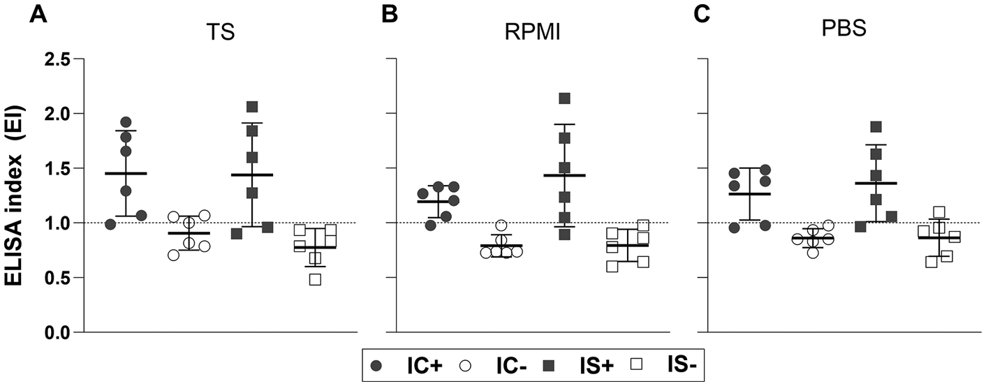
Animals from the IS+ group presented mean EPG (IC+: x̄ = 13167 ± SD = 6345 vs. IS+: x̄ = 31133 ± SD = 6303, t = 4.92, p = 0.0006) and PF (IC+: x̄ = 113 ± SD = 56.14 vs. IS+: x̄ = 335 ± SD = 90.53, t = 6.45, p = 0.0003) counts significantly higher than the IC+ group (data not shown). The detection of anti-S. venezuelensis IgG in the serum samples of rats from the experimental groups was performed by ELISA test (Figure 1). A repeated measures ANOVA with Geisser-Greenhouse correction, followed by Tukey post hoc test, showed no statistically significant differences between the IC+, IC–, and IS+ groups across all extracts (p > 0.05); in the IS– group (F = 40.93, p < 0.0001), a significantly higher EI was observed with the PBS E/S extract compared to RPMI one (x̄ = 0.86 ± SD = 0.17 vs. x̄ = 0.79 ± SD = 0.15, p = 0.046). Statistical analysis was also performed using unpaired t-tests between each infected group and its respective control, with mean EI values in infected groups significantly higher than in the non-infected groups across the three preparations (data not shown).
Detection of IgG by ELISA using Strongyloides venezuelensis L3 larvae: total saline extract (TS) – A, excretion-secretion antigens in RPMI (RPMI E/S) – B and PBS (PBS E/S) – C in rat serum samples from groups: immunocompetent and infected (IC+), immunocompetent and non-infected (IC−), immunosuppressed and infected (IS+) and immunosuppressed and non-infected (IS−). The dotted lines indicate the ELISA index cut-off.

Figure 1. Long description
A multi-panel figure with three dot plots labeled A, B, and C. All plots share a Y-axis titled E L I S A index E I ranging from 0.0 to 2.5 with a horizontal dotted cut-off line at 1.0. A legend at the bottom identifies four groups: I C plus (filled circles), I C minus (open circles), I S plus (filled squares), and I S minus (open squares).
* Panel A, titled T S: The I C plus and I S plus groups show high variability with means above the 1.0 cut-off, reaching up to 2.0. The I C minus and I S minus groups are clustered below the 1.0 cut-off.
* Panel B, titled R P M I: The I C plus group is tightly clustered just above the 1.0 line. The I S plus group shows the highest spread, with values ranging from 1.0 to over 2.0. Both non-infected groups (I C minus and I S minus) remain below the 1.0 cut-off.
* Panel C, titled P B S: Similar to Panel A, the infected groups (I C plus and I S plus) show means above the 1.0 cut-off, while the non-infected groups (I C minus and I S minus) are positioned below the cut-off line.
Regarding diagnostic parameters (Table 1), the area under the curve (AUC) demonstrated that IgG detection with the different antigenic preparations was distinguishable, with AUC > 0.92 (p ≤ 0.0005). According to the established cut-off, RPMI E/S showed better results with higher values of sensitivity and specificity, if compared to E/S PBS and TS extracts (Table 1). Even though the LR values indicate that E/S PBS and TS extract result in informative tests, RPMI E/S showed the greatest values of LR+ (LR+ > 10; LR+ undefined) and LR– (LR– closest to <0.1; LR− = 0.17). The Youden index, as a summary of the accuracy of a diagnostic test, showed that the rate of false results was lower with RPMI E/S.
Diagnostic parameters for total saline extract (TS) and excretion/secretion antigens in RPMI (RPMI E/S) or PBS (PBS E/S) in ELISA with serum samples from rats infected with Strongyloides venezuelensis

Table 1. Long description
The table consists of seven columns: Antigenic preparations, A U C, Se percent, Sp percent, L R plus, L R minus, and J. Each row contains a primary value with a confidence interval range provided below it.
* Row 1: T S. A U C is 0.92 (0.81 to 1.00). Se is 75.0 (46.8 to 91.4). Sp is 75.0 (46.8 to 91.1). L R plus is 3.00 (1.07 to 8.43). L R minus is 0.33 (0.12 to 0.94). J is 0.50.
* Row 2: R P M I E slash S. A U C is 0.97 (0.92 to 1.00). Se is 83.3 (55.2 to 97.0). Sp is 100.0 (75.8 to 100.0). L R plus is infinity. L R minus is 0.17 (0.05 to 0.59). J is 0.83.
* Row 3: P B S E slash S. A U C is 0.95 (0.87 to 1.00). Se is 75.0 (46.8 to 91.1). Sp is 91.7 (64.6 to 99.6). L R plus is 9.00 (1.34 to 60.46). L R minus is 0.27 (0.10 to 0.74). J is 0.67.
Abbreviations used: A U C is area under R O C curve, Se is sensitivity, Sp is specificity, L R plus is positive likelihood ratio, L R minus is negative likelihood ratio, and J is Youden's index.
AUC – area under ROC curve. Sensitivity – Se. Specificity – Sp. Positive likelihood ratio – LR+. Negative likelihood ratio – LR−. Youden’s index – J.
* undefined
These findings are particularly relevant because definitive diagnosis of Strongyloides infection by the detection of larvae in faeces using conventional parasitological methods may be difficult due to low or irregular parasitic load in faeces, resulting in high rates of false-negative results (Vasquez-Rios et al Reference Vasquez-Rios, Pineda-Reyes, Pineda-Reyes, Marin, Ruiz and Terashima2019). Current parasitological methods for the detection of parasites present insufficient sensitivity for accurate infection detection in the immunocompromised host (Devi et al Reference Devi, Borkakoty and Mahanta2011). Thus, alternative diagnostic tests must be developed. Different diagnostic approaches using diverse antigenic extracts have been evaluated, such as Strongyloides coproantigen and DNA detection in faecal samples (Chaves et al Reference Chaves, Goncalves, Paula, Silva, Silva, Costa-Cruz and Freitas2015) and detection of specific IgG anti-parthenogenetic females, -eggs (Gonçalves et al Reference Gonçalves, de Araújo, Carvalho, Ueta and Costa-Cruz2016), or -L3 larvae somatic extracts (Corrêa et al Reference Corrêa, do Couto, de Carvalho, de Sousa, da Silva Ribeiro, Gonzaga and Costa-Cruz2023) in immunosuppressed and infected rats.
In this study, we evaluated the use of E/S antigens for IgG detection in serum samples of rats immunosuppressed or not and experimentally infected with S. venezuelensis. Despite the high dose of dexamethasone used to induce immunosuppression, our results showed that the humoral response (IgG) remained detectable and comparable to immunocompetent animals at 8 dpi (Figure 1). This finding is crucial, as it validates the utility of ELISA as a diagnostic tool even under corticosteroid regimens that facilitate hyperinfection. These antigenic extracts can feasibly be produced in laboratories with relatively low financial resources. Our data shows RPMI E/S and PBS E/S may be used for strongyloidiasis immunodetection purposes since they achieved diagnostic parameters superior to TS.
Products of excretion and secretion of parasites are presented as promising tools in the development of more effective serological diagnostic tests. Excretory/secretory antigens are enriched with molecules actively released during host-parasite interaction, including proteases and immunomodulatory proteins, which are directly recognized by the host immune system. In contrast, crude somatic extracts contain a broader mixture of intracellular and structural proteins that may increase non-specific reactivity. This difference in antigenic composition likely contributes to the superior diagnostic performance observed for RPMI E/S antigens. Notably, the persistence of high IgG levels in the infected immunosuppressed group (IS+) contradicts the assumption that corticosteroid therapy would end antibody production in this short window. This suggests that the massive antigenic exposure driven by the high parasitic burden in the IS+ group may compensate for the drug suppressive effects on B-cell function during the acute phase.
In our results, EPG and PF counts were higher in the IS+ group, these findings are in accordance with Gonçalves et al (Reference Gonçalves, Silva, Ueta and Costa-Cruz2012), Machado et al (Reference Machado, Carlos, Sorgi, Ramos, Souza, Soares, Costa-Cruz, Ueta, Aronoff and Faccioli2011) and Corrêa et al (Reference Corrêa, do Couto, de Carvalho, de Sousa, da Silva Ribeiro, Gonzaga and Costa-Cruz2023), these authors also analysed the effects of dexamethasone on the infection with S. venezuelensis observing greater release of eggs and greater presence of parthenogenetic females in the immunosuppressed group, dexamethasone may increase the fecundity of the parasites and subsequently, increase the release of fertile eggs increasing parasitic burden. The increased parasite burden observed in our study in dexamethasone-treated animals is likely associated with suppression of Th2-mediated immune responses, reduced eosinophilic activity, and enhanced parasite fecundity. Variations reported among experimental studies may result from differences in corticosteroid regimen, infection dose, parasite strain, and host susceptibility.
Data presented in the literature describe a peak of IgG detection between 7 and 8 dpi in immunosuppressed and immunocompetent animals (Gonçalves et al Reference Gonçalves, Silva, Ueta and Costa-Cruz2012; Machado et al Reference Machado, Carlos, Sorgi, Ramos, Souza, Soares, Costa-Cruz, Ueta, Aronoff and Faccioli2011). In our ELISA using serum samples from rats after 8 dpi, all antigens were able to detect anti-S. venezuelensis IgG, and RPMI E/S and PBS E/S antigens presented better results when compared to TS. Our data from rat samples also agree with studies involving human serum samples, regarding the better diagnostic performance of E/S products (Cunha et al Reference Cunha, de Carvalho, de Sousa and Costa-Cruz2017; Roldán Gonzáles et al Reference Roldán Gonzáles, Coelho, Pimenta, de Paula and Gryschek2022). The difference in protein composition between RPMI E/S and PBS E/S of S. venezuelensis L3, observed by Roldán Gonzáles et al (Reference Roldán Gonzáles, Coelho, Pimenta, de Paula and Gryschek2022), can explain sensitivity (Se) and specificity (Sp) differences observed in this study.
Although experimental immunosuppression in rats with S. venezuelensis infection is an important model for better studying Strongyloides hyperinfection and developing diagnostic approaches applicable for strongyloidiasis, this study has some limitations. First, the evaluation was restricted to a single time point (8 dpi). While this precludes kinetic analysis, this time point was specifically chosen to represent the peak of the acute phase in this model, prior to immune expulsion in competent hosts. Second, cross-reactivity with other helminths was not evaluated in this specific dataset. Although 100% specificity was observed against non-infected controls, future studies must assess these antigens against sera from hosts with co-infections to confirm their diagnostic specificity in broader contexts. Additionally, all infected animals received the same parasite inoculum, which ensured experimental standardization but may have reduced biological variability compared with natural infections; therefore, future studies evaluating different larval inocula may be necessary.
A major challenge in translating murine results to humans lies in the temporal differences of infection; while human strongyloidiasis is predominantly chronic, the rat model is characterized by an acute phase followed by immune expulsion. However, our study leverages this acute window to demonstrate that RPMI E/S antigens can detect specific IgG as early as 8 days post-infection. This finding provides valuable preclinical evidence for veterinary research and experimental models requiring rapid confirmation of infection status. Our data suggest that the E/S antigens are sensitive enough to detect the early immunological surge associated with the onset of hyperinfection, independent of the host carrier status. Thus, the model provides valid preclinical evidence for the utility of these antigens in screening protocols for acute or reactivated strongyloidiasis.
In conclusion, the findings from this study demonstrate that excretory/secretory (E/S) antigens of Strongyloides venezuelensis L3 larvae, particularly RPMI E/S, exhibit high diagnostic specificity in this model and are effective in detecting IgG antibodies in experimental hyperinfection models in rats. These results highlight the potential of E/S antigens as a reliable and safe tool for the detection of strongyloidiasis, offering promising applications for both experimental research and the development of diagnostic approaches for this neglected tropical disease. Further exploration of these antigens could contribute to advancing translational studies and improving public health strategies for managing strongyloidiasis.
Financial support
This study was supported by grants from Fundação de Amparo à Pesquisa do Estado de Minas Gerais (FAPEMIG – N° CBB-PPM-00396-13, JMCC and FAPEMIG – N° CDS-RED-00013-14, JMCC) and Coordenação de Aperfeiçoamento de Pessoal de Nível Superior (CAPES) funding agency (CAPES – N° 3200601004M-8; EFGC, JENS). The funders had no role in study design, data collection and analysis, decision to publish or preparation of the manuscript.
Competing interests
The authors declare none.
Ethical standard
The authors assert that all procedures contributing to this work comply with the ethical standards of the relevant national and institutional guides on the care and use of laboratory animals. This study was performed in line with the principles of the Brazilian College of Animal Experimentation (COBEA). Approval was granted by the Ethics Committee on Animal Use ( Comissão de Ética na Utilização de Animais – CEUA) of Universidade Federal de Uberlândia (Protocol numbers 019/15 and 075/08).