Implications
In vitro embryo production in small ruminants provides an excellent source of low-cost embryos for basic research on developmental biology and for commercial application of the emerging biotechnologies such as nuclear transfer, transgenesis, embryo sexing and stem cells. Moreover, this technology would allow a fast multiplication of unique and expensive animals as endangered species. The use of oocytes from prepubertal female goats into breeding programs is advantageous because it can reduce the generation interval and increase the rate of genetic gain. Thus, a challenge is to establish a system that could potentially provide an unlimited source of competent oocytes for biomedical application.
Introduction
The use of prepubertal females as oocyte donors in in vitro embryo production allows for an acceleration in genetic progress by shortening the generation interval. However, reduced developmental competence in oocytes of juvenile animals was addressed in numerous studies on ruminant species, including bovine (Khatir et al., Reference Khatir, Lonergan and Mermillod1998), ovine (Ledda et al., Reference Ledda, Bogliolo, Calvia, Leoni and Naitana1997) and caprine (Romaguera et al., Reference Romaguera, Moll, Morato, Roura, Palomo, Catala, Jimenez-Macedo, Hammami, Izquierdo, Mogas and Paramio2011). The low blastocyst formation rates obtained in vitro is mainly because of insufficient cytoplasmic maturation in the oocyte or inability to maintain embryo development beyond the transition of embryonic genome activation (Sirard et al., Reference Sirard, Richard, Blondin and Robert2006).
Oocyte growth and maturation are strictly dependent on establishing functional communications with the surrounding cumulus cells through gap junctions and reciprocal interactions mediated by paracrine and endocrine signals. These processes are accurately regulated by numerous growth factors and hormones. Moreover, during mammalian development, some cytokines, including activin-A, play a functional role in the process of cellular proliferation, differentiation and morphogenesis. In early pre-implantation embryos, cytokines produced by both the female genital tract and the embryo itself act on embryonic cells as paracrine/autocrine factors (reviewed by Heyner et al., Reference Heyner, Shan, Smith, Watson and Schultz1993).
Activin is an important member of the transforming growth factor β (TGFβ) superfamily, and its roles in gonadal functions have been extensively studied in mammals (Mather et al., Reference Mather, Moore and Li1997). Activins are homodimers or heterodimers of the βA or βB subunits of inhibin linked to one another by a single disulfide bond (Hata et al., Reference Hata, Kilkuchi, Tada and Tsuiki1988). Dimerization of these β-subunits gives rise to three forms of activin referred to as activin-A (βAβA), activin-AB (βAβB) and activin-B (βBβB) (Ying, Reference Ying1988). The expression of protein and mRNA for activin-A and activin receptors in the ovary has been localized in both oocyte and granulosa cells of follicles at various developmental stages (primates: Sidis et al., Reference Sidis, Fujiwara, Leykin, Isaacson, Toth and Schneyer1998; swine: Van den Hurk and Van de Pavert, Reference Van den Hurk and Van de Pavert2001; bovine: Hulshof et al., Reference Hulshof, Figueiredo, Beckers, Bevers, Vanderstichele and Van den Hurk1997, Izadyar et al., Reference Izadyar, Dijkstra, Van Tol, van de Eljnden-Van Raaij, Van den Hurk, Colenbrander and Bevers1998; ovine: Thomas et al., Reference Thomas, Armstrong and Telfer2003; and caprine: Silva et al., Reference Silva, van den Hurk, van Tol, Roelen and Figueiredo2004). In addition to being a local regulator of folliculogenesis, activin-A is able to directly stimulate FSH synthesis and secretion, and to promote the release of the gonadotrophin-releasing hormone (GnRH) (Childs and Unabia, Reference Childs and Unabia1997). Activin-A can also stimulate the increase of FSH and LH receptors in granulosa cells, and it plays a role in progesterone production (Alak et al., Reference Alak, Coskun, Friedman, Kennard, Kim and Seifer1998; Tsuchiya et al., Reference Tsuchiya, Minegishi, Kishi, Tano, Kameda, Hirakawa, Ibuki, Mizutani and Miyamoto1999). Thus, the granulosa cells are likely to be the main source of paracrine factors and are crucial for oocyte maturation.
Activin-A and activin receptors have been detected in mouse (Lu et al., Reference Lu, Matsuyama, Nishihara and Takahashi1993) and bovine (Yoshioka et al., Reference Yoshioka, Takata, Taniguchi, Yamanaka and Sekikawa1998b) embryos from zygote to the morula stage, suggesting that the protein might play a role in embryogenesis. As activin-A is produced by oviduct epithelial cells (Gandolfi et al., Reference Gandolfi, Modina, Brevini, Passoni, Artini, Petraglia and Lauria1995), the addition of activin-A to embryo culture in vitro may reproduce the environment of the cleavage-stage embryos in the oviduct in vivo. However, the effect of activin-A on in vitro embryo culture (IVC) is still controversial. Positive effects of activin-A on embryo development were shown when the protein was added at the earliest stages of bovine (Yoshioka and Kamomae, Reference Yoshioka and Kamomae1996; Yoshioka et al., Reference Yoshioka, Suzuki and Iwamura1998a, Lee et al., Reference Lee, Bettegowda, Wee, Ireland and Smith2009) embryo culture, whereas no effects on blastocyst formation rates, depending on the timing of its addition to the culture medium, were reported in the literature either (Park et al., Reference Park, Oh, Hong, Jang, Kim and Lee2010; Trigal et al., Reference Trigal, Gomez, Diez, Caamano, Martin, Carrocera and Munoz2011).
Summarizing, although prepubertal oocytes give rise to similar rates of fertilization and cleavage to those achieved using adult oocytes, the capacity of prepubertal oocytes to develop in the blastocyst stage is relatively lower and it would seem that embryos from prepubertal oocytes are less capable of establishing pregnancies (reviewed in Armstrong, Reference Armstrong2001). Therefore, to improve the developmental competence of prepubertal oocytes after their recovery from the follicle, two approaches have been described. The first has been the addition of putative growth promoting substances to the culture medium (gonadotrophins, steroids, growth factors). The second approach has attempted to mimic the intrafollicular conditions of the oocytes. However, none of these approaches have managed to improve developmental competence from prepubertal goat oocytes. Keeping in mind that protein and mRNA for activin-A and activin receptors have been localized in both oocyte and granulosa cells of follicles at various developmental stages in a variety of species including caprine (Silva et al., Reference Silva, van den Hurk, van Tol, Roelen and Figueiredo2004 and Reference Silva, Tharasanit, Taverne, Van der Weijden, Santos, Figueiredo and Van den Hurk2006), and the potential role of activin-A in the regulation of oocyte maturation has been demonstrated to promote in vitro oocyte maturation (IVM) in the rat (Itoh et al., Reference Itoh, Igarashi, Yamada, Hasegawa, Seki, Eto and Shibai1990), cow (Stock et al., Reference Stock, Woodruff and Smith1997), rhesus monkey (Alak et al., Reference Alak, Smith, Woodruff, Stouffer and Wolf1996) and human (Alak et al., Reference Alak, Coskun, Friedman, Kennard, Kim and Seifer1998), we hypothesized that the addition of activin-A to the maturation and/or embryo culture medium could improve the in vitro potential of prepubertal goat oocytes for nuclear and cytoplasmic maturation and embryo development. Such a system could potentially provide an unlimited source of competent oocytes for biomedical application.
To the best of our knowledge, there is no study that reports the effects of activin-A on in vitro developmental potential of prepubertal goat oocytes. Therefore, the present study was conducted to evaluate the effect of adding activin-A at the IVM and IVC media on meiotic maturation, embryo development and embryo quality of in vitro-produced blastocysts.
Material and methods
Reagents
Unless specified, all chemicals, reagents and hormones were purchased from Sigma (St Louis, MO, USA). Activin-A was purchased from R&D Systems (Minneapolis, MN, USA), and it was reconstituted at 50 μg/ml in sterile phosphate-buffered saline (PBS) containing at least 0.1% bovine serum albumin (BSA) and stored in aliquots at −20°C until use.
Oocyte collection and in vitro maturation (IVM)
Prepubertal goat (1 to 2 months old) ovaries were obtained from a slaughterhouse and transported to the laboratory in PBS at 38°C within 1 h of collection. Cumulus–oocyte complexes (COCs) were recovered by the slicing technique. Only COCs with a compact cumulus and homogeneous cytoplasm were selected for IVM. The COCs were washed three times in the IVM medium (TCM199 supplemented with 275 μg/ml sodium pyruvate, 146 μg/ml l-glutamine, 50 μg/ml gentamycin, 10% (v/v) donor bovine serum (CanSera, Ontario, Canada), 10 μg/ml FSH, 10 μg/ml LH, 1 μg/ml 17 β-estradiol and 100 μM cysteamine. Groups of 25 to 30 COCs were transferred into drops of 100 μl of the IVM medium, covered with mineral oil, and incubated for 24 h at 38.5°C and 5% CO2 in humidified air.
In vitro fertilization (IVF)
Fresh semen was collected with an artificial vagina from four Murciano-Granadina bucks of proven fertility and transported to the laboratory at 35°C within 30 min. The motile sperm fraction was selected by the swim-up method (Parrish et al., Reference Parrish, Suskoparrish, Leibfriedrutledge, Cristser, Eyestone and First1986). Briefly, 90 μl of ejaculate mixture was deposited at the bottom of a conical tube containing 2 ml mDM (defined medium modified by Younis et al., Reference Younis, Zuelke, Happer, Oliveira and Brackett1991). After 1 h of incubation at 38.5°C, the upper layer of the tubes was recovered in a tube of 15 ml and centrifuged at 160×g for 3 min. The sperm pellet was resuspended in a 1 : 1 proportion (v/v) with mDM containing heparin sodium salt (final concentration of 50 μg/ml), and it was incubated for 45 min at 38.5°C and 5% CO2 in humidified air. After maturation, oocytes were washed three times in the fertilization medium (Tyrode’s medium (TALP) supplemented with 1 μg/ml hypotaurine and 0.3 mg/ml glutathione) and groups of 25 to 30 oocytes were transferred to microdrops of 100 μl IVF medium, under mineral oil and co-cultured with sperm in a final concentration of 4×10⁶ motile sperm/ml at 38.5°C and 5% CO2 in humidified air.
In vitro culture (IVC)
At 24-h post-insemination (hpi), presumptive zygotes were denuded of surrounding cumulus cells and attached spermatozoa, washed three times in synthetic oviductal fluid medium (SOF (Takahashi and First, Reference Takahashi and First1992)), and groups of 20 presumptive embryos were placed into droplets of 20 μl of SOF medium supplemented with 10% fetal calf serum (FCS) under mineral oil and cultured in a humidified atmosphere with 5% CO2, 5% O2 and 90% N2 for 8 days. The cleavage rate was evaluated at 48 hpi and blastocyst formation rates were recorded at Days 7, 8 and 9 post-insemination (pi).
Evaluation of nuclear maturation stage
After the in vitro maturation, oocytes were freed from their surrounding cumulus cells by vortexing and then washed three times in PBS. To evaluate meiotic progression, the oocytes were fixed in ethanol : acetic acid (3 : 1) overnight. The oocytes were mounted on microscope slides with vaseline, covered with a glass coverslip and stained with 1% lacmoid in 45% acetic acid solution. The stage of nuclear maturation was assessed by phase-contrast microscopy (magnification, ×40). The criteria used to determine oocyte maturation stage have been described elsewhere (Rodriguez-Gonzalez et al., Reference Rodriguez-Gonzalez, Lopez-Bejar, Velilla and Paramio2002). Oocytes were scored as: germinal vesicle (GV) stage, when their chromatin, either filamentous or slightly condensed, was enclosed in a nuclear membrane; germinal vesicle breakdown (GVBD) stage, when there was no visible nuclear membrane and chromatin was condensed; metaphase I (MI) stage, when the chromatin was distributed at the equator of a large spindle; anaphase I stage, when homologous chromosomes could be seen to move away from each other and there was a visible spindle; telophase I stage, when homologous chromosomes were apart, and metaphase II (MII) stage, when the chromatin was located at the spindle and the first polar body extrusion was detected.
Evaluation of blastocyst quality
Hoechst staining
At the end of IVC, blastocysts were stained with 1 mg/ml Hoechst 33342 (H3570; Invitrogen, Life Technologies, Burlington, Ontario, Canada) for 5 min at 38°C. Cell count was carried out under a fluorescence microscope (Olympus BX53; Olympus, Germany).
Blastocyst differential staining
The cellular composition of Day 8 pi blastocysts was assessed by differential staining of inner cell mass (ICM) and trophectoderm (TE) cells, as described by Thouas et al. (Reference Thouas, Korfiatis, French, Jones and Trounson2001), with some modifications. Briefly, embryos that reached the blastocyst stage were first incubated in PBS with 1% (v/v) Triton X-100 and 100 μg/ml propidium iodide for up to 15 s. Blastocysts were then immediately transferred into a fixative solution of 100% ethanol supplemented with 25 μg/ml Hoechst 33342 for 2 h. Fixed and stained whole blastocysts were mounted on a slide in a drop of 3 μl of glycerol and flattened with a coverslip. Cells were counted under UV illumination (Olympus BX53) and a 460 nm excitation filter. The images showed ICM and TE nuclei as blue and red/pink, respectively.
Experimental design
Two experiments were conducted in this study. Each experiment was replicated at least five times.
Experiment 1. Effect of activin-A during IVM on the nuclear stage, embryo development and blastocyst quality of prepubertal goat oocytes
The objective was to assess the effects of recombinant human activin-A at concentrations of 10 ng/ml and 100 ng/ml added at the IVM of prepubertal goat oocytes on nuclear maturation, cleavage and blastocyst formation rates, and embryo quality as compared with the control group, without activin-A. COCs were randomly assigned into one of the three IVM treatment groups: (1) A0: IVM medium without activin-A (control group); (2) A10: supplemented with 10 ng/ml activin-A and; (3) A100: supplemented with 100 ng/ml activin-A. After IVM, COCs were fertilized and the cleavage rate and blastocyst yield were assessed at Day 2 pi and Days 7, 8 and 9 pi, respectively. At the end of the culture period (Day 9 pi), and to determine the effects on embryo quality, in vitro-produced blastocysts were fixed and stained to determine the total cell number. In each repetition, a sample of COCs was obtained after IVM, denuded, washed in PBS, fixed overnight, lacmoid-stained and observed under a phase-contrast microscope to determine their nuclear status.
Experiment 2. In vitro embryo development in the presence of 10 ng/ml activin-A during IVM and IVC of prepubertal goat oocytes
The aim was to evaluate the effect of recombinant human activin-A added to the IVM and IVC media on the cleavage rate, embryo development and blastocyst quality of oocytes from prepubertal goat. COCs were randomly allocated into four groups: (1) A0A0: COCs matured in the IVM medium and cultured in the SOF medium; (2) A0A10: COCs matured in the IVM medium and cultured in the SOF medium supplemented with 10 ng/ml activin-A; (3) A10A0: COCs matured in the IVM medium plus 10 ng/ml activin-A and cultured in the SOF medium; and (4) A10A10: COCs matured in the IVM medium plus 10 ng/ml activin-A and cultured in the SOF medium plus 10 ng/ml activin-A. The cleavage rate was recorded at 48 hpi and blastocysts yield at Days 7, 8 and 9 pi, respectively. To assess the quality of blastocysts obtained from the treatment groups, cell number and the cellular composition of Day 8 pi blastocysts were evaluated by differential staining of ICM and TE.
Statistical analysis
Data were analyzed using the Statistical Analysis Systems package (SAS, v9.1; Cary, NC, USA). At least five replicates were performed in all experimental groups. χ 2 analysis was used to determine which concentration groups differed in the nuclear stage after in vitro maturation. Cleavage rates, blastocyst yield and blastocyst cell number were analyzed using ANOVA. All data were checked for normality and homogeneity of variances using the Levene and Kolmogorov–Smirnov tests. The most suitable activin-A concentration for conducting the experiments depicted in Table 1 was determined after evaluating the effects of supplementing IVM with three different concentrations of activin-A (0, 10 and 100) on cleavage and blastocyst development rates by running a one-way ANOVA (facto r=activin-A concentration in IVM) and a post-hoc Sidak test for pairwise comparisons). The effects of supplementing IVM and IVC media with 10 ng/ml activin-A on cleavage rate, number of cells per blastocyst, and accumulated number and percentages of blastocysts at Day 9 were evaluated through a two-way ANOVA where the factors were the composition of IVM (A0 v. A10) and IVC (A0 v. A10). On the other hand, the effects of 10 ng/ml activin-A on the blastocyst development parameters, evaluated as numbers and percentages of blastocysts at Days 7, 8 and 9, were tested by a three-way ANOVA for repeated measures, where the two inter-subjects factors were IVM and IVC, and the intra-subject factor was the culturing time (Days 7, 8 and 9). A post-hoc Sidak’s test was also used for multiple comparisons. In all the cases, the interaction between factors within the model was also evaluated, and the significance level was set a P<0.05.
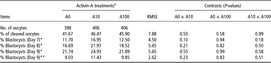
Effects of supplementing IVM medium with three different concentrations of activin-A (0, 10 and 100 ng/ml) on cleavage and blastocyst development rates from prepubertal goat oocytes

RMSE=root mean squared error.
Data are given as means. Treatments A0, A10 and A100 were: oocytes matured in TCM199 supplemented with 0, 10 and 100 ng/ml of activin-A, respectively.
a Overall treatment effect not significant for any of the measured parameters (P>0.05). Data analyzed by ANOVA with activin-A concentration in in vitro maturation medium as a treatment factor.
* Rate of blastocyst is calculated from cleaved embryos.
** Rate of blastocyst is calculated from in vitro matured oocytes.
Results
Experiment 1. Effect of activin-A during IVM on the nuclear stage, embryo development and blastocyst quality of prepubertal goat oocytes
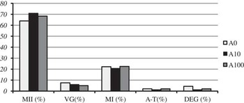
At the end of the IVM period, nuclear maturation stages were established and results are shown in Figure 1. Oocytes cultured in the presence of activin-A showed similar rates of oocytes at GV, MI, ATI and MII stage to those of the control group (A0). Indeed, the percentages of MII oocytes recorded after maturation for the 10 ng/ml and 100 ng/ml concentrations were not significantly different from those observed for the control oocytes (P>0.05). No differences according to activin-A concentration were observed (P>0.05).
Meiotic status of prepubertal goat oocytes matured in TCM 199 supplemented with various concentrations of activin-A during IVM. Treatments A0, A10 and A100: oocytes matured in TCM199 supplemented with 0, 10 and 100 ng/ml of activin-A for 24 h. GV=germinal vesicle; MI=metaphase I; A–T=anaphase–telophase; MII=metphase II; DEG=degenerated oocytes.

Table 1 shows the embryo development rates of oocytes cultured in the maturation medium alone or in the medium supplemented with activin-A (10 and 100 ng/ml). No differences in cleavage rates were detected among the different experimental groups (P>0.05). Similarly, no significant differences (P>0.05) were observed in the percentage of embryos developing to the blastocyst stage on Days 7, 8 and 9 pi. No interaction (P>0.05) between activin-A concentration and culture period were either observed.
An amount of 54 blastocysts were stained to assess the embryo quality. The total cell number of Day 8 blastocysts obtained from oocytes cultured in the maturation medium alone or in the medium supplemented with activin-A is summarized in Table 2. There were no differences among groups, irrespective of the maturation medium (P>0.05). All blastocysts showed a similar number of cells.
Effect of supplementing IVM medium with three different concentrations of activin-A (0, 10 and 100 ng/ml) on mean cell number from prepubertal goat blastocysts

RMSE=root mean squared error.
Data are given as means. Treatments A0, A10 and A100 were: oocytes matured in TCM199 supplemented with 0, 10 and 100 ng/ml of activin-A, respectively.
a Overall treatment effect not significant for any of the measured parameters (P>0.05). Data analyzed by ANOVA with activin-A concentration in in vitro maturation medium as a treatment factor.
Experiment 2. In vitro embryo development in the presence of 10 ng/ml activin-A during IVM and IVC of prepubertal goat oocytes
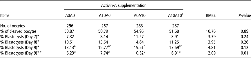
There were no significant differences in the cleavage rate among treatments. No significant differences were either observed when activin-A was added at the in vitro maturation medium (A10A0; A10A10) (P>0.05). However, embryo development, in terms of total number of blastocysts obtained at Day 9, improved significantly (P<0.05) with the addition of activin-A at the in vitro culture medium (A0A10). The highest blastocyst formation rate (19.5%) was obtained when activin-A was added at the in vitro culture medium (A0A10) but not at the in vitro maturation medium. Moreover, interaction effects between addition of activin-A at IVM medium and IVC medium were observed (P=0.001) (Table 3).
Effects of supplementing IVM and IVC media with 10 ng/ml activin-A on cleavage and blastocyst development rates from prepubertal goat oocytes

IVM=in vitro maturation; IVC=in vitro embryo culture; RMSE=root mean squared error.
Data are given as means. Treatment A0A0 and A0A10 were: oocytes matured in TCM199 and cultured in SOF medium supplemented with 0 and 10 ng/ml of activin-A, respectively. Treatments A10A0 and A10A10 were: oocytes matured in TCM199 supplemented with 10 ng/ml and cultured in SOF medium with 0 and 10 ng/ml of activin-A, respectively.
a,bValues in the same row with different superscript letters differ significantly (P<0.05).
c Interaction effects were observed only between addition of activin-A at IVM medium and IVC medium (P=0.001). Data analyzed by ANOVA where the factors were the composition of IVM (A0 v. A10) and IVC (A0 v. A10).
* Rate of blastocyst is calculated from cleaved embryos.
** Rate of blastocyst is calculated from in vitro matured oocytes.
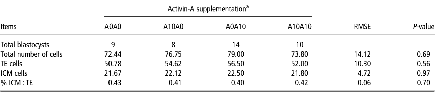
Blastocyst quality was evaluated in terms of total number of cells per blastocyst and the proportion of ICM cells and TE cells assessed by the differential staining (Table 4). No differences in any of these variables were detected among groups (P>0.05).
Number of TE and ICM cells in blastocysts developed from goat oocytes after IVM and IVC with or without 10 ng/ml activin-A

TE=trophectoderm; ICM=inner cell mass; IVM=in vitro maturation; IVC=in vitro embryo culture; RMSE=root mean squared error.
Data are given as means. Treatment A0A0 and A0A10 were: oocytes matured in TCM199 and cultured in presence of 0 and 10 ng/ml of activin-A, respectively. Treatments A10A0 and A10A10 were: oocytes matured in TCM199 supplemented with 10 ng/ml and cultured SOF medium supplemented with 0 and 10 ng/ml of activin-A, respectively.
a Overall treatment effect not significant for any of the measured parameters (P>0.05). Data analyzed by ANOVA where the factors were the composition of IVM (A0 v. A10) and IVC (A0 v. A10).
Discussion
The present study was designed to evaluate the effect of activin-A during the IVM and IVC on maturation rates, embryo development and embryo quality of prepubertal goat oocytes. In general, prepubertal oocytes show reduced developmental competence, when compared with results obtained with adult oocytes (Armstrong, Reference Armstrong2001). Indeed, the defective capacity of these oocytes to develop up to blastocyst at a rate equivalent to that of adult oocytes has been reported by several other studies in cattle (Presicce et al., Reference Presicce, Jiang, Simkin, Zhang, Looney, Godke and Yang1997), in sheep (Ledda et al., Reference Ledda, Bogliolo, Calvia, Leoni and Naitana1997), in pig (Marchal et al., Reference Marchal, Feugang, Perreau, Venturi, Terqui and Mermillod2001) and more recently in goats stimulated hormonally (Leoni et al., Reference Leoni, Succu, Satta, Paolo, Bogliolo, Bebbere, Spezzigu, Madeddu, Berlinguer, Ledda and Naitana2009).
In the present work, the presence of activin-A during IVM of cumulus-enclosed prepubertal goat oocytes did not affect nuclear maturation rates. Thus, the results obtained in Experiment 1 indicate that activin-A added to maturation media at concentrations of 10 and 100 ng/ml resulted in similar rates of oocytes in the MII stage, as compared with those oocytes that matured without activin-A. Similar results have already been reported in bovine (Vantol et al., Reference Vantol, Deloos, Vanderstichele and Bevers1994; Izadyar et al., Reference Izadyar, Zeinstra, Colenbrander, Vanderstichele and Bevers1996) and porcine (Coskun and Lin, Reference Coskun and Lin1994) oocytes, suggesting no effect of activin-A on oocyte maturation. Considering that activin-A is able to directly stimulate FSH synthesis and secretion, promote the release of the GnRH, and also stimulate the increase of FSH and LH receptors in the granulosa cells (Alak et al., Reference Alak, Coskun, Friedman, Kennard, Kim and Seifer1998; Norwitz et al., Reference Norwitz, Xu, Jeong, Bedecarrats, Winebrenner, Chin and Kaiser2002), the ineffectiveness of exogenous activin-A may be either because of activin-A production by the COCs and/or the presence of FSH and LH in our culture medium. However, this does not exclude the possibility that activin-A, secreted by the granulosa cells and present in the follicular fluid, has an effect on the cytoplasmic maturation of goat oocytes, which would affect subsequent pre-implantation embryo development. Nevertheless, our results demonstrate that activin-A added to IVM has no effect on the percentage of blastocysts obtained after IVF. This observation is consistent with other reports in bovine (Vantol et al., Reference Vantol, Deloos, Vanderstichele and Bevers1994; Izadyar et al., Reference Izadyar, Zeinstra, Colenbrander, Vanderstichele and Bevers1996) and pig (Coskun and Lin, Reference Coskun and Lin1994), which showed that adding activin-A during IVM did not result in an improvement of blastocyst development.
Regarding the effect of activin-A during the whole embryo culture period, our data clearly show that activin-A promotes embryonic development, showing an improving effect on the blastocyst yield of prepubertal goat oocytes. Given that activin-A is produced by the oviduct epithelial cells (Gandolfi et al., Reference Gandolfi, Modina, Brevini, Passoni, Artini, Petraglia and Lauria1995), addition of activin-A to embryo culture in vitro could help reproduce the environment of the cleavage-stage embryos in the oviduct in vivo. Controversial results have been reported about the effects of activin-A on embryo development depending on the timing of its addition to the culture medium. Thus, Yoshioka et al. (Reference Yoshioka, Suzuki and Iwamura1998a) observed that activin-A had an effect on blastocyst yield when it was added to IVC medium of bovine embryos before the 9- to 16-cell stage. However, a recent study on bovine (Trigal et al., Reference Trigal, Gomez, Diez, Caamano, Martin, Carrocera and Munoz2011) has demonstrated a better effect of activin-A when it is added later in culture media. The development-enhancing effect of activin-A may be associated with the timing of the activation of appropriate genes within the embryonic genome. Studies in mouse (Lu et al., Reference Lu, Matsuyama, Nishihara and Takahashi1993) and bovine (Yoshioka et al., Reference Yoshioka, Takata, Taniguchi, Yamanaka and Sekikawa1998b) showed that activin-A and activin receptors are detected in embryos from the zygote to the morula stage, suggesting that the protein might play a role in embryogenesis during the early stage of embryo development. Our data show an evident improving effect on Day 9 blastocyst yield, suggesting an effect during the late stages of embryo development in prepubertal goats. Otherwise, we should also consider the possibility of an accumulative effect of activin-A added during in vitro maturation and culture medium. Consequently, greater effects are observed as the in vitro culture is longer. Thus, further studies will be required to determine the functions of activin-A during these stages of development.
Finally, the quality of blastocysts, as determined by the evaluation of their cell number, seemed to be similar among all of the groups. The number of cells of the embryo may be informative of embryonic viability (VanSoom et al., Reference VanSoom, Ysebaert and DeKruif1997). The lower quality of embryos produced in vitro could be attributed to a lower number of cells. Cell counts were unaffected by the presence of activin-A, similarly to other authors showing no differences in total cell counts between bovine blastocysts produced in the presence or absence of activin-A (Yoshioka et al., Reference Yoshioka, Suzuki and Iwamura1998a, Trigal et al., Reference Trigal, Gomez, Diez, Caamano, Molina, Carrocera, Martin and Munoz2010). In addition, in our study, blastocyst cell counts were similar to those reported in adult and prepubertal goats (Koeman et al., Reference Koeman, Keefer, Baldassarre and Downey2003; Romaguera et al., Reference Romaguera, Casanovas, Morato, Izquierdo, Catala, Jimenez-Macedo, Mogas and Paramio2010).
Several studies have demonstrated that the expression of protein and mRNA for activin-A and activin receptors in the ovary is present in both oocytes and granulosa cells of follicles at various stages of development (pigs: (Van den Hurk and Van de Pavert, Reference Van den Hurk and Van de Pavert2001); and ruminants: (Hulshof et al., Reference Hulshof, Figueiredo, Beckers, Bevers, Vanderstichele and Van den Hurk1997; Izadyar et al., Reference Izadyar, Dijkstra, Van Tol, van de Eljnden-Van Raaij, Van den Hurk, Colenbrander and Bevers1998)), suggesting a physiological role of activin in the growth and development of follicles and/or oocytes. Although there is convincing evidence that activin signaling is important for ovarian function, information on its localization and function is mainly obtained from rodents and cows. Few studies on goats (Silva et al., Reference Silva, van den Hurk, van Tol, Roelen and Figueiredo2004 and Reference Silva, Tharasanit, Taverne, Van der Weijden, Santos, Figueiredo and Van den Hurk2006) have demonstrated that activin-A, its binding protein follistatin and its receptors are formed in all types of goat follicles, in all compartments of antral follicles and in ovarian surface epithelium. The widespread distribution pattern of the follistatin–activin–activin receptor system in goat ovaries points to a crucial role of this system in various reproductive processes, including follicle growth and differentiation, and luteal activity. However, there are limited data on goats and on prepubertal animals (Brawtal, Reference Brawtal1994; Zhao et al., Reference Zhao, Taverne, van der Weijden, Bevers and Van den Hurk2001; Patel et al., Reference Patel, Bettegowda, Ireland, Coussens, Lonergan and Smith2007) about the expression of protein and mRNA for activin-A and activin receptors. Therefore, it is necessary to determine whether activin-A and its receptors are present in the embryos from the zygote to the morula stage to improve the results on in vitro embryo development of prepubertal goat oocytes.
In conclusion, the addition of 10 ng/ml of activin-A throughout the whole embryo culture improves blastocyst yield. This is the first study that provides data of the effects of activin-A on in vitro developmental potential of prepubertal goat oocytes. Nevertheless, to the best of our knowledge, there is no study reporting the gene expression and protein localization for activin-A and activin receptors in prepubertal goats. A detailed profiling of the activin-A effects on embryonic development of prepubertal goat oocytes and determination of gene expression and protein synthesis for activin-A require further experiments.
Acknowledgments
This study was financially supported by the Spanish Ministry of Science and Innovation (Project Number: code: AGL2011-23784) and a predoctoral grant awarded to S.H. from the Spanish Ministry of Foreign Affairs and Cooperation. The authors are grateful to Dr Chuck Simmons, an English instructor of our University, for his English assistance.