Congenital coronary artery fistulas are abnormal connections between the coronary arteries and the chambers of the heart or major thoracic vessels. Many spontaneously regress. Since most patients are asymptomatic, the incidence of coronary artery fistulas is likely underestimated. However, many do develop symptoms, including dyspnoea, fatigue, and angina. Reference Loukas, Germain, Gabriel, John, Tubbs and Spicer1–Reference Lo, Lin and Hsieh4 Complications include congestive heart failure, arrhythmias, myocardial ischaemia, coronary aneurysm and rupture, endocarditis, and sudden death. Reference Loukas, Germain, Gabriel, John, Tubbs and Spicer1–Reference Luo, Kebede, Wu and Stouffer3,Reference Liberthson, Sagar, Berkoben, Weintraub and Levine5 We present two similar cases of coronary fistulas.
Case presentation
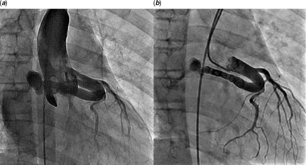
Patient 1: A 10-year-old girl, diagnosed with coronary artery fistula incidentally by echocardiogram as a newborn, was asymptomatic for many years. She was lost to follow-up, but then developed 1 month of chest pain with exertion. Echocardiogram showed a dilated left main coronary artery and a large proximal coronary fistula draining to the right atrium. Cardiac catheterisation demonstrated a mildly elevated Qp/Qs ratio of 1.2:1 and a large fistula from the proximal left main coronary artery draining into the right atrium. The diameters of the mid-fistula and narrowest part of the fistula were 7.8 and 4.6 mm, respectively. The left main coronary artery was selectively engaged with a 5F JL3 catheter, and a microcatheter was advanced around the curve to the distal end of the fistula, where a 6.5 mm Covidien® Microvascular Plug was deployed. There was still flow through the fistula, so a Concerto® 8 × 30 mm detachable coil was placed as well. Coronary flow was noticeably improved after fistula closure (Fig 1). There were no procedural complications. At follow-up 2 months later, she had no symptoms, and echocardiography showed complete occlusion of the fistula and a reduction in left main coronary artery diameter to 4.7 mm.
Coronary artery fistula in Patient 1 ( a ) with improved coronary flow after closure ( b ).

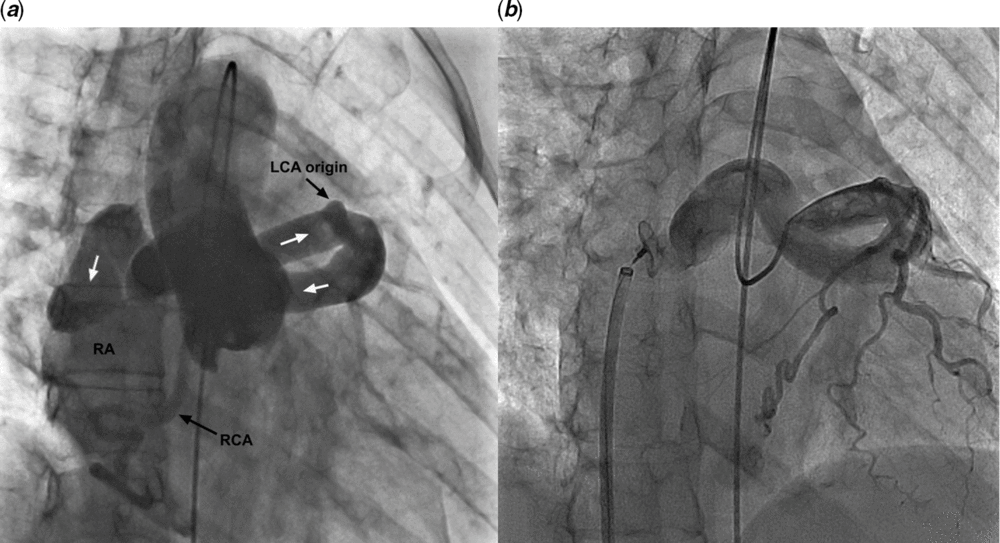
Patient 2: A 43-year-old female presented with a year-long history of recurrent extremity numbness, palpitations, and dyspnoea on exertion. Transesophageal echocardiogram showed an enlarged left main coronary artery measuring 2 cm with a large, tortuous coronary artery fistula (Fig 2). CT angiography confirmed coronary artery fistula drainage to the right atrium and also demonstrated bilateral small peripheral pulmonary emboli. The source of these was unknown but may have occurred from left to right shunt of tiny particles through the fistulous track. Cardiac catheterisation revealed a significant left-to-right shunt (Qp/Qs ratio 2.5:1), elevated pulmonary artery pressures (36 mmHg systolic and 23 mmHg mean), and a large coronary artery fistula that arose from the proximal left coronary artery and drained into the right atrium. The diameters of the mid-fistula and narrowest part of the fistula were 18.7 and 8.8 mm, respectively. The right coronary artery was mildly dilated and tortuous, suggesting a small distal fistula there as well. A 5F AL1 coronary catheter selectively engaged the left coronary artery, and a microcatheter was passed through it, along with a 0.014″ guidewire. Despite the tortuous fistula path, the wire acted as a support and allowed the microcatheter to advance through the distal connection into the right atrium. The microcatheter was then snared from a femoral venous approach, and after advancing the wire, it was exteriorised to create a continuous arteriovenous loop. A 10F Flexor sheath (Cook®) was then advanced from the venous side and its tip positioned in the distal portion of the fistula. The fistula was successfully occluded by placement of an 18 mm Amplatzer® Vascular Plug II. That evening, the patient developed atrial fibrillation, which resolved spontaneously. While compromise of sinus node artery flow could explain this rhythm, the angiograms showed this vessel to arise from the circumflex artery with no such artery arising from the right coronary artery. The next day, she developed angina and had a slight elevation of her troponin level and mild ST-segment elevation on electrocardiogram, so an urgent repeat catheterisation was performed, which showed patent coronary arteries, complete occlusion of the coronary fistula, and filling defects in the distal portion of the fistula pouch likely representing thrombus. She was placed on warfarin and aspirin for several months and then switched to aspirin and apixaban for long-term therapy. Atrial fibrillation recurred several weeks later and successfully converted to sinus rhythm with sotalol treatment. Three months after catheterisation, she had resolution of her symptoms, and echocardiography showed no residual fistula flow and a reduction in left main coronary artery diameter to 9.0 mm.
( a ) White arrows indicate flow through large coronary artery fistula in Patient 2. ( b ) After closure.

Discussion
Most coronary artery fistulas are small and patients are usually asymptomatic. While some fistulas close spontaneously, many are thought to grow with age and cause more symptoms later in life. Coronary fistulas can cause symptoms by producing a left-to-right shunt and volume overloading of the heart or by causing coronary steal where blood flows preferentially through the fistula instead of distal myocardial circulation.Reference Latson 2 It is generally agreed that all symptomatic coronary fistula should be closed. The management of asymptomatic coronary fistulas is controversial. While many advocate for closure of asymptomatic fistulas that are medium-sized or larger in children because of the risk of future complications, others advise conservative management since fistula may regress or completely resolve. Reference Latson2,Reference Sunder, Balakrishnan and Tharakan6,Reference Ceresnak, Gray, Altmann, Chen, Glickstein and Hellenbrand7 Periprocedural complications of transcatheter closure of coronary fistulas include myocardial ischaemia, arrhythmia, coronary artery dissection, or device embolisation. Late complications after closure include dilation or stenosis of coronary arteries, arrhythmias, and occlusion of coronary arteries, presumably due to thrombus propagation within the dead space area. Reference Latson2,Reference Ceresnak, Gray, Altmann, Chen, Glickstein and Hellenbrand7–Reference Jama, Barsoum, Bjarnason, Holmes and Rihal9 Older patients, as well as those with hypertension and hyperlipidaemia, have been shown to have more late complications after closure, suggesting that fistula closure may be safer at a younger age before patients develop further comorbidities that put them at higher risk.Reference Valente, Lock and Gauvreau 10
In both of our cases, the coronary fistulas were virtually identical in origin, course, and drainage. Both were proximal and distinct from the anterior descending and posterior circumflex branches, turning sharply posteriorly and rightward to course posteriorly behind the aorta and enter the right atrium superiorly. There was dilation of the proximal left main coronary artery due to the coronary fistula in both, but the fistula in our older patient was significantly larger with a greater left-to-right shunt, likely due to progressive dilation of the fistula over time. Transcatheter device closure was successful in both, but challenges occurred due to tighter catheter turns in the child and due to the large fistula diameter in the adult, who had a complicated post-procedure course. While one cannot derive a conclusion that a natural progression of this entity exists based on just these two cases, we are inclined to believe that earlier intervention may prevent fistula enlargement and greater haemodynamic significance in later life. Therefore, we recommend giving strong consideration to closure of coronary fistulas at younger ages to minimise later complications.
Conflicts of Interest
None.
Ethical Standards
This study was performed in accordance with ethical standards of our institutional research policies.