Introduction
In developing countries, 30-50% of potential production (quality and quantity) is lost because of the poor disease control (OIE, 2007; BBSRC, 2013). Molecular diagnostics are not commonly performed in developing countries because of the limited availability of sophisticated laboratory equipment to carry out these tests. This diagnosis should be adopted in small poultry laboratories, especially for fastidious micro-organisms which have prioritised detection using modern molecular techniques. Conventional diagnostic tools (serology and culture) are insufficient to effectively monitor poultry infections. Many pathogens are fastidious in nature, so it is difficult to culture them. Some need longer incubation to grow ranging from days to weeks, whereas, the presence of non-specific reactors hinders the serological diagnosis. Molecular methods such as polymerase chain reaction (PCR), multiplex PCR, and real-time PCR have been used to detect different infectious diseases in chickens (Sprygin et al., Reference SPRYGIN, ANDREYCHUK, KOLOTILOV, VOLKOV, RUNINA, MUDRAK, BORISOV, IRZA, DRYGIN and PEREVOZCHIKOVA2010). Although, PCR assays are rapid and precise, they require agarose gel electrophoresis which increases the risk of false positive results due to amplicon carry-over. The conventional assay is an end-point PCR reaction. Real-time PCR involves less handling of the products as compared to the conventional assay; however, the use of a costly thermal cycler with a fluorescence detector limits the effectiveness of this technique in poorly funded laboratories (Saito et al., Reference SAITO, MISAWA, MORIYA, KOIKE, UBUKATA and OKAMURA2005). PCR methods are further limited by doubling-by-cycle amplification and most of the time cannot be used for detection of DNA or RNA directly from a variety of crude sample preparations. PCR methods are sensitive to sample matrix inhibitors and are not amenable to visual detection of amplified product, which reduces their use for pathogen detection in field settings (Takahashi et al., Reference TAKAHASHI, FUKUTA, KUROYANAGI, MIYAKE, NAGAI, KAGEYAMA and ISHIGURO2014).
Loop-mediated isothermal amplification (LAMP) is a novel nucleic acid amplification technology, recently developed and reported by Notomi et al. (Reference NOTOMI, OKAYAMA, MASUBUCHI, YONEKAWA, WATANABE, AMINO and HASE2000). It offers simple, one step, closed-tube detection of amplification (µg yields in <15 minutes) which is compatible with multiple detection methods. The amplification is carried out under isothermal condition usually (60-65°C), in presence of Bst I or Bsm DNA polymerase which exhibits high strand displacement activity. LAMP does not require additional reverse transcription step for amplification of RNA viral gene products, and can amplify DNA with high specificity, sensitivity, and speed (Notomi et al., Reference NOTOMI, OKAYAMA, MASUBUCHI, YONEKAWA, WATANABE, AMINO and HASE2000).
Various reliable LAMP assays have been reported for detection of poultry diseases including avian influenza, Newcastle disease, infectious bronchitis, infectious bursal disease, hydropericardium syndrome (HPS), Marek's disease, chicken infectious anaemia, avian mycoplasmosis, fowl cholera, and avian coccidiosis (Eimeria spp.). Currently, there is only one published review describing LAMP assays developed for the detection of viral infections in poultry (Mansour et al., Reference MANSOUR, ALI, CHASE and CEPICA2015). The following review aims to cover the developed LAMP assays reported to date, for the detection of different types of important pathogens infecting poultry.
The principle and major advantages of LAMP
The LAMP assay incorporates a set of four to six primers recognising six to eight sites on a conserved target gene. The assay works on the principle of auto cycling strand displacement DNA synthesis carried out by Bst or Bsm DNA polymerase. The primers can easily be generated from conserved DNA sequences of poultry pathogens. Primers may be designed by selecting the parameters defaults of LAMP primer design software named ‘Primer Explorer’ V4 or V5 http://primerexplorer.jp/elamp4.0.0/index.html developed by the Eiken Chemical Co., Ltd, Tokyo, Japan. The two outer primers consist of a forward outer primer (F3) and backward outer primer (B3). The inner primers are named as the forward inner primer (FIP) and backward inner primer (BIP), as described by Notomi et al. (Reference NOTOMI, OKAYAMA, MASUBUCHI, YONEKAWA, WATANABE, AMINO and HASE2000). Loop primers (LP) are optional primers, and, if incorporated in to LAMP reaction, may further increase specificity and reduce the time of amplification (Barkway et al., Reference BARKWAY, POCOCK, VRBA and BLAKE2011).
LAMP reagents, reaction mixture and kits
The LAMP reaction mixture includes the addition of four to six sets of primers, Bst or Bsm DNA polymerase large fragment, dNTPs, reaction buffer and template DNA. The reaction mixture can be incubated in a water bath or heat block. Amplification is performed at 60-65°C for 30-60 min followed by heating at 80°C for 5 min to terminate the reaction (Peng et al., Reference PENG, XIE, LIU, PANG, DENG, XIE, XIE, FAN, FENG and KHAN2011). The whole amplification (109-1010 copies) is completed within 30 to 60 min at 63°C, isothermally. Because of the high number of DNA copies, the amplified product can be detected by visualising turbidity or fluorescence detection, thereby limiting analysis on gel.
The reagents used in LAMP assay are comparatively thermo-stable. Dried reagents fixed on the lids can save steps for preparing the reaction mixture. Such reagents are stable at room temperature for at least one year (no need to be stored in the refrigerator) as reported by Mair et al. (Reference MAIR, VILEI, WADE, FREY and UNGER2013). The lyophilised reaction mixture (LAMP amplification kit) is commercially available as customised products from Thermo Fisher Scientific, Lithuania to be stored at 2-8°C in the dark. This lyophilised reaction mixture uses FAMTM fluorescent dye as indicator to be used in a portable equipment ESEQuant tube scanner (Qiagen) or any real-time PCR detection system. Similarly, a new WarmStart® Colorimetric LAMP 2X Master Mix, (New England Bio Labs Ltd. UK) utilises pH sensitive phenol red, as an end-point indicator. This system is designed for visual detection of amplification based on production of proton from the extensive Bst 2.0 WarmStart® DNA polymerase activity or WarmStart® RTx Reverse Transcriptase activity, producing a change in solution colour from pink to yellow. A novel commercial WarmStart® LAMP Kit (DNA & RNA), NEB, UK contains a blend of Bst 2.0 WarmStart® DNA polymerase and WarmStart® RTx Reverse Transcriptase. This WarmStart LAMP Kit is compatible with multiple detection methods, including fluorescence and or turbidity (as the end-point visualisation).
Quantitative capability of the LAMP
The LAMP method is an efficient method which allows synthesis of large amount of DNA in a short time with great specificity. As the reaction progress it produces precipitates of magnesium pyrophosphate which can be detected by spectroscopic or enzymatic analysis. The loopamp real-time turbidimeter (LA-320, Teramecs, Co., Ltd., Kyoto, Japan) and LA-500, (Eiken Chemical Co., Ltd.) are specifically designed instruments for real-time monitoring of the LAMP reaction. The reaction is considered positive when the turbidity reaches 0.1 within 60 min at 650 nm. The time needed for the turbidity of each tested sample to exceed at 0.1 is referred to as the ‘threshold time’ (TT). In other words, the turbidity-versus-time curve obtained using the real-time turbidimeter represents the reaction curve of the LAMP amplification (Mori et al., Reference MORI, KITAO, TOMITA and NOTOMI2004). Such quantitative LAMP (qlamp) assays have been successfully applied for the quantitative detection of Campylobacter jejuni and C. coli in chicken carcass and water rinses using a real-time turbidimeter (Sabike et al., Reference SABIKE, UEMURA, KIRINO, MEKATA, SEKIGUCHI, OKABAYASHI, GOTO and YAMAZAKI2016; Yamazaki et al., Reference YAMAZAKI, TAGUCHI, KAWAI, KAWATSU, SAKATA, INOUE and MISAWA2009).
Another assay is the PMA-LAMP (propidium monoazide treatment) assay as reported by Youn et al. (Reference YOUN, JEONG, CHOI, JUNG and KANG2017). The PMA-LAMP assay is a rapid, accurate, and simple method for quantifying viable Salmonella spp. without pre-enrichment in raw samples, such as rinse water of chicken carcasses (e.g. treated with sanitisers). The LAMP reaction was performed with a Genie II (Optigene, UK) real-time fluorometer, which displays time to positivity and amplicon melting temperature (Tm). Youn et al. (Reference YOUN, JEONG, CHOI, JUNG and KANG2017) has reported a high correlation (R2 = 0.99 to 0.976) between LAMP time threshold (TT) values and viable Salmonella spp. with a quantification range of 1.0×10³ to 1.0×10⁸ CFU/ml in pure culture and rinse water of chicken carcasses.
LAMP assays available for detection of economically important chicken pathogens
application of lamp for detection of avian viruses
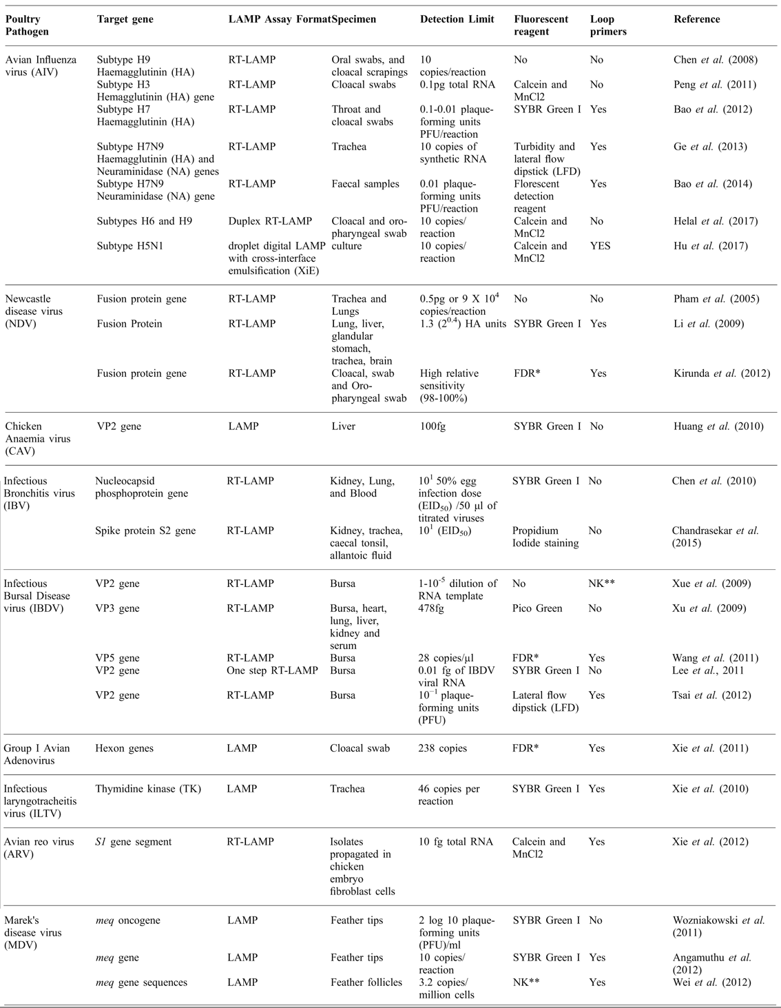
Recently, various workers have reported the development of LAMP reactions for different poultry viral pathogens. Pham et al. (Reference PHAM, NAKAJIMA, OHASHI and ONUMA2005) detected the fusion protein gene of Newcastle disease (ND) virus from tracheal and lung specimens, and its improved version has been demonstrated by Li et al. (Reference LI, XUE, QIN, ZHOU, CHEN, BI and CAO2009). Kirunda et al. (Reference KIRUNDA, THEKISOE, KASAIJA, KERFUA, NASINYAMA, OPUDA-ASIBO and INOUE2012) detected the ND fusion protein gene from less invasive samples, such as oropharyngeal and cloacal swabs, and the assay incorporated loop primers (LP). These are added to increase specificity and to reduce the time of incubation for LAMP reaction by up to several minutes. Peng et al. (Reference PENG, XIE, LIU, PANG, DENG, XIE, XIE, FAN, FENG and KHAN2011) detected avian influenza (AI) virus, hemagglutinin (HA) gene from cloacal swab samples using calcein and MnCl2 as an end-point indicator for turbidity and colour change in positive samples. Later work by Ge et al. (Reference GE, WU, QI, ZHAO, GUO, ZHU, QI, SHI, ZHOU and WANG2013) detected avian origin influenza virus from tracheal swabs on a lateral flow dipstick (LFD) format to provide ease in visual identification of reaction in field conditions. Helal et al. (Reference HELAL, LI, GIRSHICK and KHAN2017) reported duplex LAMP for the detection of subtypes H6 and H9 of AI, and Hu et al. (Reference HU, XU, LUO, HE and DU2017) used droplet digital LAMP (dLAMP) for the detection of H5N1. In the dLAMP assay, emulsification was carried out using cross-interface emulsification (XiE) platform, which generates monodisperse droplets using a vibrating capillary, which can be easily set up and is user friendly to those who lack microfabrication facilities. Xie et al. (Reference XIE, TANG, FAN, LIU, PANG, DENG, XIE, PENG, XIE and KHAN2011) detected the hexon gene of Group I avian adenovirus from a cloacal swab sample using a commercial fluorescent detection reagent (FDR; Eiken Chemical Co., Ltd, Tokyo, Japan). In addition, Xie et al. (Reference XIE, JI, PICKENS, DU, CAO, LI, WANG, MA and BI2010) developed a LAMP assay incorporating LP for detection of infectious laryngotracheitis (ILT) virus by amplifying its thymidine kinase (TK) gene. Its visual detection was made by adding SYBR Green I dye. Reverse transcriptase LAMP (RT-LAMP) for avian reo virus (S1 gene) was developed by the Xie et al. (Reference XIE, PENG, LUO, WANG, LIU, PANG, DENG, XIE, XIE and FAN2012) incorporating calcein and MnCl2 to aid in visualisation of amplified product in the reaction tube.
Marek's disease virus (meq gene sequence) has been detected by Wei et al. (Reference WEI, SHI, ZHAO, ZHANG, WANG, LIU, CUI, HU, QUAN and CHEN2012) from feather follicles using LP. Other LAMP assays for Marek's disease virus were reported by Wozniakowski et al. (Reference WOZNIAKOWSKI, SAMOREK-SALAMONOWICZ and KOZDRUN2011) and Angamuthu et al. (Reference ANGAMUTHU, BASKARAN, GOPAL, DEVARAJAN and KATHAPERUMAL2012) to specifically amplify the meq oncogene and its detection through SYBR green stain. Chen et al. (Reference CHEN, ZHANG, MA, MA, DING, LIU, CAI, MA, ZHANG and LIU2010) established a RT-LAMP assay for the detection of infectious bronchitis virus (IBV) based on the nucleocapsid phosphoprotein gene of IBV. A LAMP assay to detect chicken anaemia virus (CAV) targeting the VP2 gene of CAV was developed by (Huang et al., Reference HUANG, LAI, LEE, LIN, LIEN, HSUEH, KAO, CHANG, LU and LIN2010). Additionally, various RT-LAMP assays have been reported for the detection of infectious bursal disease (IBD) of chicken and the key features of all LAMP assays developed so far, for the detection of important viral pathogens of chickens are detailed in Table 1.
LAMP assays developed for detection of viral pathogens of chickens.

*FDR= Fluorescent detection reagent, Eiken Chemical Co. Ltd, Tokyo, Japan; **NK=Not Known; RT-LAMP= Reverse transcriptase-Loop-mediated Isothermal Amplification
application of lamp for detection of bacterial pathogens of poultry
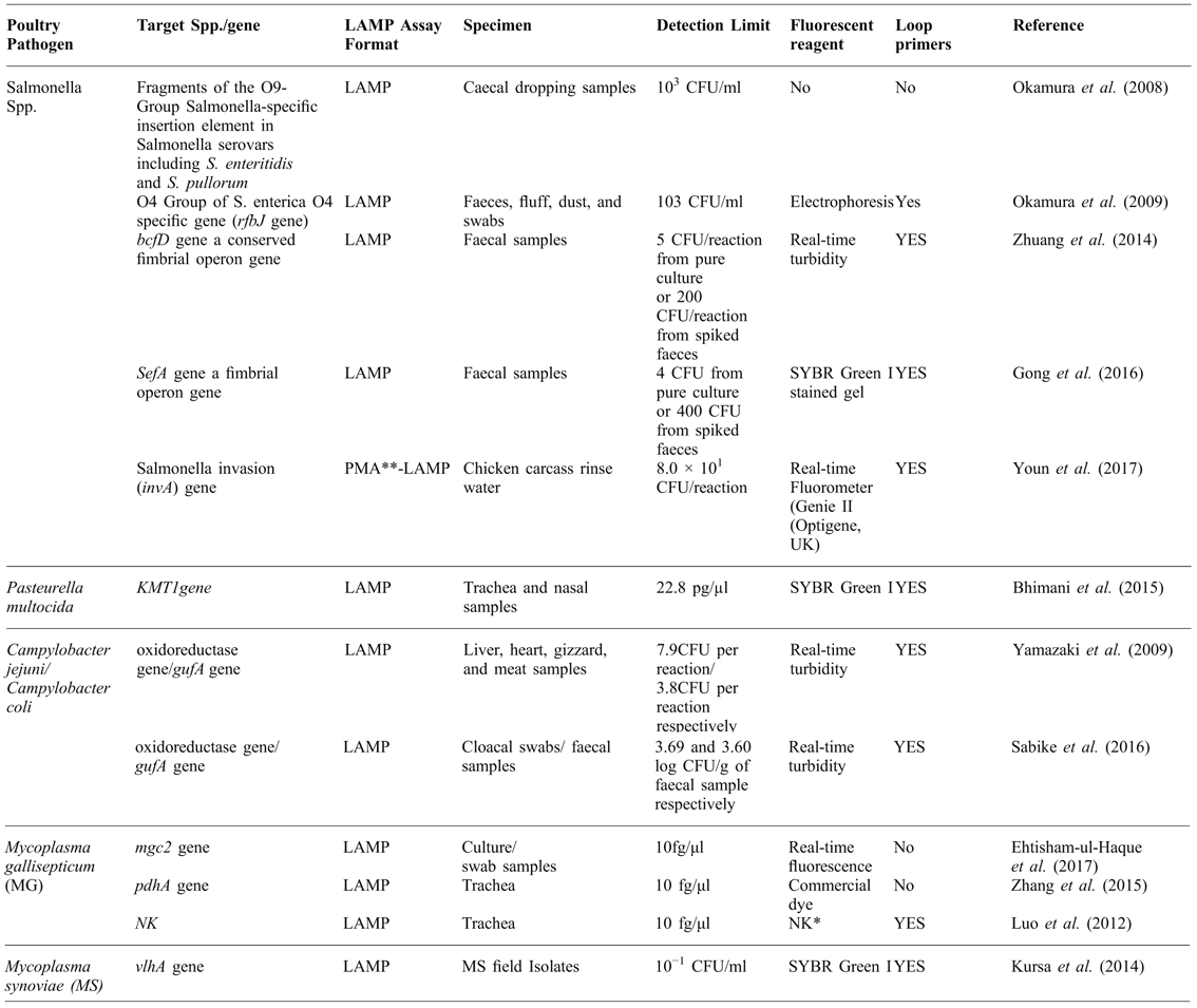
In human medicine, there is a LAMP kit available for the field diagnosis of bacterial infection caused by Mycobacterium tuberculosis (WHO, 2013). However, the use of the LAMP method for detection of bacterial pathogens of livestock and poultry is limited. To allow its widespread application in veterinary diagnostics, there has to be a recognition and promotion of LAMP technology by the world organisation for animal health (OIE) and other regulatory agencies (Mansour et al., Reference MANSOUR, ALI, CHASE and CEPICA2015). Salmonella spp. in poultry were initially detected by LAMP assays from caecal droppings (Okamura et al., Reference OKAMURA, OHBA, KIKUCHI, SUZUKI, TACHIZAKI, TAKEHARA, IKEDO, KOJIMA and NAKAMURA2008; Zhuang et al., Reference ZHUANG, GONG, LI, ZHU, YU, DOU, LIU, XU and WANG2014; Gong et al., Reference GONG, ZHUANG, ZHU, SHI, ZHANG, ZHANG, YU, DOU, XU and WANG2016; Youn et al., Reference YOUN, JEONG, CHOI, JUNG and KANG2017) (Table 2). For other pathogens, Pasteurella multocida from chickens was detected through LAMP by Bhimani et al. (Reference BHIMANI, BHENDERI and ROY2015) using SYBR green stain as an end-point indicator for visual detection of the amplification. Campylobacter jejuni (oxidoreductase gene) and C. coli (gufA gene) have been detected in form of real-time turbidity in the LAMP reaction from tissue, meat and faecal samples of chicken. Among other bacterial pathogens, avian mycoplasmas cause dramatic disease problems (chronic respiratory disease) with economical losses. Although mycoplasma are economically significant in poultry, only a limited number of LAMP assays have been reported for the detection of Mycoplasma gallisepticum (MG) and Mycoplasma synoviae (MS), as shown in Table 2. A rapid conventional LAMP assay can detect MG within 30 min at 60°C. This assay targets the highly conserved pdhA gene of MG, resulting with 10-fold higher sensitivity as compared to PCR (Zhang et al., Reference ZHANG, BAO, YU, CHENG, TAN, QIU, SONG, DAI, FEI and DING2015). Recently, Ehtisham-ul-Haque et al. (Reference EHTISHAM-UL-HAQUE, KIRAN, WAHEED and YOUNUS2017) reported a real-time LAMP assay for the detection of cytadhesin (mgc2) gene of MG using an ESEQuant tube scanner (Qiagen). For the detection of MS, Kursa et al. (Reference KURSA, WOŹNIAKOWSKI, TOMCZYK, SAWICKA and MINTA2014) developed a sensitive LAMP assay targeting variable haemagglutinin (vlhA) gene of MS.
LAMP assays developed for detection of bacterial pathogens of chickens.

*NK=Not Known; **PMA= propidium monoazide treatment
application of lamp for detection of protozoan parasites of poultry
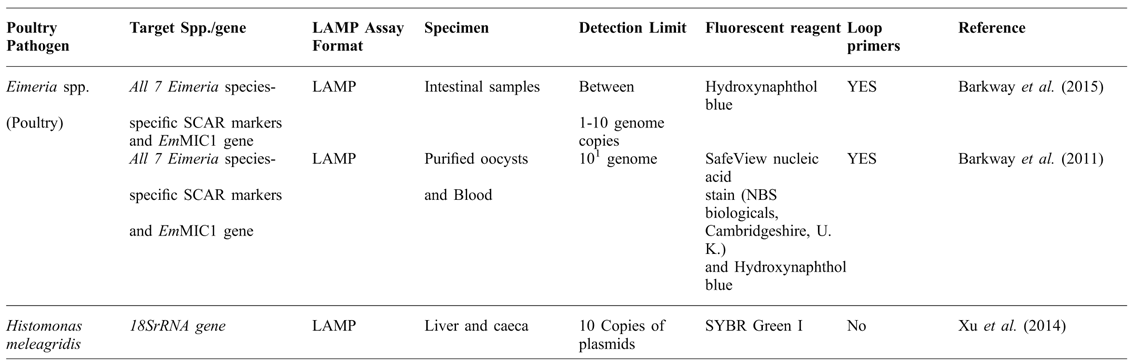
LAMP has been widely used in detecting infections caused by protozoan parasites such as Toxoplasma gondii, Trypanosoma spp., Theileria annulata, Neospora caninum, Babesia caballi, Leishmania spp, bovine babesiosis trichinellosis, bovine trichomonosis, malaria and giardiasis as described by Ramos et al. (Reference RAMOS, MUÑOZ, CORTÉS-VECINO, BARATO and PATARROYO2017) and Xu et al. (Reference XU, QU and TAO2014). In human medicine, there has been a major LAMP field success in form of a malaria detection LAMP kit approved by WHO, as reported by Patel et al. (Reference PATEL, LUCCHI, SRIVASTAVA, LIN, SUG-ARAM, ARUNCHARUS, BHARTI, SHUKLA, CONGPUONG, SATIMAI, SINGH, UDHAYAKUMAR and MESHNICK2014). Only few LAMP assays have been described for the detection of avian protozoan parasites. Barkway et al. (Reference BARKWAY, POCOCK, VRBA and BLAKE2011) and Barkway et al. (Reference BARKWAY, POCOCK, VRBA and BLAKE2015) have reported the LAMP technique for detection of all seven Eimeria spp. in chickens by amplification of their species-specific (SCAR markers and EmMIC1 gene) from purified oocysts, intestinal and blood samples using loop primers (Table 3). In this assay SafeView nucleic acid stain (NBS Biologicals, Cambridgeshire, UK) and hydroxynaphthol blue were used to visualize the final product. Xu et al. (Reference XU, QU and TAO2014) has reported the visual detection of 18SrRNA gene of Histomonas meleagridis infection in chicken.
LAMP assays developed for detection of protozoan pathogens of chickens.

Conclusions
Various LAMP assays have been developed for the molecular diagnosis of important chicken pathogens, based upon pathogen type, specimen, target genes, LAMP primers type, detection limit, fluorescent detectors and LAMP chemistry used. If the LAMP technique is properly applied, may be a useful diagnostic procedure in developing countries, as it does not require expensive equipment. Molecular diagnostics should be adopted in small microbiology laboratories, especially for fastidious pathogens like avian mycoplasma species (MG and MS) which are hard to culture via conventional techniques and are prioritised for detection using molecular techniques. It is recommended that the technique of LAMP should be adopted in a national disease control programme for avian infections in resource-limited countries. LAMP poultry disease diagnostics holds great potential for field and laboratory use. However, for its widespread application in veterinary diagnostics, there should be recognition and promotion of the LAMP technology by OIE, other animal health regulatory authorities and national poultry improvement plans (NPIP). This implementation will further pave the ways toward development of simple, cost effective, standardised, accurate and thermostable LAMP kits.