Implications
The improved knowledge of ruminal ion transport clearly underlines its physiological meaning for the whole animal. For example, magnesium absorption is markedly reduced at low Mg and high K intake and the effect of potassium is diminished at high Mg intake. This variable interaction has been quantified recently permitting the prediction of Mg absorption. Great progress has also been made in understanding the interactions between the absorptive pathways for Na, short chain fatty acids and ammonium. The new findings on structure and regulation of various ion transporters will allow a better understanding of the challenges that different diets pose to the maintenance of homeostatic conditions within the rumen and within the cells of the surrounding epithelium, with the implications for the investigation of ruminal adaptation to different diets. Future studies on transport pathways should include the barrier function of rumen epithelium and its possible impairment under harsh feeding conditions.
Introduction
An efficient digestion of plant material by ruminants requires a large community of microorganisms within the forestomachs, with an ample secretion of saliva ensuring an optimal environment. Within the large volume of saliva (>10 l in sheep and up to 300 l in cows; Kay, Reference Kay1960; Erdmann, Reference Erdmann1988; Allen, Reference Allen1997; Maekawa et al., Reference Maekawa, Beauchemin and Christensen2002) sodium (Na+) and bicarbonate (HCO3−) represent the main cation and anion, respectively. Daily salivary secretion of Na+ can exceed the Na+ content of the whole animal by a factor of 2 to 3 (Silanikove, Reference Silanikove1994), and salivary secretion is a major factor leading to a rapid reduction of plasma volume after feeding (Blair-West and Brook, Reference Blair-West and Brook1969). Secretion of HCO3− via saliva is 6 to 8 times as high as the amount in the extracellular space (Erdmann, Reference Erdmann1988) and thus poses a significant challenge to the acid-base balance of the ruminant. As the requirements of the fermentational processes in the rumen only allow a slow release of ruminal material into the lower parts of the digestive tract, efficient absorptive mechanisms across forestomach epithelia are essential to allow for an early recycling of these essential electrolytes and water from the gastrointestinal cavity to the blood. Thus, 80% of the sodium that is secreted by saliva is absorbed by the forestomachs (Dobson, Reference Dobson1959), with the rumen alone absorbing an amount that exceeds the sodium content of the body (Silanikove, Reference Silanikove1994). Fermentation releases additional cations and anions from organic matter that have to be absorbed to meet the requirements of ruminal homeostasis in terms of regulating osmotic pressure (Lang and Martens, Reference Lang and Martens1999) and pH (Gäbel and Aschenbach, Reference Gäbel and Aschenbach2006). Thus, large amounts of short chain fatty acids (SCFAs) and ammonia (NH3/NH4+) are absorbed by the ruminal epithelium. Ruminal absorption is also essential for the uptake of minerals such as Mg2+. Other substrates can be absorbed across forestomach epithelia if delivered in high amounts (Ca2+, Pi and K+), with movements of Cl− and HCO3– via anion exchange depending on the gradients present. Although the presence of transport mechanisms for these electrolytes has been described earlier, our knowledge about their nature, regulation and crosstalk has increased greatly in the last years (Leonhard-Marek et al., Reference Leonhard-Marek, Stumpff, Brinkmann, Breves and Martens2005 and Reference Leonhard-Marek, Breves and Busche2006; Abdoun et al., Reference Abdoun, Stumpff and Martens2006; Ali et al., Reference Ali, Shen, Tietjen and Martens2006; Gäbel and Aschenbach, Reference Gäbel and Aschenbach2006; Schweigel et al., Reference Schweigel, Park, Etschmann and Martens2006; Etschmann et al., Reference Etschmann, Suplie and Martens2009; Stumpff et al., Reference Stumpff, Martens, Bilk, Aschenbach and Gäbel2009a).
The transport mechanisms generally seem to be the same in the rumen and omasum. Although the large size of the rumen facilitates a higher total absorptive capacity than that of the omasum, both the highly specialized anatomy of the omasum with its many laminae and the electrophysiological properties allow a greater efficiency of absorption by this organ. A major difference between the two organs is the handling of Cl− and HCO3−. In the rumen, Cl− is absorbed and HCO3− is secreted, while the movements of these anions are reversed in the omasum (see below). By the time the ingesta reach the abomasum, almost 50% of the water and 75% of the Na+ have been absorbed against an electrochemical gradient, while Cl− has almost completely replaced SCFA− and HCO3− as the major anion present (Yang and Thomas, Reference Yang and Thomas1965; Edrise et al., Reference Edrise, Smith and Hewitt1986). The function of the forestomachs has thus to be seen not only in terms of a chamber for fermentation of ingested matter, but also as a very major player in the systemic acid-base and electrolyte balance of the ruminant.
Anatomy and histology of the forestomachs
The macroscopic anatomy of the forestomachs underlines their important absorptive function. The surface of the reticulum is enlarged by a network-like system of ridges, which may serve to separate material, but which also increases the surface area. The surface of the rumen is coated by numerous papillae, the length of which greatly increases in response to dietary challenges (Gäbel et al., Reference Gäbel, Martens, Suendermann and Galfi1987; Simmons et al., Reference Simmons, Chaudhry, Graham, Scriven, Thistlethwaite, Smith and Stewart2009), thus showing a clear correlation with absorptive function. Omasal papillae are smaller, but macroscopic inspection of the organ shows an exquisite anatomy with multiple leaflets, between which ingesta with a high dry matter (DM) content can be found.
Rumen, reticulum and omasum are covered by a stratified epithelium, consisting of four cell layers: the stratum corneum, granulosum, spinosum and basale, respectively. The initial description of the rumen epithelium as a multilayered syncytium in which an apical epithelial barrier communicates with lower basal layers via gap-junctional pathways (Henrikson, Reference Henrikson1971) has recently been re-investigated and confirmed with more specific immunohistological methods (Graham and Simmons, Reference Graham and Simmons2005). In these studies, the occluding junction proteins claudin-1 and zonula occludens 1 were predominantly expressed at the cell border between stratum granulosum and stratum corneum, while the expression of connexin 43 (consistent with intercellular communication) showed membrane staining at the level of the stratum granulosum, spinosum and more punctate at the stratum basale. Transepithelial transport of ions therefore depends on their uptake across the apical membrane of the stratum granulosum cells and on their exit across the basolateral membranes of deeper cell layers.
Absorption of sodium
All forestomach epithelia show a considerable absorptive capacity for Na+, which is mediated by active and secondary active transport mechanisms (Gäbel and Martens, Reference Gäbel and Martens1991).
In lactating cows, total daily secretion of saliva can be as high as 310 l/day (Cassida and Stokes, Reference Cassida and Stokes1986; Erdmann, Reference Erdmann1988; Bowman et al., Reference Bowman, Beauchemin and Shelford2003). As salivary concentration of Na+ is about 150 to 165 mmol/l saliva (Silanikove and Tadmor, Reference Silanikove and Tadmor1989), secretion of Na+ can easily exceed 50 mol/day Na+ (or more than 1 kg Na+, Erdmann, Reference Erdmann1988). Of this amount, less than 200 g/day (or less than 10 mol/day) reaches the duodenum (Rahnema et al., Reference Rahnema, Wu, Ohajuruka, Weiss and Palmquist1994; Khorasani et al., Reference Khorasani, Janzen, McGill and Kennelly1997). Even if the amount of Na+ secreted by the abomasum is neglected (Yang and Thomas, Reference Yang and Thomas1965; Edrise et al., Reference Edrise, Smith and Hewitt1986), this suggests that 80% of the total amount of sodium secreted with saliva has been absorbed by the forestomachs. The amount actively absorbed by the epithelium will be even higher, because a backflow of Na is very likely according to the in vitro data, but the exact magnitude is unknown.
Functional studies with isolated epithelia have shown that Na+ transport across the rumen, reticulum and omasum exhibits electroneutral and electrogenic components. At high concentrations and low pH, Na+ is mainly transported via the electroneutral pathway, whereas at low Na+ concentrations and alkaline pH the electrogenic pathway predominates (Gäbel et al., Reference Gäbel, Vogler and Martens1991b; Martens, Reference Martens1994; Abdoun et al., Reference Abdoun, Stumpff, Wolf and Martens2005).
Active transepithelial Na+ transport implies an assymetric distribution of different sodium transport proteins to the apical and basolateral membranes. As, in other epithelia, the driving force for active transport across the forestomachs is generated by the basolaterally localized Na+/K+-ATPase. Immunohistochemical staining for the α-subunit of this protein was most prominent at the basal membrane of cells within the stratum basale with decreasing staining intensity toward stratum spinosum and granulosum (see also Schnorr, Reference Schnorr1971; Graham and Simmons, Reference Graham and Simmons2005). Blocking the Na+/K+-ATPase by the basolateral addition of ouabain abolishes the net Na+ absorption across reticulum, rumen and omasum (Harrison et al., Reference Harrison, Keynes, Rankin and Zurich1975; Martens and Gäbel, Reference Martens and Gäbel1988; Gäbel and Martens, Reference Gäbel and Martens1991).
Electroneutral Na+ absorption
Electroneutral absorption of Na+ is mainly performed via Na+/H+ exchange (NHE) as suggested by the inhibitory effect of high doses (1 mmol/l) of amiloride on Na+ absorption across ruminal, reticulular and omasal epithelia (Gäbel and Martens, Reference Gäbel and Martens1991). To date, nine isoforms of NHE have been identified in different tissues (Lam et al., Reference Lam, Wise and O’Donnell2009). Experiments with isolated sheep rumen epithelial cells (RECs) have shown that the specific inhibitors HOE 694 and S3226 of NHE-1 and NHE-3, respectively, are able to inhibit an acid-induced pHi recovery in these cells (Schweigel et al., Reference Schweigel, Freyer, Leclercq, Etschmann, Lodemann, Böttcher and Martens2005). In the same study, the mRNA of NHE-3 and NHE-1 was detected in samples of sheep rumen epithelium by reverse transcription-PCR. Studies of bovine rumen epithelium (and attached smooth muscle) showed the presence of mRNA from NHE1, NHE2, NHE3 and NHE8 isoforms (Graham et al., Reference Graham, Gatherar, Haslam, Glanville and Simmons2007). Immunohistochemical experiments revealed a prominent staining for Na+/H+ exchanger type 1 at the lumen facing membrane of the stratum granulosum with decreasing intensity across the stratum spinosum and stratum basale (Graham et al., Reference Graham, Gatherar, Haslam, Glanville and Simmons2007). The unusual finding of NHE1 in the apical membrane of the bovine rumen could not be confirmed in functional studies of Na+ transport across the sheep omasum (Dölle, Reference Dölle2008) and sheep rumen (H. Martens, unpublished data). Functionally, at least, expression of NHE isoforms by sheep forestomachs resembles that found in other gastrointestinal epithelia, with NHE3 localized apically, while NHE1 is basolaterally expressed (Zachos et al., Reference Zachos, Tse and Donowitz2005; Donowitz and Li, Reference Donowitz and Li2007).
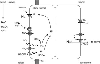
Ruminal electroneutral Na+ absorption via NHE (Figure 1) can be stimulated by acidifying intracellular pH, with protons being provided by dissociation of protonated SCFA (HSCFA), NH4 and conversion of CO2 (Gäbel et al., Reference Gäbel, Vogler and Martens1991b; Abdoun et al., Reference Abdoun, Stumpff, Wolf and Martens2005). Conversely, electroneutral Na absorption is minimized by the removal of stimulatory anions such as Cl−, HCO3−, and SCFA− (Chien and Stevens, Reference Chien and Stevens1972; Henseleit, Reference Henseleit1991).
Model of ruminal Na+ transport. Large amounts of sodium (up to 50 mol/day in cows) enter the rumen via saliva and are absorbed locally for recirculation. Apical uptake of Na+ occurs via two pathways. The major proportion of Na+ is taken up in exchange for protons via sodium-proton exchange. This pathway is stimulated by fermentation products and ensures cellular pH homeostasis by a rapid extrusion of protons entering the cytosol with short chain fatty acids, CO2 and – to a lesser extent – NH4+. The pathway is blocked by amiloride but not affected by voltage. A smaller proportion of Na+ enters via a non-selective cation channel that is blocked by divalent cations (Ca2+ and Mg2+) in a voltage dependent manner. At the negative potentials normally found between the cytosol and the rumen, these positively charged ions are pushed into the mouth of the channel from the outside, driven by the potential, reducing channel conductance. Depolarization of the tissue alleviates the block and thus, stimulates channel mediated, electrogenic uptake of Na+. After crossing the multilayered functional syncytium, sodium is driven out basolaterally by the ATP dependent sodium pump (Na+/K+-ATPase), which provides the energy for the net uptake of Na+ against sizable electrochemical gradients. Depending on both the conductance of the apical non-selective channel and the activity of the pump, a transepithelial potential is generated that can be measured both in vivo and in vitro. Fluxes of Na+ from the serosal to the mucosal side of the epithelium are much smaller, and may involve both a paracellular route and a basolateral cotransport.

Interestingly, in ruminants, the amount of Na+ absorbed by the rumen in vitro shows a very robust correlation to the amount of SCFA absorbed, as has been shown in the rumen not just of sheep (Gäbel et al., Reference Gäbel, Vogler and Martens1991b), but also of cows (Sehested et al., Reference Sehested, Diernaes, Moller and Skadhauge1993 and Reference Sehested, Diernaes, Moller and Skadhauge1999) and reindeer (Storeheier et al., Reference Storeheier, Sehested, Diernaes, Sundset and Mathiesen2003). This suggests an important function of Na+ as a counterion accompanying the absorption of SCFA− from the rumen.
In omasum, Na+ absorption is not only blocked by amiloride (1 mmol/l mucosal). The mucosal addition of hydrochlorothiazide reduced Na+ and Cl− absorption in parallel, suggesting the presence of an apically localized Na+Cl− cotransport in addition to NHE (Ali, Reference Ali2005; Ali et al., Reference Ali, Shen, Tietjen and Martens2006).
However, even in the presence of amiloride (and hydrochlorothiazide), Na+ absorption cannot be reduced to 0. The persisting net transport of Na+ in the presence of amiloride is still slightly higher than the short circuit current (Isc) which can be measured across isolated forestomach epithelia under all experimental conditions and, together with unequal K+ diffusion potentials across apical and basolateral membranes, gives rise to a transruminal potential difference of 20 to 60 mV (blood side positive) that can be observed not only in vitro but also in vivo (Dobson, Reference Dobson1959; Ferreira et al., Reference Ferreira, Harrison and Keynes1966a and Reference Ferreira, Harrison, Keynes and Nauss1966b). The presence of this Na+ current points to an (additional) electrogenic pathway for the uptake of Na+.
At alkaline pH or high ruminal NH3, an uptake via NHE is low (Martens et al., Reference Martens, Krützfeld and Wolf2004; Abdoun et al., Reference Abdoun, Stumpff, Wolf and Martens2005), which also shows that absorption of Na+ must be maintained by additional pathways under these conditions to prevent an alkalinization of the cytosol.
Electrogenic Na+ absorption
Ruminal electrogenic Na+ transport differs from that of classical Na+ absorbing epithelia, as it is not blocked by low doses of amiloride (<0.1 mmol/l; Martens et al., Reference Martens, Gäbel and Strozyk1991), but varies with the luminal concentration of divalent cations (Figure 1). Both in the rumen and in the omasum, net Na+ absorption and Isc as a measure of total electrogenic ion transport across a tissue are markedly increased when the mucosal concentrations of free divalent cations such as Ca2+ and Mg2+ are decreased (Leonhard et al., Reference Leonhard, Smith, Martens, Gäbel and Ganzoni1990; Rübbelke, Reference Rübbelke1998; Schultheiss and Martens, Reference Schultheiss and Martens1999). In further experiments with isolated rumen epithelia, a divalent sensitive current and conductance could also be shown when Na+ was replaced by K+ or Rb+, but not in the presence of the bigger cation n-methyl-d-glucamine+ (Leonhard-Marek, Reference Leonhard-Marek2002). In omasum, K+, Rb+, Cs+ and Li+ ions carried a divalent cation sensitive current, which reached about half the magnitude of the respective Na+ current. (Schultheiss and Martens, Reference Schultheiss and Martens1999). Microelectrode experiments revealed that the removal of divalent cations from the mucosal solution depolarized the apical membrane and reduced its fractional resistance (Lang and Martens, Reference Lang and Martens1999; Schultheiss and Martens, Reference Schultheiss and Martens1999). All these observations pointed to a non-selective cation conductance (NSCC) in the apical membrane of ruminal and omasal epithelium that may resemble certain amiloride insensitive conductances found in esophagus (Awayda et al., Reference Awayda, Bengrine, Tobey, Stockand and Orlando2004) or lung (Kemp and Kim, Reference Kemp and Kim2004), but clearly distinct from the classical epithelial Na+ channel found in most mammalian epithelia as distal colon and collecting tubule of the kidney (Rossier, Reference Rossier2003).
The development of a divalent cation sensitive Isc in Na+ buffer requires an active Na+/K+ ATPase and K+ recycling through Ba2+ sensitive K+ conductances on the basolateral side (Figure 1). Interestingly, the Na+ current was not only blocked by extracellular Ca2+ and Mg2+ ions, but also by intracellular Mg2+ ions, which points to a regulatory function of Mg2+ (Mgi) in rumen. Thus, manœuvres that reduce intracellular Mg2+ could be shown to open the conductance both on the level of the tissue (Leonhard-Marek, Reference Leonhard-Marek2002), and in isolated cells (Leonhard-Marek et al., Reference Leonhard-Marek, Stumpff, Brinkmann, Breves and Martens2005). Data demonstrate a NSCC with P K > P Cs > P Na, sensitive to the removal of Mg2+, or both Ca2+ and Mg2+ from the external solution. Conductance increased and inward rectification decreased when internal Mg2+ was removed, allowing more influx of Na+ at higher potentials. The conductance could also be blocked by the divalent cation Ba2+. Elevation of cAMP opened the conductance, both via effects on a basolateral Na+/Mg2+ exchanger (Leonhard-Marek et al., Reference Leonhard-Marek, Stumpff, Brinkmann, Breves and Martens2005), and via direct mechanisms on the channel (Brinkmann, Reference Brinkmann2006).
An important feature to emerge from the patch clamp studies was the voltage dependence of the channel block by divalent cations (Leonhard-Marek et al., Reference Leonhard-Marek, Stumpff, Brinkmann, Breves and Martens2005; Stumpff and Martens, Reference Stumpff and Martens2007a and Reference Stumpff and Martens2007b). As the cytosol becomes depolarized, the external divalent cations (Ca2+ and Mg2+) blocking the external mouth of the channel are pushed out and the permeability of the channel for Na+ rises. This explains the observation that depolarization of the ruminal epithelium in vitro leads to an increase in the electrogenic conductance for sodium across the tissue (Lang and Martens, Reference Lang and Martens1999).
The increase of ruminal electrogenic Na+ transport after a depolarization of the apical membrane may be of practical importance for the ruminant in vivo. The release of high amounts of potassium from plant material after an increase of dietary K intake results in an increased Na+ absorption from the rumen (Scott, Reference Scott1967; Warner and Stacy, Reference Warner and Stacy1972) and keeps the sum of Na+ and K+ concentrations within the rumen almost constant. As ruminal K+ concentration rises, the potential across the ruminal tissue in toto increases (Dobson, Reference Dobson1959; Scott, Reference Scott1966; Martens et al., Reference Martens, Gäbel and Strozyk1987), while the apical membrane of the ruminal epithelium depolarizes (Leonhard-Marek and Martens, Reference Leonhard-Marek and Martens1996). Albeit the electric driving force for Na+ uptake is reduced under these conditions, the conductance of the electrogenic pathway opens and mediates the influx of Na+ necessary to maintain ruminal osmolarity (Lang and Martens, Reference Lang and Martens1999). It should be noted that an increase in electrogenic Na+ transport across the tissue will lead to a depolarization and thus, to a decrease in the driving force for the efflux of potassium from the rumen, as will be discussed in more detail below.
Recent structural studies have given evidence for the expression of the channels transient receptor potential vanilloid channel ((TRPV6); Wilkens et al., Reference Wilkens, Kunert-Keil, Brinkmeier and Schröder2009) and TRPM7 (Schweigel et al., Reference Schweigel, Kolisek, Nikolic and Kuzinski2008) in rumen epithelium. Although these channels are discussed in the context of Ca2+ and Mg2+ absorption, respectively (see below), it appears quite likely that they might also be responsible for the electrogenic uptake of Na+ or the permeation of other monovalent cations when the concentration of divalents is reduced or when other mediators such as cAMP are present.
Given the decreasing Na+ uptake via NHE at alkaline pH, the effect of pH on electrogenic Na+ transport was especially interesting. A higher mucosal pH decreases the stimulatory effects of SCFA and CO2 on ruminal NHE (Gäbel et al., Reference Gäbel, Vogler and Martens1991b) and enhances the inhibitory effects of ammonia on NHE (Abdoun et al., Reference Abdoun, Stumpff, Wolf and Martens2005). However, a higher pH also reduces the concentration of free Ca2+ and Mg2+ ions in rumen fluid (Johnson and Aubrey Jones, Reference Johnson and Aubrey Jones1989). This might relieve the blocking effect of these cations on the electrogenic Na+ transport. Interestingly, increasing mucosal pH from 7.5 to 8.0 in vitro increased the Na+ current through the apical conductance in the absence of Ca2+ and Mg2+ (Leonhard-Marek et al., Reference Leonhard-Marek, Breves and Busche2006).
In this context, it should be noted that the stratum corneum forms an apical microclimate that presents a barrier between the pH of the bulk solution and that adjacent to the stratum granulosum where the apical Na+-conductance is localized (Leonhard-Marek et al., Reference Leonhard-Marek, Breves and Busche2006). This pH microclimate can be measured with the aid of a fluorescent probe incorporated in the apical membrane of the outermost epithelial cells and inside epithelial cells that belong to the stratum corneum (Leonhard-Marek et al., Reference Leonhard-Marek, Breves and Busche2006), and has recently been confirmed using ion-selective microelectrodes (Abdoun et al., Reference Abdoun, Stumpff, Rabbani and Martens2010). An increase in luminal Cl− induces a visible increase in rumen surface pH in the presence as well as in the absence of bicarbonate, pointing to an action via apical Cl−/HCO3− or Cl−/OH− exchange (Leonhard-Marek et al., Reference Leonhard-Marek, Breves and Busche2006). As seen after direct alkalinization of mucosal pH, this manœuvre had impacts on Isc depending on the mucosal presence or absence of Ca2+ and Mg2+.
As for luminal pH, the effects of hormones and neurotransmitters acting via cAMP also seem to have different effects on electroneutral and electrogenic Na transport respectively. While an intracellular increase in cAMP or the addition of prostaglandins decreased Na absorption via NHE (Gäbel et al., Reference Gäbel, Butter and Martens1999), cAMP stimulated the transepithelial Na+ current via a stimulation in Mg2+/Na+ exchange and a subsequent decrease in intracellular Mg2+, thereby relieving the intracellular block of the unspecific cation conductance (Leonhard-Marek et al., Reference Leonhard-Marek, Stumpff, Brinkmann, Breves and Martens2005), possibly augmented by direct effects of cAMP on the selectivity of the channel for Na+ (Brinkmann, Reference Brinkmann2006). Like many other tissues, the ruminal epithelium shows endogenous secretion of prostaglandins (Gäbel et al., Reference Gäbel, Butter and Martens1999), which may inter alia serve to switch between the electroneutral and electrogenic form of Na transport across the tissue, with implications for acid-base balance.
In summary, water and electrolyte balance of the ruminant cannot be understood without taking the ruminal-portal-salivary exchanges into account. These have to be regulated to ensure adequate recirculation of sodium through the tissue under multiple different feeding conditions (Silanikove, Reference Silanikove1994). Thus, ingestion of readily fermentable carbohydrates with an increase in the concentration of ruminal SCFA requires an increase in the uptake of Na+ in exchange for H+ via NHE. This mechanism serves to limit influx of protons into the epithelium, to recirculate Na+ for secretion with salivary buffering anions such as HCO3− and H2PO4−, and to provide Na+ as a counterion for the absorption of SCFA across the rumen. In situations where intraruminal potassium concentration rises greatly (Hyden, Reference Hyden1961; Stacy and Warner, Reference Stacy and Warner1966 and Reference Stacy and Warner1972; Martens and Hammer, Reference Martens and Hammer1981), an increase in the uptake of Na+ via electrogenic mechanisms is essential for both the maintenance of ruminal osmolarity and to prevent an excessive and rapid influx of K+ into the plasma space. Research on intracellular signaling pathways that may regulate the relative contributions of electrogenic, apical and basolateral uptake of Na+ with implications for ruminal homeostasis has only just begun.
Absorption of magnesium
The forestomachs are the main site of magnesium (Mg2+) absorption in ruminants (Tomas and Potter, Reference Tomas and Potter1976), which is essential for maintaining the normal blood Mg concentration (Pfeffer and Rahman, Reference Pfeffer and Rahman1974). Although older studies have shown that Mg can be absorbed across all the three forestomach compartments (Martens and Rayssiguier, Reference Martens and Rayssiguier1980), attempts to characterize the underlying mechanisms have only been performed with ruminal tissues. Within the rumen epithelium at least two mechanisms ensure an efficient Mg2+ uptake into RECs (Figure 2). One of these is electrogenic in nature, so that the uptake of Mg2+ depends on the potential difference across the apical cell membrane (Martens et al., Reference Martens, Gäbel and Strozyk1987; Leonhard-Marek and Martens, Reference Leonhard-Marek and Martens1996). This interdependance explains the negative impact of high ruminal K+ concentrations on Mg2+ absorption observed in vivo (e.g. Care et al., Reference Care, Brown, Farrar and Pickard1984; Grace et al., Reference Grace, Caple and Care1988) and in vitro (Martens et al., Reference Martens, Gäbel and Strozyk1987; Leonhard-Marek and Martens, Reference Leonhard-Marek and Martens1996) as well as part of the negative effects of NH4+ (Martens and Rayssiguier, Reference Martens and Rayssiguier1980). The uptake of K+ depolarizes the apical membrane potential and thus reduces the driving force for electrogenic uptake of Mg2+ (Leonhard-Marek and Martens, Reference Leonhard-Marek and Martens1996). A comparable interaction might explain the negative effect of luminal NH4+ on Mg2+ absorption, as ruminal potassium channels also show permeability for ammonium in its protonated form (Abdoun et al., Reference Abdoun, Stumpff, Wolf and Martens2005). Structural equivalents for electrogenic Mg2+ uptake seem to be TRPM7 and MagT1 whose expression and functional activity have recently been shown in RECs (Schweigel et al., Reference Schweigel, Kolisek, Nikolic and Kuzinski2008).
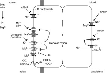
Model of ruminal K+ and Mg2+ transport. Ruminants ingest large amounts of potassium with the diet (approximately 12 mol/day in cows). Both dietary intake and salivary flow contribute to the influx of K+ into the rumen, which can accumulate to reach levels of 100 mmol/l in sodium-depleted animals. Both apical uptake and basolateral efflux of K+ is mediated by channels, and accordingly, transport of K+ across the rumen is passive and follows the electrochemical gradient. Transport is highest when the concentration gradient is high and drops when the transruminal potential rises. At low concentrations in the rumen, apical uptake of Mg2+ follows the electrochemical gradient across the apical membrane and is thought to occur via a channel. At higher concentrations, a second, transporter mediated pathway contributes to the total uptake. This pathway is stimulated by short chain fatty acids and CO2. Basolaterally, efflux of Mg2+ occurs coupled to the influx of Na+ via a pathway that can be stimulated by cAMP. The channel-mediated uptakes of Na+, K+ and Mg2+ are driven by potential and interact with each other in a complex fashion. Normally, the inside of the ruminal epithelium is negative v. the rumen. As the concentration of Mg2+ in the cell is similar to that of the rumen, this potential is essential to drive the uptake of this element into the epithelium. When ruminal K+ rises, the potential difference across the apical membrane drops and the channel mediated uptake of Mg2+ is reduced. The pore of these channels is now free for the passage of Na+ into the cytosol, driven by the concentration gradient, which is maintained by the basolateral Na+ pump. The rising electrogenic transport of Na+ will raise transruminal potential and thus, limit the efflux of K+ from the rumen.

However, even at high luminal K+ concentrations, absorption of Mg2+ ions across the rumen remains possible and can be increased by an elevation of the luminal Mg2+ concentration (Martens and Blume, Reference Martens and Blume1986; Martens et al., Reference Martens, Heggemann and Regier1988; Ram et al., Reference Ram, Schonewille, Martens, Van’t Klooster and Beynen1998; Jittakhot et al., Reference Jittakhot, Schonewille, Wouterse, Uijttewaal, Yuangklang and Beynen2004a and Reference Jittakhot, Schonewille, Wouterse, Yuangklang and Beynen2004b; Holtenius et al., Reference Holtenius, Kronqvist, Briland and Spörndly2008). This variable interaction between K+ intake and Mg2+ absorption has been quantified, permitting the prediction of Mg absorption (Weiss, Reference Weiss2004). The K+-insensitive Mg2+ absorption in the presence of high ruminal potassium is due to a second, electroneutral mechanism of Mg2+ absorption (Leonhard-Marek and Martens, Reference Leonhard-Marek and Martens1996; Leonhard-Marek et al., Reference Leonhard-Marek, Marek and Martens1998b). The fact that Mg2+ absorption across rumen epithelium or Mg2+ uptake into RECs can be stimulated by the presence of different anions like Cl−, CO2/HCO3− or SCFA has led to the suggestion of Mg2+/anion− cotransporters or Mg2+/H+ exchangers coupled to anion exchangers as underlying mechanisms (Figure 2, Leonhard-Marek et al., Reference Leonhard-Marek, Gäbel and Martens1998a, Leonhard-Marek, Reference Leonhard-Marek1999; Schweigel and Martens, Reference Schweigel and Martens2003). More recently, a vacuolar-type H+-ATPase (vH+-ATPase) has been found in RECs which is involved in regulation of pHi and transport of Mg2+ (Schweigel and Martens, Reference Schweigel and Martens2003; Etschmann et al., Reference Etschmann, Heipertz, von der Schulenburg and Schweigel2006).
The extrusion of Mg2+ across the basolateral membrane to the blood side is a secondary active process and depends on the presence of sodium (Figure 2). Reducing the Na+ concentration on the basolateral side of rumen epithelia decreases transepithelial Mg2+ absorption (Leonhard-Marek and Martens, Reference Leonhard-Marek and Martens1994; Leonhard-Marek et al., Reference Leonhard-Marek, Stumpff, Brinkmann, Breves and Martens2005); while the Mg2+ efflux from Mg2+-loaded RECs increases with the extracellular Na+ concentration and can be partly blocked with imipramine (Schweigel et al., Reference Schweigel, Park, Etschmann and Martens2006). These observations point to Mg2+/Na+ exchange as Mg2+ extruding mechanism on the basolateral membranes of rumen epithelium. In line with this assumption a monoclonal antibody raised against porcine red blood cell Mg2+/Na+ exchanger was able to inhibit Mg2+ extrusion from preloaded RECs and detected a specific immunoreactive band in protein lysates of these cells (Schweigel et al., Reference Schweigel, Park, Etschmann and Martens2006). Studies with isolated cells and sheets of rumen epithelium have shown that cAMP stimulates Mg2+/Na+ exchange with indirect consequences for electrogenic Na+ absorption via a reduction in intracellular Mg2+ concentration (Figure 2, Leonhard-Marek et al., Reference Leonhard-Marek, Stumpff, Brinkmann, Breves and Martens2005; Schweigel et al., Reference Schweigel, Park, Etschmann and Martens2006), pointing to a regulation of Mg2+/Na+ exchange via the PKA pathway.
Absorption of potassium
Physiologically speaking, a rapid efflux of K+ from the gastrointestinal tract of the ruminant should be undesirable, as under most dietary conditions, potassium intake is so ample that energy consuming redistribution into the cytosolic compartment with subsequent renal secretion is necessary to maintain potassium homeostasis (Rabinowitz et al., Reference Rabinowitz, Sarason and Yamauchi1984; Giebisch, Reference Giebisch1998; Stumpff and Martens, Reference Stumpff and Martens2007b). Correspondingly, in ruminants on low intakes of K+, a net secretion of K+ occurs before the duodenum. Thus, a small net excretion of 18 g/day of K+ could be observed in cows on a K+ intake of about 200 g/day (5 mol/day, Khorasani et al., Reference Khorasani, Janzen, McGill and Kennelly1997), with similar results observed in lambs (with an excretion of 0.72 g/day at a K+ intake of 5.5 g/day or 140 mmol/day, Greene et al., Reference Greene, Webb and Fontenot1983b). In cows on an somewhat higher K+ intake of 322 g/day (or 8 mol/day), net K+ absorption can be observed before the duodenum, but it is small (10%, Rahnema et al., Reference Rahnema, Wu, Ohajuruka, Weiss and Palmquist1994). The small intestine is thus the main site of net K+ absorption from the diet under these feeding conditions (Pfeffer et al., Reference Pfeffer, Thompson and Armstrong1970; Rahnema et al., Reference Rahnema, Wu, Ohajuruka, Weiss and Palmquist1994; Khorasani et al., Reference Khorasani, Janzen, McGill and Kennelly1997), and efflux of K+ from the rumen is slow considering the gradients driving the element into the blood.
However, to gain a more complete understanding of the role that the rumen plays in K+ metabolism, recirculation of K+ via saliva has to be taken into account. Even in sodium replete animals, in which K+ concentration in saliva is usually around 5 mmol/l, contributions of up to 1.5 mol/day (cows) or 80 mmol/day (sheep) of K+ to the total balance are to be expected. When animals are Na+-depleted (Scott, Reference Scott1966) and under stimulation by aldosterone (Blair-West et al., Reference Blair-West, Coghlan, Denton, Goding and Wright1963), both the salivary and the ruminal concentration of K+ may rise to 100 mmol/l (Kay, Reference Kay1960; Blair-West et al., Reference Blair-West, Coghlan, Denton, Goding and Wright1963; Scott, Reference Scott1966 and Reference Scott1967) Under these conditions, K+ replaces (scarce) Na+ in saliva, and not surprisingly, perhaps, a sizable reabsorption of K+ from the rumen can be observed.
Provided that ruminal K+ is sufficiently high, a number of in vivo and in vitro studies have shown that preintestinal regions can contribute substantially to total recirculation of K+ in the gastrointestinal tract of ruminants. Using the technique of the isolated reticulorumen in sheep, Dobson (Reference Dobson1959) observed a K+ absorption rate of 67 mmol/day at a ruminal K+ concentration of 23 mmol/l in sheep. When the potassium level in the diet was increased from 0.6% to 2.4% and 4.8% in steers or lambs and from 1.5% to 2.6% in lactating cows, the animals absorbed between 40% and 72% of the additional K+ intake in preintestinal regions (Greene et al., Reference Greene, Fontenot and Webb1983a and Reference Greene, Webb and Fontenot1983b; Khorasani et al., Reference Khorasani, Janzen, McGill and Kennelly1997), thus recirculating not only the entire amount of K+ secreted with saliva, but also absorbing a certain proportion of dietary K+.
Scott (Reference Scott1967) calculated ruminal K+ absorption with the aid of marker infusions, and found a linear increase in absorption with ruminal K+ concentrations through the range of 35 to 100 mmol/l. Conversely, Warner and Stacy (Reference Warner and Stacy1972) calculated positive K+ absorption rates only when ruminal concentration exceeded 100 mmol/l, suggesting that the absorption of K+ may be regulated in a complex manner. Interestingly, the omasum showed a lower K+ absorption rate (19 mmol/day) at higher concentrations in sheep (43 mmol/l, von Engelhardt and Hauffe, Reference Von Engelhardt and Hauffe1975) and a K+ absorption rate of about 81 mmol/day in young steers (Edrise et al., Reference Edrise, Smith and Hewitt1986).
Studies with isolated forestomach epithelia of sheep have shown a small net K+ secretion between 0.04 and 0.36 μeq/cm2h across the rumen when the tissues were incubated in the absence of electrical and chemical gradients (Ferreira et al., Reference Ferreira, Harrison, Keynes and Zurich1972; Harrison et al., Reference Harrison, Keynes, Rankin and Zurich1975; Wolffram et al., Reference Wolffram, Frischknecht and Scharrer1989; Leonhard-Marek and Martens, Reference Leonhard-Marek and Martens1996). A small K+ absorption of 0.04 to 0.13 μeq/cm2h was observed in the omasum (Harrison et al., Reference Harrison, Heynes and Zurich1970; Harrison, Reference Harrison1971). However, when the luminal K+ concentration was increased from 4 to 100 mmol/l, net K+ absorption across rumen epithelia increased by 2.5 μeq/cm2h (Kronshage and Leonhard-Marek, Reference Kronshage and Leonhard-Marek2009), corresponding to an amount of 0.7 mol/day (or 27 g/day) across the estimated 1.2 m2 of ruminal surface in sheep (Ferreira et al., Reference Ferreira, Harrison, Keynes and Zurich1972). This equals the amount of K+ absorption observed in vivo by Scott (Reference Scott1967), but exceeds that observed by Warner and Stacy (Reference Warner and Stacy1972) at the same ruminal potassium concentration of 100 mmol/l. Although the efflux of K+ from the rumen is passive, in that it follows the electrochemical gradient, the permeability of the rumen to the K+ ion is thus clearly regulated in a complex fashion that is yet poorly understood. However, outlines are beginning to emerge.
Uptake of K+ occurs via a transcellular route while the paracellular permeability of the rumen to K+ seems to be lower (Figure 2). The use of different blockers has revealed the contribution of both apical and basolateral K+ channels to ruminal K+ transport (Harrison et al., Reference Harrison, Keynes, Rankin and Zurich1975; Bödeker and Kemkowski, Reference Bödeker and Kemkowski1996; Leonhard-Marek and Martens, Reference Leonhard-Marek and Martens1996). Apical K+ channels could be blocked by quinidine or verapamil, basolateral K+ channels were sensitive to inhibition by barium (Figure 2).
Luminal K+ channels are responsible for the effect of a change in luminal K+ concentration on the apical membrane potential (PDa), which is depolarized with increasing ruminal K+ concentrations (Leonhard-Marek and Martens, Reference Leonhard-Marek and Martens1996). As mentioned, the K+-dependent depolarization of PDa decreases ruminal Mg2+ absorption particularly in animals on a low Mg2+ diet. Both the reduction in intracellular Mg2+ and the relief of the voltage dependent block by extracellular divalent cations as outlined above should increase the NSCC of the rumen (Figure 2). This mechanism might in part be responsible for the increased Na+ absorption seen at high ruminal K+ concentrations (Scott, Reference Scott1967; Warner and Stacy, Reference Warner and Stacy1972).
The increase in the active, electrogenic transport of Na+ across the tissue under high K+ conditions also has implications for the transport of K+: as the transepithelial potential across the rumen increases, the gradient driving the efflux of K+ decreases. Although the potentials observed in vivo (Scott, Reference Scott1967; Warner and Stacy, Reference Warner and Stacy1972) and in vitro (Leonhard-Marek and Martens, Reference Leonhard-Marek and Martens1996) under high ruminal K+ conditions are induced by the high ruminal K+ concentrations, they serve to limit the actual efflux of this element by decreasing the electrochemical driving force (Stumpff and Martens, Reference Stumpff and Martens2007a and Reference Stumpff and Martens2007b).
In summary it can be said that efflux of K+ from the rumen is passive and determined by the electrochemical gradient. The activity of the Na+/K+ ATPase in conjunction with the regulatory control of the permeabilities of the apical, basolateral and paracellular conductances for Na+ and K+ should ultimately determine the amount of K+ distributed into the ruminal cavity or released into plasma.
Absorption of calcium
To meet the Ca2+ requirements of milk production, the mechanisms of gastrointestinal Ca2+ absorption are of great interest especially in high yielding dairy cows. Although the small intestine is regarded as the main absorption site for Ca2+ in monogastric animals, ruminants can absorb Ca2+ from all gastrointestinal regions (Breves et al., Reference Breves, Goff, Schröder and Horst1995). Balance studies show that preintestinal Ca2+ absorption gains significance at higher Ca2+ intake. When Ca2+ intake rises to 8 g/day in sheep or to 120 g/day in cattle, absorption of Ca2+ is shifted to preintestinal regions (Grace et al., Reference Grace, Ulyatt and MacRae1974; Khorasani et al., Reference Khorasani, Janzen, McGill and Kennelly1997; Schröder and Breves, Reference Schröder and Breves2006). A survey of different balance studies showed that a high preintestinal Ca2+ absorption is usually accompanied by a low intestinal Ca2+ absorption or even by intestinal Ca2+ secretion (Schröder and Breves, Reference Schröder and Breves2006). It is worth to mention in this context that in a recent meta-analysis Lean et al. (Reference Lean, Degaris, McNeil and Block2006) have shown that prevention of milk fever seems to be possible not only at low Ca intake (<0.5% of DM), but also at high Ca intake (>2% of DM), which may indicate that at high Ca2+ intake, absorption of Ca2+ from the forestomachs could help to cover increased Ca2+ requirement at parturition.
Ruminal and omasal Ca2+ absorption are at least partly active and involve electrogenic and electroneutral components (Höller et al., Reference Höller, Breves and Dubberke1988a and Reference Höller, Breves, Kocabatmaz and Gerdes1988b; Schröder et al., Reference Schröder, Rittmann, Pfeffer and Breves1997; Wadhwa and Care, Reference Wadhwa and Care2000). Active ruminal Ca2+ absorption can be increased by concentrate feeding (Uppal et al., Reference Uppal, Wolf and Martens2003), in the presence of SCFAs (Schröder et al., Reference Schröder, Rittmann, Pfeffer and Breves1997 and Reference Schröder, Vössing and Breves1999; Wadhwa and Care, Reference Wadhwa and Care2000; Uppal et al., Reference Uppal, Wolf and Martens2003, Leonhard-Marek et al., Reference Leonhard-Marek, Becker, Breves and Schröder2007a) or by increasing the amounts of luminal chloride (Leonhard-Marek et al., Reference Leonhard-Marek, Becker, Breves and Schröder2007a), whereas sulfate ions seem to have stimulatory and depressing effects with no net change of Ca flux rates in vitro (Leonhard-Marek et al., Reference Leonhard-Marek, Busche and Schröder2007b). An increased absorption of undissociated SCFA will provide protons to the cell interior, while the ruminal uptake of SCFA− or Cl− via anion exchange mechansims is accompanied by a secretion of bicarbonate (Gäbel et al., Reference Gäbel, Bestmann and Martens1991a) and an increase in surface pH (Leonhard-Marek et al., Reference Leonhard-Marek, Breves and Busche2006). The stimulating effects of SCFA and amiloride (increased availability of protons because of blocked NHE) have therefore suggested that Ca2+/H+ antiporters contribute to apical Ca2+ uptake (Schröder et al., Reference Schröder, Rittmann, Pfeffer and Breves1997 and Reference Schröder, Vössing and Breves1999). However, in the light of the presence of a ruminal vH+-ATPase (Schweigel and Martens, Reference Schweigel and Martens2003; Etschmann et al., Reference Etschmann, Heipertz, von der Schulenburg and Schweigel2006), these findings might be additionally explained as a stimulation of other Ca2+ transport proteins by an apical pH microclimate.
An increased electrogenic Na+ absorption in the absence of divalent cations as shown for rumen and omasum (see above), is a feature of many Ca2+ channels (Sather and McCleskey, Reference Sather and McCleskey2003; Hoenderop et al., Reference Hoenderop, Nilius and Bindels2005). Ca2+ channels might thus represent the functional basis for a divalent sensitive Na+ absorption as well as for an electrogenic Ca2+ absorption across forestomach epithelia. In the rumen, the Ca2+ channel agonist Bay K 8644 (50 nM) was able to increase Ca2+ absorption (Wadhwa and Care, Reference Wadhwa and Care2000), which could indicate the presence of l-type Ca2+ channels in rumen epithelium. Although many epithelia use TRPV-type Ca2+ channels for Ca2+ absorption (Hoenderop et al., Reference Hoenderop, Nilius and Bindels2005), l-type Ca2+ channels have been suggested to contribute additionally to intestinal Ca2+ absorption (Morgan et al., Reference Morgan, Mace, Helliwell, Affleck and Kellett2003). Both the epithelial TRPV-type Ca2+ channels and l-type Ca2+ channels increase their conductance with alkaline pH (Pietrobon et al., Reference Pietrobon, Prod’hom and Hess1989; Vennekens et al., Reference Vennekens, Prenen, Hoenderop, Bindels, Droogmans and Nilius2001), so that an increased uptake of Cl− or SCFA− in exchange for bicarbonate could stimulate a luminal Ca2+ conductance via an increase in surface pH. TRPV6 has recently been shown to be expressed in rumen tissues. In situ hybridization, immunohistochemistry and Western blot analysis, however, indicated only a weak TRPV6 signal, which was observed along the entire epithelium and not just localized in the luminal cell layers. The actual significance of this channel for ruminal Ca2+ uptake remains questionable (Wilkens et al., Reference Wilkens, Kunert-Keil, Brinkmeier and Schröder2009).
Transepithelial Ca2+ absorption depends on the activity of the basolateral Na+/K+-ATPase and on the presence of sodium, suggesting the contribution of Na+/Ca2+ exchangers (NCXs) to basolateral Ca2+ extrusion (Höller et al., Reference Höller, Breves and Dubberke1988a and Reference Höller, Breves, Kocabatmaz and Gerdes1988b; Schröder et al., Reference Schröder, Rittmann, Pfeffer and Breves1997 and Reference Schröder, Vössing and Breves1999). Indeed, the expression of NCX-specific mRNA has been shown in rumen epithelium (Wilkens, Reference Wilkens2006). In contrast, the plasma membrane Ca2+-ATPase, although equally shown to be expressed on mRNA level with no further information about expression patterns (Wilkens, Reference Wilkens2006), does not seem to contribute to ruminal Ca2+ absorption as the Ca2+ pump inhibitor vanadate had no significant effect on net Ca2+ absorption (Schröder et al., Reference Schröder, Vössing and Breves1999).
Absorption of ammonia
Within the forestomachs, the microbial degradation of proteins, peptides, amino and nucleic acids taken up with the food as well as the hydrolysis of recycled urea delivers substantial amounts of ammonia, which is mostly re-used for the synthesis of microbial protein. An optimal growth rate of ruminal microbes, however, requires ammonia concentrations between 3.5 and 6 mmol/l (Satter and Slyter, Reference Satter and Slyter1974; Kang-Meznarich and Broderick, Reference Kang-Meznarich and Broderick1980), while a diet rich in rapidly degradable proteins can easily raise ruminal ammonia concentrations up to 20 mmol/l (Wernli and Wilkins, Reference Wernli and Wilkins1980). According to Nolan and Strachin (Reference Nolan and Strachin1979) between 35% and 65% of the generated ammonia is absorbed across rumen epithelium. A smaller quantity of ammonia (about 10% of the generated amount, Nolan and Strachin, Reference Nolan and Strachin1979) flows into the omasum and can be absorbed there (von Engelhardt and Hauffe, Reference Von Engelhardt and Hauffe1975).
Ruminal absorption of ammonia can be raised by increases in pH and is depressed to a constant value when pH is decreased to values below 6, suggesting that most of it is absorbed in the lipid soluble form of NH3 (Hogan, Reference Hogan1961; Abdoun et al., Reference Abdoun, Stumpff, Wolf and Martens2005 and Reference Abdoun, Stumpff and Martens2006). However, as ammonia is a base with a pK value around 9, the majority of it will be present in the form of NH4+ at physiological rumen pH. Abdoun et al. (Reference Abdoun, Stumpff, Wolf and Martens2005) measured ammonia fluxes across rumen epithelium at different pH values and found a strong correlation between total ammonia absorption and the concentration of mucosal NH3 (in mmol/l): J (NH3, NH4+) = 4.1 μeq/cm2h [NH3]+0.7 μeq/cm2h (r = 0.99). In this equation the intercept of 0.7 μeq/cm2h (at [NH3] = 0) should represent electrogenic absorption of NH4+ (Abdoun et al., Reference Abdoun, Stumpff, Wolf and Martens2005 and Reference Abdoun, Stumpff and Martens2006). Assuming a pH independent absorption rate of NH4+, the absorption of NH4+ would explain nearly 100% of the total ammonia absorption measured by Abdoun et al. (Reference Abdoun, Stumpff, Wolf and Martens2005) at pH 6.4 and about 20% of the ammonia absorption at pH 7.4. Adding NH4+ or K+ salts to the luminal side of isolated sheep rumen epithelia induced increases in transepithelial Isc, which could both be blocked by quinidine (Bödeker and Kemkowski, Reference Bödeker and Kemkowski1996). In the same study, absorption of ammonia was reduced by the addition of quinidine or by imposing a positive potential difference across the epithelia, suggesting the permeation of NH4+ through epithelial K+ channels (Figures 1 and 2). Quinidine sensitive NH4+ and K+ permeable channels have also been demonstrated in isolated RECs using the patch clamp technique (Abdoun et al., Reference Abdoun, Stumpff, Wolf and Martens2005).
In rumen and omasum, increasing amounts of ammonia interfere with Na+ and Cl− absorption in a manner that depends on luminal pH. While an increase in ammonia concentration raised ruminal Na+ absorption at pH 6.4, it had no effect at pH 6.9 and depressed Na+ absorption at pH 7.4 (Abdoun et al., Reference Abdoun, Stumpff, Wolf and Martens2005). In omasal epithelia, ammonia only showed depressing effects on Na+ absorption at pH 7.4 that vanished when the sheep had been kept on a concentrate-based diet instead of a hay diet before the experiments (Martens et al., Reference Martens, Krützfeld and Wolf2004). In the rumen of sheep that had been fed concentrate or urea, ammonia even had stimulating effects on Na+ fluxes at pH 7.4 (Abdoun et al., Reference Abdoun, Wolf, Arndt and Martens2003). A predominant absorption of NH4+ at pH 6.4 would provide protons to the cell interior and could thereby stimulate apical Na+/H+ exchangers (Figure 1), which is underlined by the observation that this ammonia dependent increase in Na+ absorption was completely blocked in the presence of amiloride (Abdoun et al., Reference Abdoun, Stumpff, Wolf and Martens2005). A predominant absorption of NH3 at a more alkaline pH will, conversely, increase intracellular pH (Müller et al., Reference Müller, Aschenbach and Gäbel2000) and thereby reduce NHE activity (Aronson et al., Reference Aronson, Nee and Suhm1982). An alkaline load is followed by HCO3− extrusion from RECs (Bilk et al., Reference Bilk, Huhn, Honscha, Pfannkuche and Gäbel2005). In line with this connection, an increase in luminal NH3 concentration enhanced ruminal Cl− absorption in a CO2/HCO3−containing buffer solution (Abdoun et al., Reference Abdoun, Stumpff, Wolf and Martens2005). A diet rich in nitrogen seems to change the relative permeabilities of rumen and omasum for NH3 and NH4+ resulting in altered ammonia dependent effects on the absorption of other ions (Abdoun et al., Reference Abdoun, Wolf, Arndt and Martens2003; Martens et al., Reference Martens, Krützfeld and Wolf2004).
Ammonia also interferes with ruminal Mg2+ absorption (Martens and Rayssiguier, Reference Martens and Rayssiguier1980), with effects on both the electrogenic and the electroneutral component (S. Leonhard-Marek and H. Martens, unpublished data). In the light of the pathways of ruminal ammonia absorption discussed above, an electrogenic uptake of NH4+ should depolarize the apical membrane potential and thereby reduce the driving force for electrogenic uptake of Mg2+. At the same time, however, absorption of NH4+ should increase the intracellular provision of protons and thus increase a vH+-ATPase dependent stimulation of Mg2+ absorption and/or an uptake via Mg2+/H+ exchange. Conversely, uptake of NH3 would reduce these electroneutral mechanisms of Mg2+ absorption while increasing electrogenic absorption via pH effects on TRPM or MagT1 (Goytain and Quamme, Reference Goytain and Quamme2005; Kozak et al., Reference Kozak, Matsushita, Nairn and Cahalan2005). It thus seems difficult to hypothesize mechanisms of adaptation that would explain why the effect of ammonia on ruminal Mg absorption disappears within some days as observed by Gäbel and Martens (Reference Gäbel and Martens1986).
Absorption and secretion of chloride
The concentration of chloride within the reticulorumen varies between 10 and 40 mmol/l with the highest values measured 1 h and the lowest ones from 8 to 14 h after feeding, reflecting the concentration in saliva at this latter time point (Bailey, Reference Bailey1961). In vitro – in the absence of electrochemical gradients – chloride is absorbed across rumen epithelia, an absorption that depends on the presence of sodium, an active Na+/K+-ATPase and on the presence of bicarbonate (Chien and Stevens, Reference Chien and Stevens1972; Harrison et al., Reference Harrison, Keynes, Rankin and Zurich1975; Martens et al., Reference Martens, Gäbel and Strozyk1991). These dependencies have suggested the involvement of Cl−/HCO3− exchangers coupled to NHE via intracellular pH in analogy to other epithelia of the gastrointestinal tract on the apical side (Figure 3).
When monitoring ruminal surface pH with the aid of a fluorescent microprobe inserted into the outer membranes of rumen epithelium, an increase in luminal Cl− concentration resulted in an increase in surface pH (Leonhard-Marek et al., Reference Leonhard-Marek, Breves and Busche2006), in line with a secretion of HCO3− in exchange for Cl− absorption. Recently, Bilk et al. (Reference Bilk, Huhn, Honscha, Pfannkuche and Gäbel2005) have shown that isolated RECs express mRNA for the anion transporters PAT, DRA and AE2, all of which might contribute to Cl− transport across rumen epithelium and have been shown to transport Cl− in exchange for HCO3− in other tissues (Mount and Romero, Reference Mount and Romero2004; Romero et al., Reference Romero, Fulton and Boron2004).
Studies aimed at elucidating the effect of bicarbonate showed differences between the various forestomach epithelia. The elimination of bicarbonate, which reduced absorptive and secretory Cl− flux rates across the rumen (Gäbel et al., Reference Gäbel, Vogler and Martens1991b), had no effect in the reticulum. However, the inhibition of the carbonic anhydrase with azetazolamide reduced Cl− absorption across the reticulum (Vogler, Reference Vogler1991), suggesting a role for an anion exchange mechanism in this tissue.
A Cl− dependent base (HCO3−) extrusion should increase the intracellular availability of protons. This context was used repeatedly to explain the stimulatory effects of Cl− on the electroneutral absorption of Na+, Mg2+ and Ca2+ across rumen epithelium (Martens and Gäbel, Reference Martens and Gäbel1988; Leonhard et al., Reference Leonhard, Martens and Gäbel1991; Sehested et al., Reference Sehested, Diernaes, Moller and Skadhauge1996; Leonhard-Marek et al., Reference Leonhard-Marek, Becker, Breves and Schröder2007a). Additional effects of Cl− on electrogenic cation absorption raised the hypothesis that apical Cl− channels might mediate a Cl− dependent change in membrane potential and thereby increase the electric driving force for cation uptake (Leonhard-Marek and Schröder, Reference Leonhard-Marek and Schröder2002; Leonhard-Marek et al., Reference Leonhard-Marek, Breves and Busche2006). In contrast to this expectation, however, an increase in luminal Cl− concentration did not hyperpolarize, but instead depolarized the apical membrane of rumen epithelium (Leonhard-Marek and Schröder, Reference Leonhard-Marek and Schröder2002; Leonhard-Marek et al., Reference Leonhard-Marek, Breves and Busche2006), which suggested the presence of Cl− conductances not in the apical but in the basolateral membranes of rumen epithelia (Figure 3).
These data are supported by other deliberations. Net electroneutral transport of NaCl (Gäbel et al., Reference Gäbel, Vogler and Martens1991b) requires not only an apical uptake pathway as outlined above via NHE coupled to an anion exchanger: efflux, too, has to be electroneutral. However, sodium leaves the tissue via the Na+/K+-ATPase. As potassium is recirculated, the basolateral efflux of Na+ would result in an increasingly negative cytosol unless Cl−, too, leaves the tissue electrogenically (Abdoun et al., Reference Abdoun, Stumpff, Wolf and Martens2005; Stumpff et al., Reference Stumpff, Martens, Bilk, Aschenbach and Gäbel2009a).
Accordingly, studies with isolated RECs have shown the expression of a large conductance anion channel sensitive to 4,4'-diisothiocyano-2,2'-disulfonic stilbene (DIDS). This channel was shown to be permeable not only to Cl−, but also to HCO3− and the anions of SCFA (Figures 3 and 4, Stumpff et al., Reference Stumpff, Martens, Bilk, Aschenbach and Gäbel2009a), and it has been proposed that Cl− efflux occurs through this channel, coupled via its charge to the efflux of Na+ via the Na+/K+-ATPase.
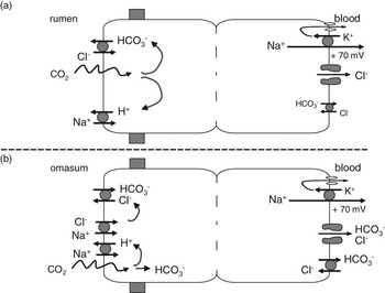
Model of Cl− and HCO3− transport across the forestomachs. (a) Extrusion of protons by the Na+/H+ exchange increases the cytosolic production of HCO3− from CO2. Secretion of HCO3− in exchange for chloride follows. Basolaterally, Cl− leaves via a large conductance anion channel coupled to the basolateral extrusion of Na+ by the Na+/K+-ATPase, resulting in a net electroneutral transport of NaCl across the tissue. There is also evidence for a basolateral anion exchanger in addition to the channel, which may mediate a (small) flux of Cl from the serosal to the mucosal side. (b) The omasum expresses an apical cotransporter for Na+ and Cl− in addition to the pathways present in the rumen. This apical cotransporter generates higher levels of cytosolic Cl−, which reverse the direction of the anion exchanger. Secretion of Cl− and absorption of HCO3− follows. Basolaterally, bicarbonate can leave either via an anion channel, coupled to the extrusion of Na+ by the sodium pump, or without Na+ in exchange for chloride via an anion exchanger.

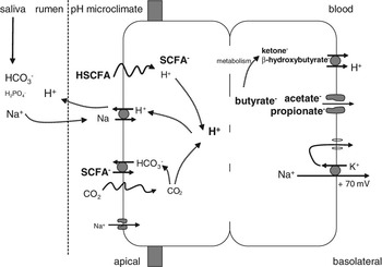
Model of ruminal short chain fatty acid (SCFA) transport. SCFA enter the apical membrane via different pathways. At low pH, uptake involves a diffusive pathway with SCFA coupled to a proton. Influx is regulated by an apical pH microclimate within the stratum corneum. In addition, the anion of SCFA can be taken up in exchange for HCO3−. Both pathways are electroneutral and transfer a proton to the cytosol, which is removed in exchange for the apical uptake of Na+ via Na+/H+ exchange. As HSCFA dissociate rapidly at the near neutral pH of the cytosol (pK≈4.8), basolateral efflux cannot occur via diffusion of HSCFA but has to involve specific transport proteins. Larger SCFA such as butyrate are metabolized into products that leave via a basolateral monocarboxylate transporter. The anions of the lower chain SCFA leave the tissue unmetabolized and coupled to the efflux of Na+. The model suggests that basolateral efflux of SCFA anions is driven by the potential induced by the Na+/K+-ATPase and occurs through a large-conductance anion channel.

Chloride transport rates measured across isolated epithelia from reticulum and omasum are higher than those across the rumen. In the absence of electrochemical gradients, both epithelia exhibit a small net Cl− absorption. In the reticulum, absorptive and secretory flux rates of Cl− were partly reduced by the addition of bumetanide or the stilbene derivatives SITS and DIDS (Vogler, Reference Vogler1991). Omasal Cl− transport was affected by DIDS (absorptive and secretory flux) and furosemide (secretory flux, Tiling, Reference Tiling1997), with absorption of Cl− most likely occuring via an apical anion exchanger and a basolateral anion channel, as described for the rumen. Studies currently underway suggest that the omasum expresses an anion channel similar to that observed in the rumen (F. Stumpff and M. Georgi, unpublished data).
The extraordinarily high serosal to mucosal Cl− fluxes observed in the omasum suggest a potentially more prolific role for an additional basolateral Cl−/HCO3− exchanger in this tissue (Figure 3, Tiling, Reference Tiling1997). In vivo studies of the omasum have shown a net secretion of chloride (von Engelhardt and Hauffe, Reference Von Engelhardt and Hauffe1975), leading to a measurable increase in Cl− concentration from proximal to distal parts within the omasum of cattle (Ekman and Sperber, Reference Ekman and Sperber1953; Edrise et al., Reference Edrise, Smith and Hewitt1986). This secretion can also be shown in vitro, but only when the mucosal Cl− concentration is lowered as would be the case under in vivo conditions (Tiling, Reference Tiling1997). Subsequent studies with epithelia from the omasum tested for Cl− transport under physiological gradients (Cl− serosal > mucosal, HCO3− mucosal > serosal). Under these conditions, anion exchange should allow for a net secretion of Cl− and a net absorption of HCO3−.
In the absence of any gradient, the small net absorption of Cl− is coupled to Na+ absorption and can be blocked by hydrochlorothiazide, suggesting the involvement of apical Na+-Cl−-cotransporters (Figure 3, Ali, Reference Ali2005). This Cl− transporter is thought to be necesssary for the provision of sufficient Cl− ions into subapical domains to allow for high transport rates of the apical Cl−/ HCO3− exchanger (see below).
In summary, both the rumen and the omasum allow the passage of sizable amounts of chloride, with the direction of transport differing. While the pronounced ability of the ruminal epithelium to absorb chloride in vitro correlates with the low chloride concentrations observed in the rumen in vivo, the pronounced ability of the omasum to secrete chloride leads to rising levels of this anion as the ingesta move toward the abomasum.
Secretion and absorption of bicarbonate
Bicarbonate transport across the rumen
Although salivary production of HCO3− is considerable, corresponding to some 40 mol/day (Bailey and Balch, Reference Bailey and Balch1961; Erdmann, Reference Erdmann1988; Allen, Reference Allen1997), bicarbonate (HCO3−) is also secreted in large amounts by the rumen epithelium, thereby contributing to an optimally buffered environment within the reticulorumen in general and in the microclimate adjacent to the transporting layer of cells in particular. Part of this bicarbonate is converted to CO2 in the course of acid buffering and exhaled by the ructus of the animals or taken up diffusively into the blood. The remaining bicarbonate passes to the omasum and is absorbed there in exchange for chloride (Figure 3). The removal of bicarbonate from the ingesta is essential to prevent an immediate and huge liberation of CO2 upon entry into the abomasum with its high concentration of hydrochloric acid (HCl).
Experiments with the temporarily isolated and washed reticulorumen of sheep have shown that bicarbonate secretion can be observed in the presence of SCFA (Gäbel et al., Reference Gäbel, Bestmann and Martens1991a). Changing from hay to concentrate diets raised net bicarbonate secretion as well as net SCFA absorption, while the replacement of SCFA by gluconate and mannitol reversed net bicarbonate secretion into net absorption, which was further enhanced by the additional removal of luminal chloride. These studies gave rise to the concept of bicarbonate secretion via anion exchangers that would also accept SCFA− or Cl− anions (Figures 3a and 4, Kramer et al., Reference Kramer, Michelberger, Gürtler and Gäbel1996).
Under physiological, low chloride conditions, secretion of HCO3− is apparently driven by the uptake of SCFA anions (Aschenbach et al., Reference Aschenbach, Bilk, Tadesse, Stumpff and Gäbel2009). In line with this, experiments on isolated cells of the ruminal epithelium have clearly demonstrated the existance of functional Cl−/HCO3− transport. As in other preparations, the direction of exchange depended on the gradients present (Huhn et al., Reference Huhn, Müller, Honscha, Pfannkuche and Gäbel2003; Bilk et al., Reference Bilk, Huhn, Honscha, Pfannkuche and Gäbel2005). Isolated RECs that were alkalized by a switch in the bathing solution from a CO2/HCO3− to a HEPES buffer, were not able to recover from this alkalized state in the absence of Cl− and SCFA on the extracellular side. In contrast, the recovery from acidification (after switching from HEPES to CO2/HCO3− in the bath) was partly dependent on extracellular Na+ (Na+/HCO3− basolaterally located cotransport, Huhn et al., Reference Huhn, Müller, Honscha, Pfannkuche and Gäbel2003) and could be enhanced by the reduction of extracellular Cl− (Bilk et al., Reference Bilk, Huhn, Honscha, Pfannkuche and Gäbel2005).
A semiquantative PCR analysis of anion transporters on the mRNA level (Bilk, Reference Bilk2008) revealed a high presence of DRA in SCFA exposed rumen epithelia, while no DRA expression could be detected in isolated RECs cultivated in the absence of SCFA. In contrast, PAT1 showed a higher presence in cultivated rumen cells compared with the native epithelium. On the basis of these differences, Bilk (Reference Bilk2008) postulated that DRA and PAT1 might be the structural equivalents of SCFA−/HCO3− and Cl−/HCO3− exchange, respectively.
Bicarbonate transport across the omasum
As mentioned above, removal of HCO3− from the ingesta before the abomasum seems essential to prevent the formation of gas (CO2 (Reference SvendsenSvendsen, 1975)) in the abomasum. In addition, presence of powerful buffers with a high pK value require the secretion of large amounts of HCl by the abomasum to reduce pH to a level at which gastric enzymes are known to work. Both formation of gas and accumulation of Cl (with cations and water following) should lead to a distension of the organ with possible subsequent displacement. For this reason, the transport of bicarbonate across the omasum has been systematically studied and clearly depends on the transepithelial and transmembranal concentration gradients for Cl− and HCO3−. Interestingly and in contrast to what is observed in the rumen, Cl− and HCO3− do not seem to interfere directly with the transport of SCFA across the omasum, suggesting that the anion exchangers expressed by this epithelium show very low affinity to the larger and more polar anions of SCFA than to those of Cl− and HCO3−.
Depending on the gradients present, bicarbonate can be absorbed or secreted across in vitro preparations of omasal epithelia. Transport rates are, however, not symmetrical at identical (opposed) gradients. The same relative gradients allowed for higher absorptive than secretory fluxes of bicarbonate (Wegeler, Reference Wegeler2008). While apical influx of HCO3− is clearly linked to the extrusion of Cl− (reversed to the rumen), bicarbonate absorption was significantly reduced when the Cl− concentration on the serosal side was lowered (Niebuhr, Reference Niebuhr2003). However, bicarbonate absorption could not be reduced by an increase in luminal Cl− concentration from 25 to 90 mmol/l in the presence of 50 mmol/l bicarbonate (Wegeler, Reference Wegeler2008). This has been suggested to represent a higher affinity for bicarbonate at the external binding site of an apical anion exchanger. An alternate hypothesis for these findings is to suggest that under the conditions studied, basolateral extrusion of HCO3− sets the pace for the transport across the entire tissue. This would certainly be beneficial in terms of maintaining cytosolic buffer capacity. In contrast, reducing Cl− absorption via Na+Cl− cotransport by addition of hydrochlorothiazide led to a significant reduction of bicarbonate absorption (Beisele, Reference Beisele2008), which is discussed as a consequence of a reduced Cl− availability in subapical domains.
In contrast to the observations in the rumen, chloride transport in the omasum did not interact with SCFA (Tiling, Reference Tiling1997). Interestingly, an inhibition of NHE with amiloride reduced bicarbonate absorption (Wegeler, Reference Wegeler2008). This may involve an allosteric regulation of the anion exchanger (which has evolved to extrude HCO3− and thus acidify tissues at the high chloride concentrations observed in most physiological situations), or a direct coupling of NHE and the anion exchanger via scaffolding proteins (Seidler et al., Reference Seidler, Singh, Cinar, Chen, Hillesheim, Hogema and Riederer2009). As the mucosal presence of SCFA increases Na+ absorption via NHE (Ali et al., Reference Ali, Shen, Tietjen and Martens2006), an increase in bicarbonate absorption after exposure to SCFA is to be expected. In line with this argument, an increase in the concentration of acetate was found to stimulate the absorption of Na+ more strongly than that of acetate or chloride, raising the question of the ‘missing’ anion, which may well be HCO3−.
On the molecular level, the expression of mRNA for DRA and AE2 has been shown for the omasum (Wegeler, Reference Wegeler2008), as for the rumen, while there is no information on the intraepithelial localization of these transporters. The opposed transport rates observed in vivo for bicarbonate movements across rumen and omasum, might thus depend on different anion affinities in differently localized anion exchangers, and/or on different transmembranal gradients for the transported ions. This latter assumption is underlined by the observed Cl− absorption via a Na+-Cl− cotransporter, which can be observed in the omasum, but not in the rumen.
In summary, the omasum shows a considerable absorptive capacity for bicarbonate, resulting in a net absorption of this anion from the forestomachs. High individual variations in basal and gradient induced HCO3− absorption may explain why ruminants on the same diet show different susceptibility to abomasal gas accumulation and displacement.
Absorption of SCFAs
Transport of SCFA across the rumen
Microbial fermentation of carbohydrates delivers 4 to 6 mol SCFA per kg of organic matter (Allen, Reference Allen1997) to the forestomachs of ruminants, corresponding to a production of 55 to 100 mol/day in cows. The majority of these SCFA (which consist mainly of acetate (∼60%), propionate (∼20%) and butyrate (∼10%) are absorbed across the forestomachs and cover a substantial part of the animals energy requirements (Bergman, Reference Bergman1990). The major fraction of SCFA crosses the forestomach epithelia unmetabolized (Kiddle et al., Reference Kiddle, Marshall and Phillipson1951; Gäbel et al., Reference Gäbel, Aschenbach and Müller2002), although SCFA, in particular butyrate, are also used by the epithelial cells themselves for cell metabolism, which could support active transport of other electrolytes (Weigand et al., Reference Weigand, Young and McGilliard1975; Sehested et al., Reference Sehested, Diernaes, Moller and Skadhauge1999; Kristensen et al., Reference Kristensen, Gäbel, Pierzynowski and Danfaer2000; Gäbel et al., Reference Gäbel, Aschenbach and Müller2002; Gäbel and Aschenbach, Reference Gäbel and Aschenbach2006).
Apical uptake mechanisms for SCFA
Previous studies and reviews have focussed on two possible mechanisms of SCFA uptake across the apical membrane: diffusion of the protonated, undissociated form through the lipid bilayer and exchange of the dissociated SCFA− anion for bicarbonate via a transport protein that exchanges SCFA− for HCO3− anions under physiological conditions (Figure 4, Gäbel et al., Reference Gäbel, Bestmann and Martens1991a; Kramer et al., Reference Kramer, Michelberger, Gürtler and Gäbel1996; Gäbel et al., Reference Gäbel, Aschenbach and Müller2002; Aschenbach et al., Reference Aschenbach, Bilk, Tadesse, Stumpff and Gäbel2009).
In assessing the relative importance of these two uptake mechanisms, it should be noted that permeabilities of SCFA to lipid bilayers are so high (>10−3 cm s−1) that diffusion through the aqueous unstirred layers adjacent to the membrane is the rate-limiting step for diffusion across the membrane system (Walter et al., Reference Walter, Hastings and Gutknecht1982). Given the high surface area available for transport processes, only small amounts of undissociated SCFA are necessary to maintain transport, and accordingly, widely diverse preparations such as snail neurons (Szatkowski and Thomas, Reference Szatkowski and Thomas1989), ciliary epithelial cells of the eye (Helbig et al., Reference Helbig, Korbmacher, Stumpff, Coca-Prados and Wiederholt1989) or isolated cells of the ruminal epithelium (Bilk, Reference Bilk2008) are acidified by small amounts of SCFA.
Given the high lipophility of butyrate, an uptake as undissociated acid might represent a major pathway for butyrate absorption. When measured at equal concentrations of 10 mmol/l in vitro, the absorptive flux of butyrate is about twice as high as the respective flux rates of propionate and acetate (Rechkemmer et al., Reference Rechkemmer, Gäbel, Diernæs, Sehested, Møller and von Engelhardt1995), which has been attributed to both a higher lipophility and a higher metabolic breakdown, thereby ensuring a permanent butyrate gradient across the apical membrane. This latter fact is supported by the observation that absorption of n-butyrate largely depends on the availability of ATP (Gäbel et al., Reference Gäbel, Müller, Pfannkuche and Aschenbach2001), although effects on the absorption of acetate and propionate were also observed.
Without SCFA gradients, however, a small net absorption of butyrate across bovine rumen could also be measured, but only in the presence of bicarbonate, and tended to be reduced by the presence of Cl− (Sehested et al., Reference Sehested, Diernaes, Moller and Skadhauge1999). In the reticulorumen of sheep in vivo, that is in the presence of SCFA gradients, the disappearance of butyrate was not affected by Cl−, while butyrate slightly reduced the disappearance of chloride (Aschenbach et al., Reference Aschenbach, Bilk, Tadesse, Stumpff and Gäbel2009), pointing to a small additional contribution of anion exchangers to butyrate absorption and a possible competition between butyrate and Cl− absorption.
The absorptive flux of propionate across sheep rumen epithelium could be stimulated by a decrease in pH – in line with uptake of undissociated acid via lipid diffusion or via a specific transport protein – but also by a reduction in mucosal Cl− concentration – in line with uptake via anion exchange (Kramer et al., Reference Kramer, Michelberger, Gürtler and Gäbel1996). In vivo, the negative effects of Cl− on SCFA disappearance from the washed reticulorumen were most pronounced for propionate, which vice versa had a high negative impact on the disappearance of Cl− (Aschenbach et al., Reference Aschenbach, Bilk, Tadesse, Stumpff and Gäbel2009).
Acetate, the least lipophilic SCFA, is present in the forestomachs in the highest amounts under most feeding conditions. A decrease in pH was able to stimulate the uptake of acetate into rumen epithelia. In the presence of bicarbonate, however, the same decrease in pH induced an additional gradient for bicarbonate across the apical membrane and resulted in a much greater stimulation of acetate uptake (Aschenbach et al., Reference Aschenbach, Bilk, Tadesse, Stumpff and Gäbel2009). In vivo studies showed negative impacts of acetate or chloride on the disappearance of the respective other anion. Nitrate that had been shown to block ruminal Cl− and propionate absorption in the presence of bicarbonate (Würmli et al., Reference Würmli, Wolffram and Scharrer1987; Kramer et al., Reference Kramer, Michelberger, Gürtler and Gäbel1996) blocked ruminal acetate absorption in the presence and in the absence of bicarbonate to almost the same final level (Aschenbach et al., Reference Aschenbach, Bilk, Tadesse, Stumpff and Gäbel2009). From these observations the investigators conclude the presence of bicarbonate dependent and independent anion exchangers in addition to lipophilic uptake as mechanisms of acetate absorption.
Given the high capacity of monocarboxylate transporters (MCTs) for the transport of lactate, and the low transport rates for lactate observed in forestomach epithelia in vivo and in vitro, the role of an apical MCT has to be marginal. In line with this suggestion, the MCT inhibitors pCMBS, pHMB and phloretin had no effect on acetate uptake by sheep rumen (Aschenbach et al., Reference Aschenbach, Bilk, Tadesse, Stumpff and Gäbel2009). Thus, although apical MCT4 seems to be expressed by the cell membranes of the stratum corneum and stratum granulosum of the goat (Kirat et al., Reference Kirat, Matsuda, Yamashiki, Hayashi and Kato2007), functional data do not support a role in apical monocarboxylate transport for the species of sheep, at least. The bicarbonate insensitive mechanisms of non-diffusive acetate uptake into sheep rumen (Aschenbach et al., Reference Aschenbach, Bilk, Tadesse, Stumpff and Gäbel2009) must be performed via another hitherto unknown transport protein.
Whether apical uptake of SCFA occurs via cotransport with a proton, or in exchange for HCO3−, all of the uptake routes outlined above are stimulated by low ruminal pH and lead to intracellular acidification. This has been shown in vitro both in isolated cells of the ruminal epithelium (Bilk, Reference Bilk2008) and directly in the intact tissue using pH sensitive microelectrodes (Abdoun et al., Reference Abdoun, Stumpff, Rabbani and Martens2010). This acidification is limited by the stimulation of NHE and by proton extrusion via vH+-ATPases in the upper layers of the stratified rumen epithelium (see above). However, the severe epithelial lesions seen in ruminal acidosis suggest that the capacity of the tissue to deal with the influx of SCFA is limited.
The challenge to cellular homeostasis by influx of SCFA
It may be argued that if SCFA enter the epithelium via lipid diffusion as HSCFA and leave the epithelium the same way, no problem should occur once steady state sets in.
Some deliberations can show why a model assuming classical lipid diffusion of HSCFA across the ruminal epithelium runs into problems. If both influx and efflux rate are equal and determined by the concentration gradient, substrates can be expected to accumulate to a level halfway between the apical and basolateral concentration. In the best-case scenario, with ruminal pH at cytosolic pH (∼7.2), SCFA will thus accumulate in the ruminal epithelium to a concentration around half that found in the rumen, or some 50 mmol/l. An equimolar amount of cations will have to enter the cytosol to maintain electroneutrality, with osmolarity rising by a staggering 100 mosmol/l. At lower ruminal pH, the amount of protonated HSCFA available for influx will rise dramatically, diffuse into the cell and dissociate within so that a further accumulation of SCFA is to be expected before efflux equals influx at a cytosolic pH compatible with cell survival (see Stumpff et al., Reference Stumpff, Martens, Bilk, Aschenbach and Gäbel2009a, for detailed calculation). Basolateral efflux thus has to be more efficient than influx. Of course, the challenge of SCFA to cell survival becomes even more formidable if additional mechanisms for the apical uptake of SCFA are present (Aschenbach et al., Reference Aschenbach, Bilk, Tadesse, Stumpff and Gäbel2009; Penner et al., Reference Penner, Aschenbach, Gäbel, Rackwitz and Oba2009a). Efflux across the basolateral membrane can thus not occur via diffusion of the undissociated form driven by a concentration gradient (Stumpff et al., Reference Stumpff, Martens, Bilk, Aschenbach and Gäbel2009a).
Basolateral efflux mechanisms for SCFA
In light of what has been outlined above, a rapid efflux of SCFA is essential for tissue homeostasis and requires efficient, protein-mediated pathways. One candidate that has been discussed as a basolateral efflux pathway for SCFA is the monocarboxylate transporter MCT1 (Figure 4). Although MCT does not seem to be a probable candidate for sizable amounts of SCFA influx into the ruminal epithelium, isolated cells of the ruminal epithelium posses an MCT1 which is able to extrude ketone bodies and protons and plays a role in SCFA transport in these cells, while immunohistochemical studies have shown a plasma membrane staining for MCT1 in cells of the stratum basale of the intact tissue (Müller et al., Reference Müller, Huber, Pfannkuche, Aschenbach, Breves and Gäbel2002; Kirat et al., Reference Kirat, Masuoka, Hayashi, Iwano, Yokota, Taniyama and Kato2006; Graham et al., Reference Graham, Gatherar, Haslam, Glanville and Simmons2007). Accordingly, serosal application of pCMB has been found to block the transport of SCFA across ruminal tissue (Kirat et al., Reference Kirat, Masuoka, Hayashi, Iwano, Yokota, Taniyama and Kato2006). However, extrusion of SCFA via MCT requires protons for cotransport, and the protons that enter the epithelium apically are rapidly extruded via NHE and v-HTPase. One possible explanation for this discrepancy could be that the missing protons for MCT are delivered via entry of CO2 into the epithelium from the basolateral side. HCO3− could then leave apically via SCFA−/HCO3− exchange, resulting in a net exchange of SCFA− for HCO3− across the tissue. In net terms, this would correspond to an absorption of SCFA− and H+ on a 1 : 1 basis.
However, studies across the rumen epithelium in sheep, cows and reindeer show a very consistent coupling of SCFA transport with Na+ transport (Danielli et al., Reference Danielli, Hitchcock, Marshall and Phillipson1945; Gäbel et al., Reference Gäbel, Vogler and Martens1991b; Sehested et al., Reference Sehested, Diernaes, Moller and Skadhauge1993 and Reference Sehested, Diernaes, Moller and Skadhauge1999; Storeheier et al., Reference Storeheier, Sehested, Diernaes, Sundset and Mathiesen2003), suggesting that Na+ is the counterion for transepithelial transport of SCFA− and not H+. As outlined above, the major fraction of Na+ is taken up electroneutrally via NHE and leaves electrogenically via the Na+/K+-ATPase. As the major part of K+ is recycled basolaterally (Leonhard-Marek and Martens, Reference Leonhard-Marek and Martens1996), a corresponding flow of anions must leave basolaterally through a conductance to balance the flow of charge across the tissue. Accordingly, the net electroneutral flux of Na+ is closely correlated with the net flux of Cl−. However, a difference between these fluxes emerges when SCFA concentrations rise, suggesting that SCFA− anions can serve as counterions for the basolateral efflux of Na+ (Gäbel et al., Reference Gäbel, Vogler and Martens1991b; Diernaes et al., Reference Diernaes, Sehested, Moller and Skadhauge1994; Abdoun et al., Reference Abdoun, Stumpff, Wolf and Martens2005).
Recent patch clamp studies have shown that the anion conductance of RECs is also permeable to acetate and might thus contribute to basolateral extrusion of SCFA (Figure 4, Stumpff et al., Reference Stumpff, Martens, Bilk, Aschenbach and Gäbel2009a). Interestingly, this conductance could also be blocked by pCMB and DIDS, which implies that some of the SCFA transport rates previously attributed to anion exchangers or monocarboxylate transporters because of blocker sensitivity (Sehested et al., Reference Sehested, Diernaes, Moller and Skadhauge1999; Kirat et al., Reference Kirat, Masuoka, Hayashi, Iwano, Yokota, Taniyama and Kato2006) might additionally or alternatively be explained by a contribution of anion channels. The permeability of the channel decreases with increasing chain length, which might explain why butyrate is retained and metabolized by the ruminal epithelium (F. Stumpff, unpublished data). Driven by the potential generated by the Na+/K+-ATPase, efflux of the anions of SCFA through such a channel would certainly contribute greatly to cellular homeostasis.
In summary, a solid body of evidence supports the notion that SCFA are taken up apically with a proton that is recirculated to the ruminal cavity and has to be buffered by salivary and ruminal secretion of HCO3− to prevent ruminal acidosis. Basolaterally, the SCFA leaves the tissue coupled to an equimolar amount of Na+. There can be no doubt about the fact that at least part of ruminal SCFA absorption is mediated by proteins, the exact nature of which is currently being investigated.
Transport mechanisms for SCFA across the omasum
The rumen absorbs the major part of the SCFA produced by fermentation, but by the time the ingesta reach the abomasum, almost the total amount of SCFA (Yang and Thomas, Reference Yang and Thomas1965; Edrise et al., Reference Edrise, Smith and Hewitt1986) has been absorbed. The capacity of the omasum for the absorption of SCFA may be of particular importance in animals on high concentrate diets. Both an increase in DM intake (Tamminga and van Vuuren, Reference Tamminga and van Vuuren1988), and increases in the uptake of concentrate (Penner et al., Reference Penner, Taniguchi, Guan, Beauchemin and Oba2009b) increase the absolute amounts of SCFA flowing out of the rumen into the omasum. Additionally, SCFA can also be produced by ongoing fermentation in the omasum itself and might contribute about 15% of total SCFA production (Giesecke and von Engelhardt, Reference Giesecke and von Engelhardt1975; Smith, Reference Smith1984).
Of the SCFA, only acetate transport has been investigated in vitro with epithelia from omasum. In the absence of electrochemical gradients, acetate absorption equals acetate secretion across epithelia from omasum, suggesting a primarily passive mechanism under these (admittedly artificial) conditions (Ali et al., Reference Ali, Shen, Tietjen and Martens2006). Acetate absorption can be stimulated by an increase in the concentrations of acetate or protons on the luminal side, while propionate, bicarbonate or luminal DIDS had no effects on acetate transport (Ali et al., Reference Ali, Shen, Tietjen and Martens2006). This suggested the predominant absorption of acetate in its undissociated form across omasum rather than by anion exchange mechanisms. Possibly, the anion exchangers expressed by the omasum show poor binding with the larger and more polar SCFA− anions than those expressed by the rumen. It should be noted that competition between the transport of SCFA and bicarbonate in binding to the same sites of an anion exchanger would seem to imperil the function of the organ in absorbing both.
Interestingly, blocking NHE with amiloride at low values of pH (6.4) decreased acetate fluxes across sheep omasum (Ali et al., Reference Ali, Shen, Tietjen and Martens2006). Previous attempts to observe a corresponding drop in SCFA fluxes across ruminal epithelia were unsuccessful, which may be related to the higher pH of 7.4 at which these studies were performed (Kramer et al., Reference Kramer, Michelberger, Gürtler and Gäbel1996; Sehested et al., Reference Sehested, Diernaes, Moller and Skadhauge1999), but may also reflect more profound differences between the two tissues. A major difference between the omasum and the rumen is the direction of transport of the apical Cl−/HCO3− exchanger, mediated by the presence of an apical NaCl cotransporter in the omasum that is absent in the rumen. In the rumen, the anion exchanger delivers HCO3− to the pH microclimate under the stratum corneum (Leonhard-Marek et al., Reference Leonhard-Marek, Breves and Busche2006; Abdoun et al., Reference Abdoun, Stumpff, Rabbani and Martens2010), and thus, balances the effect of NHE on this microclimate. A drop in NHE activity (as after amiloride) should result in a drop in cytosolic pH and thus, reduce the activity of the anion exchanger with little or no effect seen on apical pH. Conversely, in the omasum, both transporters work in parallel to acidify the microclimate either by adding protons or by removing bicarbonate. A block of the NHE should thus alkalinize the microclimate with negative impact on SCFA transport.
Studies suggest that as in the rumen, efflux of SCFA from the omasum occurs in parallel to the absorption of Na+ (Ali et al., Reference Ali, Shen, Tietjen and Martens2006) while protons are recirculated to the apical side. As argued above for the rumen, additional pathways for the basolateral efflux of SCFA are required if accumulation of SCFA and/or excessive acidification of the cytosol is to be prevented. A patch clamp study currently under way suggests that a SCFA permeable anion channel may serve as an efflux route similar to the situation in the rumen (Stumpff et al., Reference Stumpff, Martens, Bilk, Aschenbach and Gäbel2009a, Reference Stumpff, Georgi, Martens, Aschenbach and Gäbel2009b and Reference Stumpff, Georgi and Martens2009c).
In summary, influx of SCFA into the omasum is thought to occur primarily via lipid diffusion, possibly enhanced by an apical microclimate that is acidified both by NHE and by the absorption of bicarbonate via an anion exchanger. Within the cytosol, HSCFA taken up dissociate. The basolateral efflux may occur as the anion via a large conductance anion channel. Absorption of SCFA in the omasum removes a potent buffer substance from the ingesta, prevents possible disturbances of the integrity of the abomasal epithelium (Bödeker et al., Reference Bödeker, Lamy, Mahler and Höller1994) and prevents inhibition of abomasal motility (Bolton et al., Reference Bolton, Merritt, Carlson and Donawick1976).
Absorption of phosphate
Almost all balance studies show that the flow of inorganic phosphate (Pi) at the duodenum of ruminants exceeds phosphate intake. This difference ranged between 1 and 5 g/day in sheep (see Breves et al., Reference Breves, Höller, Packheiser, Gäbel and Martens1988), and amounted to about 20 g/day in cattle (Yano et al., Reference Yano, Yano and Breves1991). However, sheep with an adequate P supply of 4 g/day showed a salivary phosphate secretion of about 6 g/day (Breves et al., Reference Breves, Rosenhagen and Höller1987), which implies that stomach regions have to contribute to phosphate absorption. In line with this assumption, net disappearance of inorganic phosphate could be observed from the reticulorumen in vivo when luminal phosphate concentrations exceeded 3 or 5 mmol/l and was further stimulated when intraruminal phosphate concentrations were increased up to 17 mmol/l in the range of more physiological values (Breves et al., Reference Breves, Höller, Packheiser, Gäbel and Martens1988; Beardsworth et al., Reference Beardsworth, Beardsworth and Care1989). In vitro, no net phosphate transport was apparent in the absence of electrochemical gradients across epithelia from rumen or omasum. However, phosphate transport in either direction (absorptive or secretory) could be increased when a positive potential difference was imposed across the tissues in the direction of the flux rate (Breves et al., Reference Breves, Höller, Packheiser, Gäbel and Martens1988; Höller et al., Reference Höller, Breves and Dubberke1988a). These observations point to a mainly passive transport of phosphate across rumen and omasum epithelium, which is further underlined by the observation that unidirectional flux rates across rumen epithelium increased linearily with the transepithelial conductance (Breves et al., Reference Breves, Höller, Packheiser, Gäbel and Martens1988).
However, in both, rumen and omasum, the correlation of the transepithelial phosphate flux rates to the underlying electric driving forces revealed electrogenic and electroneutral components of phosphate transport in both directions (Breves et al., Reference Breves, Höller, Packheiser, Gäbel and Martens1988; Höller et al., Reference Höller, Breves and Dubberke1988a) and subsequent studies have tried to elucidate if phosphate absorption might be stimulated by the presence of other ions. In vivo studies showed that the disappearance of Pi and Ca2+ from the reticulorumen could both be raised by an increase in the luminal concentration of Pi or Ca2+ (Höller et al., Reference Höller, Breves, Kocabatmaz and Gerdes1988b; Beardsworth et al., Reference Beardsworth, Beardsworth and Care1989; Dua et al., Reference Dua, Leonhard, Martens, Abbas and Care1994; Wadhwa and Care, Reference Wadhwa and Care2002). In vitro studies, however, found no (or even a small decreasing) effect of Pi on ruminal Ca2+ absorption (Hohls, Reference Hohls1990). A reduction in ruminal Na+ concentration decreased Pi absorption in vivo (Wadhwa and Care, Reference Wadhwa and Care2002), which might suggest a Na+ dependent Pi transport. There is, however, no evidence for the presence of a NaPi type IIb transporter in caprine rumen epithelium (K. Huber, personal communication). As the presence of SCFA and a reduction in ruminal pH also increased the disappearance of Pi from the reticulorumen, Wadhwa and Care (Reference Wadhwa and Care2002) suggested a combined action via NHE and H+/Pi cotransport for the Na+ effect on Pi disappearance. A pH effect toward an increased availability of Pi at lower pH might likewise be possible. So, while there is clear evidence that Pi can be absorbed across forestomach epithelia, the underlying mechanisms have not yet been elucidated.
Concluding remarks
Forestomach epithelia exhibit powerful absorptive and secretory mechanisms with mutual interactions between the transport rates of the different ions. This interdependence changes with the animals needs and with the specific localization within the forestomachs. New transport pathways have recently been added to our picture of epithelial transport across rumen and omasum, including an apical NSCC, a basolateral anion conductance, an apical H-ATPase, differently expressed anion exchangers and a monocarboxylate transporter. In terms of regulation, specific transport rates have been shown to be regulated by an apical pH microclimate in the stratum corneum or by the concentration of intracellular Mg ions. Although the ruminal anion exchangers show considerable binding to SCFA−, those of the omasum display a high preference for Cl− and HCO3−, with the direction of transport depending strictly on the gradients present.
Acknowledgements
Studies of the authors were supported by the German Research Foundation (DFG), the German Academic Exchange Service (DAAD), the Alexander von Humboldt Foundation and the Margarete-Markus-Charity.