Unilateral pulmonary artery atresia is a rare congenital defect.Reference Ten Harkel, Blom and Ottenkamp 1 The affected lung in unilateral pulmonary artery atresia usually receives its blood supply from many collateral vessels including the coronary artery;Reference Ten Harkel, Blom and Ottenkamp 1 , Reference Bouros, Para, Panagou, Tsinitiris and Siafakas 2 however, a few data exist on the effect of coronary collateral artery flow on myocardial perfusion in relation to the coronary steal phenomenon. In the present study, we evaluated the role of a coronary collateral artery in relation to ischaemia with the combined use of a fractional flow reserve test and myocardial scintigraphy.
Case presentation
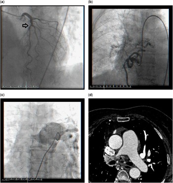
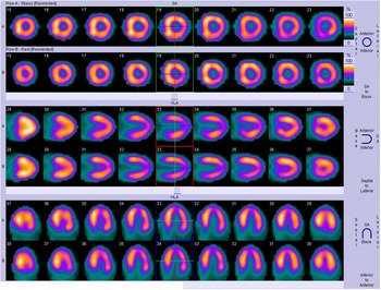
A 74-year-old woman was admitted for exertional dyspnoea and chest discomfort that had persisted for 5 years. She had diabetes and hypertension. She underwent coronary angiography for angina; thereby, a significant atherosclerotic lesion was detected in the mid-left anterior descending coronary artery (Fig 1a, indicated with an arrow), without significant stenosis of the right coronary artery; however, a well-developed coronary artery fistula, which arose from the right coronary artery and supplied the entire right lung, was detected (Fig 1b; Supplementary material 1-a). To examine the causal relationship of this lesion to ischaemia, a fractional flow reserve test was performed. The fractional flow reserve values of the mid-left anterior descending artery and right coronary artery were 0.76 and 0.93, respectively. Therefore, we deferred any intervention for the right coronary artery lesion and performed stenting only for the mid-left anterior descending stenotic lesion. Pulmonary angiography (Fig 1c; Supplementary material 1-b) and cardiac computed tomography (Fig 1d) revealed a truncated right main pulmonary artery, suggestive of a unilateral pulmonary artery atresia. Further evaluation with adenosine stress Tl-201 single-photon emission computed tomography revealed no perfusion defects of the myocardium during resting and stress conditions (Fig 2). Consistent with the pulmonary angiographic findings, the lung perfusion scan revealed a perfusion defect of the right lung field (Supplementary material 2, indicated with an arrow). Transthoracic and transoesophageal echocardiography revealed no combined congenital defects. The patient was discharged with a prescription of dual anti-platelet agents and low-dose diuretics. She continued to do well clinically without angina symptoms or functional limitations.
Left coronary angiogram showing an atherosclerotic lesion in the mid-left anterior descending artery ( a ). Right coronary angiogram showing no significant atherosclerotic lesion; however, a large fistula, which originated from the proximal right coronary artery and supplied the entire right lung was detected ( b ). Pulmonary angiogram ( c ) and cardiac computed tomographic image ( d ) showing the unilateral absence of the right pulmonary artery.

Adenosine stress Tl-201 single-photon emission computed tomographic image showing no perfusion defects of the myocardium in either resting or stress conditions.

Discussion
Unilateral pulmonary artery atresia is a rare congenital disease with a prevalence of 1 case per 200,000 individuals.Reference Ten Harkel, Blom and Ottenkamp 1 Unilateral pulmonary artery atresia frequently accompanies other congenital defects such as tetralogy of Fallot, septal defect, right aortic arch, or patent ductus arteriosus.Reference Ten Harkel, Blom and Ottenkamp 1 , Reference Bouros, Para, Panagou, Tsinitiris and Siafakas 2 Thus, echocardiography is needed to identify the combined anomaly; however, it may also occur in isolation as in our case.
Symptoms are usually vague, such as recurrent respiratory tract infections and decreased exercise tolerance, and remain unrecognised until an advanced age. Therefore, a high index of suspicion is greatly important and multimodality imaging is often needed.Reference Griffin, Mansfield and Redmond 3
The affected lung usually receives collateral blood supply from the bronchial, intercostal, subclavian, and/or subdiaphragmatic arteries.Reference Ten Harkel, Blom and Ottenkamp 1 In addition, collateral blood flowing from the coronary arteries has also been reported in several cases. Studies on whether these coronary collateral arteries induce ischaemia via the steal phenomenon are limited.Reference Soliman, Jelani, Eid and AlQaseer 4 Recently, three reports indicated that a coronary collateral artery to the unilateral pulmonary artery atresia impaired the myocardial perfusion. In those cases, myocardial scintigraphy revealed perfusion defects.Reference Soliman, Jelani, Eid and AlQaseer 4 , Reference Kadi, Kurtoglu and Karadag 5 On the contrary, one remarkable study that used a flow wire demonstrated that the shunt flow occurs mainly during systole, whereas the main coronary flow occurs during diastole.Reference Kochiadakis, Chrysostomakis, Igoumenidis, Skalidis and Vardas 6 Our case is the first to delineate the role of coronary collateral artery in relation to ischaemia by using the fractional flow reserve test. Also, one might argue the reliability of fractional flow reserve in the presence of a fistula; however, in our case, both the fractional flow reserve and cardiac scintigraphic findings supported the observation that the coronary collateral artery was not related to ischaemia. Indeed, the coronary steal issue is very complex, and thus further evaluation measuring the coronary flow reserve might be helpful. Furthermore, the therapeutic implications of confirmed coronary steal to the affected lung in this setting are challenging.Reference Nakwan 7 Although therapeutic occlusion – interventional occlusion or surgical re-implantation – of coronary artery fistulae that contribute to ischaemia is needed, this might cause infarction of the affected lung. Therefore, accurate and dedicated evaluation to determine its role in the coronary steal phenomenon is crucial before making a therapeutic decision.
In conclusion, the role of the coronary collateral artery in relation to ischaemia with a unilateral pulmonary artery atresia should be examined in each case and appropriate management should be considered accordingly.
Acknowledgements
None.
Financial Support
This research received no specific grant from any funding agency, commercial, or not-for-profit sectors.
Conflicts of Interest
None.
Ethical Standards
The study was approved by the Institutional Review Board.
Supplementary Material
For supplementary material/s referred to in this article, please visit http://dx.doi.org/10.1017/S1047951115002681