Introduction
Labyrinthitis ossificans is a condition involving pathological new bone formation within the membranous labyrinth. The causes of this disease include trauma, surgery insult, otosclerosis, malignant infiltration, autoimmune inner-ear disease, sickle cell disease and, most commonly, infection through meningogenic, haematogenic or tympanogenic routes.Reference Benson1–Reference Page and Kearns3
This study aimed to estimate the incidence of tympanogenic labyrinthitis ossificans in patients who underwent a mastoidectomy for various aetiologies, and to identify the area exhibiting the most advanced ossification in this rare condition, as revealed by computed tomography (CT) scans.
Materials and methods
We retrospectively reviewed the medical records of patients treated with mastoidectomy by a single otologist for various tympanogenic aetiologies at the Mackay Memorial Hospital from January 2007 to December 2011. High-resolution CT scans of the temporal bone were included in the routine pre-operative examinations of patients who had undergone a mastoidectomy. Patients whose high-resolution CT scans presented evidence of calcification in the inner ear, and who were reviewed by the same radiologist, were enrolled in the study. Patients with a history of head and neck cancer, meningitis, and otosclerosis, patients with cochlear implants, and patients without high-resolution CT scans, were excluded from the study.
Results
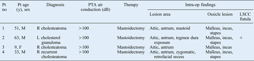
A total of 195 patients were enrolled in the study. The aetiologies for mastoidectomy were: acquired middle-ear cholesteatoma, in 163 cases; congenital cholesteatoma, in 11 cases; cholesterol granuloma, in 4 cases; and mastoiditis, in 17 cases. The high-resolution CT scans showed evidence of labyrinthitis ossificans in 4 patients (3 males and 1 female) aged between 9 and 63 years. Therefore, the incidence of tympanogenic labyrinthitis ossification in patients who underwent a mastoidectomy was 2 per cent. Data from the patients are shown in Table I. Cholesteatoma was evident in three out of the four patients, and one patient had a cholesterol granuloma.
Clinical characteristics and intra-operative findings*

* For the four patients with tympanogenic labyrinthitis ossificans. Pt = patient; no = number; y = years; PTA = pure tone audiogram; intra-op = intra-operative; LSCC = lateral semicircular canal; M = male; R = right; L = left; F = female
The intra-operative findings revealed: (1) attic and antrum involvement in all four patients; (2) obvious ossicular chain destruction in three of the four patients (three with malleus, incus and stapes involvement, and one with malleus and incus involvement); and (3) a lateral semicircular canal fistula in only one patient.
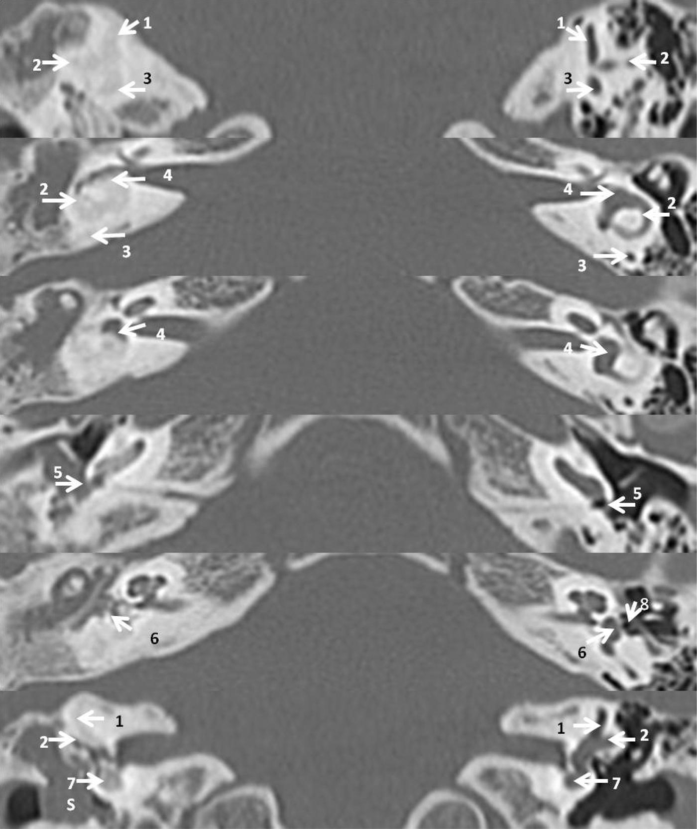
The high-resolution CT findings (Figure 1 and Table II) revealed: (1) a lateral semicircular canal fistula in one patient; (2) stapes destruction in three patients; (3) cochlear calcification of the basal and middle turn in two patients; and (4) vestibular, superior semicircular canal, posterior semicircular canal and lateral semicircular canal calcification in one, four, three and two patients, respectively.
Axial, high-resolution computed tomography scans of the temporal bone, showing ossification of the basal turn of the cochlea, the vestibule and the three semicircular canals. 1 = superior semicircular canal; 2 = lateral semicircular canal; 3 = posterior semicircular canal; 4 = vestibule; 5 = round window; 6 = oval window; 7 = basal turn of cochlea; 8 = stapes; S = soft tissue
Computed tomography findings*
* For the four patients with tympanogenic labyrinthitis ossificans. Pt = patient; no = number; LSCC = lateral semicircular canal; SSCC = superior semicircular canal; PSCC = posterior semicircular canal
Discussion
In animal and human studies, Paparella and Sugiura divided labyrinthitis ossificans into three histopathological stages, from purulent labyrinthitis to ossification: acute, fibrous and ossification.Reference Paparella and Sugiura4 Labyrinthitis ossificans is a pathological bone formation within the membranous labyrinth. The process of bone formation comprises three steps: osteoid production, mineralisation and remodelling. Osteoid mineralisation occurs in the later stage of bone formation, and can be detected on a high-resolution CT scan.
Previously, labyrinthitis ossificans was diagnosed using a post-mortem histopathological examination.Reference deSouza, Paparella, Schachern and Yoon5 The diagnosis of advanced labyrinthitis ossificans can now be made in live patients because of the development of high-resolution CT scanners, which enable analysis of the bony structure of the inner ear. In this study, the incidence of tympanogenic labyrinthitis ossificans in patients who underwent mastoidectomy was 2 per cent. The specificity of the high-resolution CT scan in detecting labyrinthitis ossificans was 100 per cent. However, Frau et al. stated that this modality is limited because it does not enable identification of labyrinthitis ossificans in its early stages.Reference Frau, Luxford, Lo, Berliner and Telischi6 In contrast, magnetic resonance imaging (MRI) is well known for its capability to detect the fine detail of the fluid spaces in the cochlea.Reference Trimble, Blaser, James and Papsin7 By retrospectively reviewing 25 meningogenic labyrinthitis ossificans cases, Isaacson et al. reported that 0.75 mm slice thickness, T2-weighted MRI is sufficiently sensitive to detect more than 94 per cent of fibrous and osseous labyrinthitis ossificans cases.Reference Isaacson, Booth, Kutz, Lee and Roland8 However, MRI is not part of routine pre-operation examinations in our hospital; hence, MRI data were not available. Accordingly, the incidence of tympanogenic labyrinthitis ossificans was likely to have been underestimated in this study.
The spread of infections from the middle to the inner ear may occur through bony destruction, along vascular channels, or directly through a break in the round or oval window.Reference Torok9 In other words, it is reasonable to assume that the most common ossification areas, the basal turn of the cochleaReference deSouza, Paparella, Schachern and Yoon5 or the lateral semicircular canal close to the mastoid cavity, are the areas exhibiting the most advanced ossification. In this study, we unexpectedly observed that the vestibular portion, especially the superior semicircular canal, was the area exhibiting the most advanced ossification.
• Labyrinthitis ossificans is a pathological bone formation within the membranous labyrinth
• The most common aetiologies are meningogenic, haematogenic or tympanogenic infections
• The incidence of tympanogenic labyrinthitis ossification in patients who underwent a mastoidectomy was 2 per cent
• The basal turn of the cochlea is the most common ossification area according to histopathology
• According to computed tomography scans, the most common ossification area is the superior semicircular canal
In a previous report of tympanogenic labyrinthitis ossificans, an eight-year-old girl had developed left-sided labyrinthitis ossificans and severe to profound sensorineural hearing loss after persistent otitis media with effusion. The CT scans showed ossification of the middle and apical turn of the cochlea, vestibule, and superior semicircular canal.Reference Page and Kearns3 In another three cases of tympanogenic labyrinthitis ossificans associated with chronic otitis media, the CT scans showed varied patterns of ossification, including: (1) more severe ossification in the semicircular canal than in the cochlea; (2) complete ossification of the inner ear, involving the cochlea, vestibule and semicircular canal; and (3) ossification of only the cochlea.Reference Swartz, Mandell, Faerber, Popky, Ardito and Steinberg10 Overall, the CT scans of the eight cases of tympanogenic labyrinthitis ossificans (i.e. the four cases identified in the current study and the four reported in previous studiesReference Page and Kearns3, Reference Swartz, Mandell, Faerber, Popky, Ardito and Steinberg10) revealed that the vestibular portion of the inner ear, especially the superior semicircular canal, was the area exhibiting the most advanced ossification. The underlying mechanism for this phenomenon remains unknown. One possible explanation for this is the small number of patients observed in this review study.
In conclusion, the incidence of tympanogenic labyrinthitis ossification in patients who underwent mastoidectomy was 2 per cent in the current study. A larger prospective study is necessary to identify the area exhibiting the most advanced ossification in this rare condition.