Introduction
Developmental orthopaedic disorders seen in the growing foal, usually designed as developmental orthopaedic disease (DOD), include several entities such as osteochondritis dissecans (OCD), subchondral bone cysts (SBC), angular limb deformities, physitis and cuboidal bone abnormalities, resulting from dysregulation of the metaphyseal growth plate (McIlwraith, Reference McIlwraith1986). DOD affects young horses with recognisable radiographic findings (RF) developing within the 1st year of life (Hoppe, Reference Hoppe1984; Olstad et al., 2007). Fetlock, hock, carpus, stifle, shoulder and cervical vertebra appear to be the most frequently affected joints (Jeffcott, Reference Jeffcott2004).
DOD is a common cause of pain and lameness for horses in sports, which limits their performance (Hoppe, Reference Hoppe1984; Kane et al., Reference Kane, Park, McIlwraith, Rantanen, Morehead and Bramlage2003). Therefore, DOD depreciates the commercial and breeding value of affected horses and is recognised as a major economic problem in the horse-breeding industry (Jeffcott, Reference Jeffcott1991). X-ray screening for DOD is becoming more commonly used in young horses, between 6 and 18 months of age. Results of X-ray screening enable breeders to decide early about the future career of horses (public or private sales, surgical treatment, etc.) and to prevent them from any further degenerative processes, in order to limit economic losses (Jeffcott, Reference Jeffcott1991 and Reference Jeffcott1996; Schougaard et al., Reference Schougaard, Falk Ronne and Phillipson1990). However, in most stud farms, this practice remains limited to a restricted number of horses with a high commercial value, because X-ray screening is expensive and time-consuming.
Aetiological factors of DOD are still not well known, but the disease appears to be of multifactorial origin. Genetic predisposition (Hoppe and Philipsson, Reference Hoppe and Philipsson1985; Schougaard et al., Reference Schougaard, Falk Ronne and Phillipson1990), body size (Sandgren et al., Reference Sandgren, Dalin and Carlsten1993), growth rate (Donabedian et al., Reference Donabedian, Fleurance, Perona, Robert, Lepage, Trillaud-Geyl, Leger, Ricard, Bergero and Martin-Rosset2006; van Weeren et al., Reference van Weeren, Sloet van Oldruitenborgh-Oosterbaan and Barneveld1999), nutritional excesses or imbalances (Jeffcott, Reference Jeffcott1991) and exercise modalities (van Weeren and Barneveld, Reference van Weeren and Barneveld1999) are considered to influence the prevalence of DOD. Thus, we hypothesised that usual differences in management practices, as well as genetic selection for many decades, may have led to between-breed variations in the prevalence and severity of DOD. A thorough knowledge of between-breed variations for their prevalence, their distribution in the various joints, and their severity, at earlier stages in the disease process is needed in order to improve the relevance and cost-effectiveness of DOD screening protocols. So, breed-specific protocols could possibly focus on the most frequently affected joints or be limited to certain joints if associations of disorders in several joints exist, depending on the between-breed variations.
Many papers have reported high prevalence of these developmental disorders. However, most of these studies (i) were performed on one unique breed, mainly Standardbreds (Alvarado et al., Reference Alvarado, Marcoux and Breton1989; Grondahl and Engeland, Reference Grondahl and Engeland1995; Storgaard Jørgensen et al., Reference Storgaard Jørgensen, Proschowsky, Falk-Rønne, Willeberg and Hesselholt1997), or (ii) were restricted to one entity, such as osteochondrosis (OC), OCD, or both (Grondahl and Engeland, Reference Grondahl and Engeland1995; Storgaard Jørgensen et al., Reference Storgaard Jørgensen, Proschowsky, Falk-Rønne, Willeberg and Hesselholt1997), or (iii) were based on a limited number of X-rayed joints, usually the hock and fetlock (Grondahl and Engeland, Reference Grondahl and Engeland1995; Storgaard Jørgensen et al., Reference Storgaard Jørgensen, Proschowsky, Falk-Rønne, Willeberg and Hesselholt1997). Moreover, in these studies, prevalence is mainly reported at joint level and associations of disorders in several joints at individual level were not explored to any great extent in foals. So, no prevalence study of DOD, simultaneously performed on several breeds and based on RF on quite a large number of joints and views, has been reported earlier.
Therefore, the objective was to describe variations in the prevalence, location and severity of DOD at weaning between Warmbloods, Standardbreds and Thoroughbreds with similar farming systems. DOD assessment was based on RF on the limb joints.
Material and methods
Data
The present study used data produced by a larger data collection and research programme conducted on DOD in Normandy, the main horse-breeding area in France. The longitudinal survey began in 2002 and is still ongoing. Due to repeated implementation of observations, questionnaires, body measurements and X-ray examinations for more than 4 years on the same farms, only convenience sampling was deemed feasible. Twenty-one stud farms owned by volunteer and motivated farmers were involved. Data here analysed concerned the three cohorts of foals born in 2002, 2003 or 2004, which were checked at least three times between birth and weaning to obtain information on their body development, feeding management, rearing conditions, locomotion and DOD affection. These foals belonged to the three main sport and racehorse breeds: Warmbloods (Wb), Standardbreds (St) and Thoroughbreds (Tb).
Feeding and housing conditions of horses could depend of several parameters such as season, weather, housing facilities of the stud farm, etc. However, all foals included in the study came from the same area of Basse-Normandie, France. Thus, rearing and housing conditions were rather similar among horses included. These data are further described in the results section and some characteristics concerning growth, rearing conditions, and feeding and management practices of foals and mares are reported according to the breed.
To assess the presence of DOD, its distribution among joints, entities and severity levels, foals were sedated and X-rayed at weaning. X-ray examination included all joints that could be X-rayed on the stud farms with a mobile unit. Five bilateral views were performed: the dorsopalmar view of the carpus, the lateromedial view of the front- and hind-limb digits (including the interphalangeal and fetlock joints) and of the hock and stifle. These joints were chosen because they are known to be frequently affected by DOD (Watkins, Reference Watkins1992).
X-ray data were analysed considering 14 bilateral anatomical sites: the distal sesamoid bone, distal phalanx, distal interphalangeal joint, fore proximal interphalangeal joint and fore fetlock (front-digit view); the carpus (carpus view); the hind foot, hind proximal interphalangeal joint, dorsal part of the hind fetlock and plantar part of the hind fetlock (hind-digit view); the proximal part of the hock and the distal part of the hock (hock view); the femoro-tibial and femoro-patellar joints (stifle view).
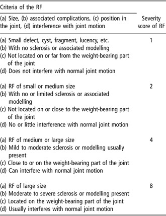
Depending on RF characteristics, three experienced equine veterinarians gave a common assessment about the status of foals (affected or not by DOD), the entity, and the severity of each RF on each of the 14 bilateral anatomical sites. RF were classified into five entities: osteochondral fragmentation (OCF) (including what is generally named OC and OCD), SBC, physitis, degenerative joint disease (DJD) and juvenile enthesopathy (JEP). The severity of RF was scored on a four-grade scale from 1 to 8 according to four criteria: the size, position in the joint, associated complications and interference with joint motion (Table 1). A non-linear scoring system was used in order to give more weight to abnormal RF corresponding to DOD with certain clinical relevance and to limit the weight of RF of little significance.
Severity score given to radiographic finding (RF) of developmental orthopaedic disease (DOD) according to four criteria

For each of the five entities, a foal was considered affected by one of them if the given entity was identified on at least one of the 14 bilateral anatomical sites; the severity score associated with the given entity for the foal was the highest severity score observed on the 14 bilateral anatomical sites. For each of the 14 bilateral anatomical sites, a foal was considered affected by DOD if at least one RF was identified on the right or left side; the severity score associated with the site for the foal was the highest one observed from either side.
Strategy of analysis and statistical methods
A two-level approach was performed. The first step was implemented for each of the 14 anatomical sites. χ 2 and Fisher’s exact tests were used to assess between-breed variations in the prevalence and in distribution for the RF severity scores. In the second step, only foals affected by DOD were considered to explore RF association patterns on the affected sites and their between-breed variations, at foal level. Both a binary status of the sites (RF presence) and the RF severity score for the affected sites were considered successively. Hierarchical clustering analyses (HCA) were performed to classify foals depending on their similarities for RF presence or severity scores. Given the high number of variables, correspondence factorial analyses (CFA) were used before HCA to represent data in a more convenient space with a reduced number of dimensions. The retained number of axes in CFA were determined using the empirical method, which reports that the explained variance percentage by each axis should be higher than the mean (i.e. 1/n, where n is the number of classifying variables) (Lebart et al., Reference Lebart, Morineau and Piron2000). To prevent scaling effects, CFA variables were standardised prior to HCA (Roux, Reference Roux1991). In the end, we chose the number of classes that combined parsimony in their number and maximisation of the coefficient of determination R 2, which represents the between-class variance. Due to very low prevalence of RF with a severity score of 4 or 8, the three following scales with, respectively, three and two grades were used for CFA: 1/2/4–8; 1/2–4–8; 1–2/4–8, instead of the initial four-grade scale. Due to very low prevalence of RF on five sites, between-breed variations were not tested and these sites were considered illustrative variables for CFA (the distal sesamoid bone, distal phalanx, distal interphalangeal joint, hind foot and femoro-tibial joint).
Results
Study sample characteristics
Out of the 21 stud farms included, six were farms with sales objectives. Their size was comprised of between 15 and 80 mares with at least 12 births a year. The remaining farms were more traditional ones, with breeding and training activities. Their size was comprised of between 4 and 40 mares with at least three births a year.
The data set retained for analysis included 392 foals born between 2002 and 2004. Wb, St and Tb accounted for 25.0%, 41.1% and 33.9%, respectively, in this number. There were slightly more fillies than colts with 52.5%. The mean age of foals at X-ray examination was 5.7 months (172 ± 38 days (mean ± s.d.)).
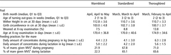
Global management of the foals and feeding practices for the mare were quite similar among breeds (Table 2). In winter and early spring, during the last 4–6 months of pregnancy and until approximately 2 weeks of age of the foal, the mares were kept in box stalls from approximately 16h00 on the afternoon until 08h00 on the following day and were group-housed in pastures during the day. Afterwards, the foals and their mares were group-housed in larger pastures for an increasing duration until 24 h/day at approximately 3 or 4 months of age of the foals. The foals were weaned at approximately 6 months of age. Management of the foals and thus rearing and housing conditions, could differ mainly depending on the date of birth, the age when first grass was offered, and the age at weaning (Table 2). Most of the foals were born in March or in April. All foals were turned out grass from early 2 or 3 weeks of life. The size of foals seemed to be the same at 30 days with a slightly higher wither height for Wb foals. Approximately 10% of foals were weaned at X-ray examination with slightly more Tb, that were 4 days older in average at weaning than Wb and St. The mean daily amount of concentrates distributed to the mares was quite similar among breeds with a slightly higher level for Tb mares during pregnancy. Distribution of a mineral and vitamin supplement (MVS) was frequent for St and Tb with more than 65% of the mares supplemented during pregnancy and lactation periods, whereas only 20% of Wb mares were supplemented during these periods.
Parameters characterising management practices of the foals, their rearing conditions, their growth, and their age at X-ray, and characterising feeding practices of the mares, depending on the breed

†MVS = mineral and vitamin supplement.
Prevalence
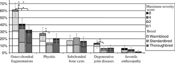
DOD was present in 66.3% of the foals (95% confidence interval (CI) = 61.6% to 71.0%). The number of foals affected, the number of RF, and their severity score, were significantly higher for Wb than for St and Tb (Table 3). OCF, then physitis and SBC were the three most frequent entities for the three breeds (Figure 1). There were more Wb affected by OCF than St and Tb, but St were more severely affected than Wb.
Prevalence (%) and 95% confidence interval of limb developmental orthopaedic disease (DOD) in 392 foals depending on the breed

a,bValues with different superscripts differ significantly (P < 0.05).
Between-breed variations in the prevalence (a) and severity score (#) of limb Developmental Orthopaedic disease (DOD) depending on the DOD entity on 392 foals (a, #: P < 0.05).

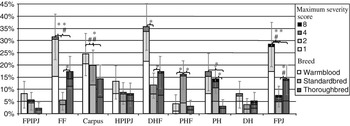
The prevalence of foals affected by DOD and the distribution of the RF severity score on the nine frequently affected sites differed depending on the breed (Figure 2). For the five remaining sites, in each breed, the prevalence of foals affected by DOD was lower than 3%. Apart from the plantar part of the hind fetlock, there were more Wb foals affected by DOD than St and Tb on every site. Wb and Tb foals were mainly affected on the dorsal part of the hind fetlock, the fore fetlock, the femoro-patellar joint, and the carpus, whereas St foals were mainly affected on the carpus, the plantar part of the hind fetlock and the proximal part of the hock.
Between-breed variations in the prevalence (a) and severity score (#) of limb Developmental Orthopaedic disease (DOD) depending on the site affected, on 392 foals (Fore proximal interphalangeal joint (FPIPJ), fore fetlock (FF), hind proximal interphalangeal joint (HPIPJ), dorsal part of the hind fetlock (DHF), plantar part of the hind fetlock (PHF) proximal part of the hock (PH), distal part of the hock (DH), femoro-patellar joint (FPJ)) (a, #: P < 0.05).

RF graded 4 and 8 were mainly located on the hind limbs and concerned few foals. St and Tb foals had more RF graded 2 than the Wb on the carpus, whereas St foals had more RF graded 4 or 8 than the Wb and Tb on the femoro-patellar joint. The most severely affected sites were the proximal part of the hock and the femoro-patellar joint for Wb and St foals, and the fore fetlock and the distal part of the hock for Tb foals.
Foal profiles for DOD
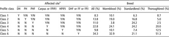
Analyses regarding the binary DOD status of the sites required six principal factors from CFA to explain 65% of the total variation. Six classes of foals were identified by HCA, corresponding to a between-class variance of 64%. The first five classes identified by HCA included foals affected on one particular site: the first class comprised all foals affected on the distal part of the hock; the second class comprised foals unaffected on the distal part of the hock but affected on the proximal part of the hock, etc. The sixth class included all remaining foals unaffected on the sites corresponding to the previous five classes (Table 3). No particular association between sites affected by DOD was observed in this class yet. This class mainly included Wb and Tb foals, whereas St foals were preferentially distributed over the previous five classes (Table 4).
Classification of 260 foals into profiles of the developmental orthopaedic disease (DOD) distribution on nine anatomical sites according to the breed

†Abbreviations are: DH = distal part of the hock; PH = proximal part of the hock; PHF = plantar part of the hind fetlock; FPIPJ = fore proximal interphalangeal joint; HPIPJ = hind proximal interphalangeal joint; DHF = dorsal part of the hind fetlock; FF = fore fetlock; FPJ = femoro patellar joint; Y = yes; N = no.
Analyses regarding distribution of the RF severity score on the sites (results not presented) did not show any strong association between sites affected either, and classes were quite similar to those described in Table 4. At least 9 to 15 principal factors from CFA were necessary to explain at least 60% of the total variations. At least nine classes of foals were necessary by HCA to get a between-class variance of at least 50%. Thus, 30% to 50% of the classes counted less than seven foals (sometimes only one foal).
Discussion
In the scientific literature, DOD prevalence in young horses has been covered extensively by studies performed on one breed and very few joints. Our study focused on between-breed variations for the prevalence and distribution of DOD in limbs of foals with similar farming systems, which had never been reported so far. The three main French breeds (Warmblood, Standardbred and Thoroughbred) were studied. Foals were X-rayed at 6 months approximately. We know that OC, and more generally DOD, start to develop before birth (Carlsten et al., Reference Carlsten, Sandgren and Dalin1993). The time of onset of DOD varies between 1 and 24 months of age, depending on the site of occurrence and on the entity (Jeffcott, Reference Jeffcott2004). Thus, some DOD lesions may develop and then disappear after visualisation at X-ray examination (Dik et al., Reference Dik, Enzerink and van Weeren1999). Dik et al. (Reference Dik, Enzerink and van Weeren1999) showed that there is a peak at about 6 months of age for the prevalence of DOD lesions in the stifle and in the hock, which can explain the high prevalence estimates that we found. They also showed that RF of DOD in the stifle tend to regress and resolve later, but not after 8 months. The first RF can be detected before 3 months of age (Carlsten et al., Reference Carlsten, Sandgren and Dalin1993). Hence, X-ray examination at 6 months of age appear to be a good compromise to detect visible changes at X-ray when most of them have developed and early enough to set conservative or curative treatments when appropriated. X-ray examinations performed here were not exhaustive. Other joints could have been X-rayed and other views could have been obtained to better document the osteo-articular status of foals. However, for economic and radiation-safety reasons, it was chosen to X-ray more foals instead of multiplying views per foal. Moreover, our approach at the foal level with a summary of the DOD profiles of foals depending on the patterns of RF distribution on the sites, had never been reported either. Because the current study used a procedure of convenient sampling,the external validity of its results is limited to foals at weaning that share the same main characteristics (e.g. breed, feeding and management practices, etc.).
Our study showed two striking results. Firstly, the site distribution of RF representing DOD was specifically designed for St, whereas it was more similar for Wb and Tb. Secondly, Wb foals were more frequently and severely affected by DOD than St and Tb foals. This difference between Wb foals and St and Tb foals can be due to genetic predisposition of Wb foals to DOD or can be due to differences among breeds in exposure to the multiple risk factors of DOD. The description of the rearing conditions and growth parameters of the foals, and of the feeding practices of the mares showed that the three breeds were managed quite similarly. Some small differences were observed for Wb foals which were born slightly later and which were 2 cm higher in average at 30 days than St and Tb. The main difference was observed for the distribution of MVS to the mares, which was less frequent for Wb than for St and Tb. The differences of birth month and distribution of MVS for Wb foals correspond to a lower farming intensity with middle term objective and lower profitability for this breed. Mineral imbalances are considered to influence the prevalence of DOD (Knight et al., Reference Knight, Weisbrode, Schmall, Reed, Gabel, Bramlage and Tyznik1990; Savage et al., Reference Savage, McCarthy and Jeffcott1993). Thus, between-breed variations observed in the prevalence of DOD may be possibly partly explained by the difference observed in the MVS distribution to the mares. However, all the assessed differences in the prevalence cannot be attributed to this factor because some not-supplemented Wb mares had foals not affected by DOD while some supplemented Tb mares had affected foals. Only an individual risk-factor analysis would make it possible to know if the observed differences in the prevalence of DOD is due to differences in MVS distribution to the mares.
Because no simultaneous prevalence study on several breeds with similar farming systems has been reported earlier, and also due to differences in X-raying protocols, comparisons to previously published estimates for the prevalence of DOD in young horses should be made with caution. Entities taken into account were often limited to OCF. Moreover, previous prevalence estimates were assessed on a particular population such as horses coming from only one stud farm, or horses genetically predisposed to DOD, or horses presented for public auction or clinical investigations. Therefore, these horses are expected to be more or less affected than a broader horse population, and thus providing possibly biased prevalence estimates. Keeping in mind all these limits in validity of any comparison, only a short overview of apparent similarities and discrepancies was deemed relevant here.
Our results showed that OCF was the most frequent entity for the three breeds which is in accordance with the particularly large distribution of OC recognised by van Weeren and Barneveld (Reference van Weeren and Barneveld1999). They estimated that the incidence of the OC including all joints and radiographically occult lesions is at least 20% to 25% in north-western Europe. In our sample of St foals, 41% were affected by OCF which is slightly higher than the 31.5% reported by Alvarado et al. (Reference Alvarado, Marcoux and Breton1989) on older yearlings. For Wb, our prevalence estimate of 61% of foals affected by OCF was higher than the 31.4% from Stock et al. (Reference Stock, Hamann and Distl2006). However, this difference can be explained by the differences in methodology as Stock et al. (Reference Stock, Hamann and Distl2006) studied horses selected for sales, aged from 3 to 7 years, and did not X-rayed the stifle and carpus. Moreover, Dik et al. (Reference Dik, Enzerink and van Weeren1999) showed that the prevalence of DOD lesions in the stifle and in the hock reach a peak at about 6 months of age, which could explain the high prevalence estimates that we found in our sample of foals X-rayed at 5.7 months on average.
For St foals, the site distribution of RF was different from those of Wb and Tb foals. Our prevalence estimate for St affected foals is only slightly lower than that reported by Alvarado et al. (Reference Alvarado, Marcoux and Breton1989) from a study involving only one stud farm of 73 St yearlings X-rayed with the same views as ours: 62.7% v. 75.3%.
Our prevalence estimates on the carpus and the femoro-patellar joint of St foals are consistent with theirs. For the distal part of the hock, our result of 3.7% of St foals affected by DOD is not in keeping with that of Hartung et al. (Reference Hartung, Münzer and Keller1983), who found 49.7% for German St yearlings affected by spavin on the tarsus.
On the fetlocks of St foals, our prevalence estimates of DOD (6%, 12%, 16%, respectively, on the fore fetlock, and on the dorsal part and plantar part of the hind fetlock) appear quite consistent with those of several studies. Storgaard Jørgensen et al. (Reference Storgaard Jørgensen, Proschowsky, Falk-Rønne, Willeberg and Hesselholt1997) reported 16.0% of 243 Danish St yearlings affected by OCF. Carlsten et al. (Reference Carlsten, Sandgren and Dalin1993) reported between 18.2% and 23.4% foals affected by OCF in 71 St foals from a Swedish stud farm. In the same way, our results are very close to the estimates of Sandgren et al. (Reference Sandgren, Dalin and Carlsten1993) from a random sample of 674 Swedish St yearlings (2.5% and 3.3% affected by OCF on the fore fetlock, and between 13.7% and 15.7% of yearlings affected on the proximal part of the hind fetlock). However, our results are in sharp contrast to the 31.9% prevalence (limited to OCF) in a study involving 753 Norwegian St X-rayed between 6 and 21 months (Grondahl and Engeland, Reference Grondahl and Engeland1995).
For the proximal part of the hock of St foals, the prevalence in our study (14.0%) is consistent with the OCF prevalence reported for Norwegian and Danish St yearlings (Grondahl and Engeland (Reference Grondahl and Engeland1995) 14.3%; Storgaard Jørgensen et al. (Reference Storgaard Jørgensen, Proschowsky, Falk-Rønne, Willeberg and Hesselholt1997) 14.0%). Our results are also quite consistent with those of Swedish St aged 5 months, which showed a prevalence comprised between 19.5% and 23.4% according to their precise location on the site (Carlsten et al., Reference Carlsten, Sandgren and Dalin1993).
Considering the severity of DOD in affected St, our results compared well with those of Alvarado et al. (Reference Alvarado, Marcoux and Breton1989). In the fetlocks of St yearlings, they found that 54.5% of OCF were graded 1. Comparatively, in the fore fetlock and in the dorsal part of the hind fetlock, we found that approximately 70% of affected St foals had only RF graded 1; in the plantar part of the hind fetlock, we found that 25% of affected St foals had only RF graded 1. In the hock and in the stifle, Alvarado et al. (Reference Alvarado, Marcoux and Breton1989) found that 60% of OCF were graded 2 on St adults and offspring yearlings, whereas we found that in the hock and in the femoro-patellar joint respectively, 70% and 40% of affected St foals had at least one RF graded 2.
Wb foals were the most affected by DOD. The number of foals, the number of RF per foal, and their severity score as well were higher than in St and Tb. Comparisons with previous studies were difficult because no study of DOD prevalence globally on the limb joints had been performed on Wb foals or yearlings. Limited to OCF and from sales-selected horses aged between 3 and 7 years, Stock et al. (Reference Stock, Hamann and Distl2006) reported prevalence estimates on 3127 Hanoverian Wb. They found lower estimates than ours on the proximal interphalangeal joint, the fore fetlock, the hind fetlock and the hock. They reported 4.5% of horses affected on the distal interphalangeal joints, which is concordant with our results. From 167 young German Coldbloods, Wittwer et al. (Reference Wittwer, Hamann, Rosenberger and Distl2006) found higher prevalence estimates than ours on the fetlocks and the hock. One reason for the high prevalence in German Coldbloods compared with Wb, or in Wb compared with St and Tb foals, could be seen in weight growth rates or in the body format conducting to a higher weight for Coldbloods and Wb than for St and Tb horses. Sandgren et al. (Reference Sandgren, Dalin and Carlsten1993) and Pagan and Jackson (Reference Pagan and Jackson1996) showed that foals with OC lesions were heavier at birth and had a faster weight growth than foals without OC indeed. Another explanation for the high prevalence in German Coldbloods when compared with our prevalence estimate in Wb can be the differences in management practices between our two studies.
For Tb foals, the most affected sites were quite similar to those of Wb foals with lower prevalence estimates. Likewise Wb foals, comparisons with previous studies were difficult because few prevalence studies of DOD had been performed globally on the limb joints of young Tb horses. Through a very detailed examination including 34 views performed on 1130 Tb yearlings at pre- or post-sale in a private veterinary practice on the same joints as in our study, Kane et al. (Reference Kane, Park, McIlwraith, Rantanen, Morehead and Bramlage2003) described all RF with more precise and detailed locations than us. For the fetlock, the hock, and the stifle, they reported a large range of prevalence in RF, which is in accordance with our results. For the carpus, they reported a very smaller occurrence of RF than in our protocol, comprised of between 0.1% and 1.7% depending on the precise location.
To explore the DOD profiles of foals according to patterns of site distribution of RF, factorial analyses and classifications were used. These two methods are complementary, because of the difficulty to interpret results from factorial analyses, which is offset by classifications. For factorial analyses, the sites with low prevalence were considered here, illustrative to avoid effects due to a nature-specific high contribution of such sites, if they had been introduced as classifying variables. For the same reason, some grades of RF severity score with a low prevalence were regrouped.
In the two ways explored (distribution of RF and distribution of RF severity score), no clear natural class structure with associated affected sites was observed. In fact, a wide variety of classes existed and corresponded to a low R 2. Most classes were characterised by one particular site affected without any typical association with other affected sites. To obtain a valid R 2, a larger number of axes and classes was required. However, results from CFA and HCA were concordant with the results obtained in the first part of this study concerning the between-breed variations in the site distribution of foals (affected by DOD based on a binary definition). Indeed, Wb and Tb foals were preferentially classified in the same class because they were preferentially affected on the same sites, whereas St foals were distributed in other classes.
Conclusion
This is the first epidemiological study reporting between-breed variations of the DOD distribution and of the severity of corresponding RF, on the limb joints of foals with similar farming systems. However, few differences were observed in the feeding practices of the mares among breeds, which need to be confirmed by further research. The main challenge for DOD epidemiology is to simultaneously study all the individual risk factors in order to quantify the roles of respective genetic predispositions and management practices and to make recommendations to breeders to reduce the prevalence and incidence of DOD.
The results of this study are consistent with those obtained by previous studies performed separately on one breed. Our results showed that the site distribution of DOD is specifically designed for Standardbreds compared with Warmbloods and Thoroughbreds, which are more similar. The most severe RF are mainly located on the hind limbs, plus the fore fetlock for Thoroughbreds. Moreover, our results suggests that young Warmblood horses are likely more affected than Standardbreds and Thoroughbreds. This study provides a better knowledge about between-breed variations in the distribution of DOD. These results are of great interest to improve DOD screening by adapting X-ray protocols to the breed regarding distribution of prevalence and severity of DOD among joints. Consequently, the X-ray DOD screening could be adapted to the breed and to the screening objectives, which could be to assess the most frequently affected joints or the most severely affected ones. In the same way, the results can help practitioners to focus their clinical examinations towards the most common findings.
Acknowledgements
This work was financially supported by the Haras Nationaux, the Conseil Régional de Basse Normandie, the Institut National de la Recherche Agronomique and the French Ministère de l’Agriculture.