INTRODUCTION
Schistosomiasis is a neglected tropical disease (NTD) caused by blood flukes of the genus Schistosoma. Schistosoma haematobium, S. japonicum and S. mansoni are the 3 main species parasitizing humans (Utzinger and Keiser, Reference Utzinger and Keiser2004; Gryseels et al. Reference Gryseels, Polman, Clerinx and Kestens2006). It has been estimated that there are more than 207 million people infected worldwide, with 779 million people at risk of infection (Steinmann et al. Reference Steinmann, Keiser, Bos, Tanner and Utzinger2006). The annual mortality rate in sub-Saharan Africa is estimated to be as high as 280 000 (van der Werf et al. Reference van der Werf, de Vlas, Brooker, Looman, Nagelkerke, Habbema and Engels2003). Estimates of the global burden of schistosomiasis range from 1·7 to 4·5 million disability-adjusted life years (DALYs) (WHO, 2002, 2004); the latter figure is likely to underestimate the true burden, as it does not consider chronic disability (Hotez et al. Reference Hotez, Molyneux, Fenwick, Ottesen, Ehrlich Sachs and Sachs2006). Revised estimates for schistosomiasis are at 3–70 million DALYs (King and Dangerfield-Cha, Reference King and Dangerfield-Cha2008).
Despite schistosomiasis being one of the most prevalent and debilitating NTDs, a devoted drug discovery and development programme does not exist (Ridley and Kita, Reference Ridley and Kita2007). Since the late 1970s, the treatment and control of schistosomiasis relies on a single drug, namely praziquantel (PZQ) (Gönnert and Andrews, Reference Gönnert and Andrews1977; Cioli and Pica-Mattoccia, Reference Cioli and Pica-Mattoccia2003; Utzinger and Keiser, Reference Utzinger and Keiser2004; Doenhoff et al. Reference Doenhoff, Cioli and Utzinger2008). However, the heavy reliance and application of PZQ within the frame of large-scale preventive chemotherapy treatment programmes might select for drug-resistant parasites (Botros and Bennett, Reference Botros and Bennett2007; Caffrey, Reference Caffrey2007). In addition, PZQ has only moderate activity against juvenile worms, which is an important deficiency in its spectrum of activity, as it requires administration of a second PZQ dose after a short interval to eliminate the parasites that have since matured (Doenhoff et al. Reference Doenhoff, Cioli and Utzinger2008).
Nonetheless, in spite of the lack of dedicated product development partnerships for helminth diseases, a number of compounds with interesting anti-schistosomal properties have been identified by various research groups in recent years; for example, peroxidic compounds (the semi-synthetic artemisinins and the synthetic trioxolanes (OZs)), 4-phenyl-1,2,5-oxadiazole-3-carbonitrile-2-oxide and the cysteine protease inhibitor K11777 (Abdulla et al. Reference Abdulla, Lim, Sajid, McKerrow and Caffrey2007; Keiser and Utzinger, Reference Keiser and Utzinger2007; Utzinger et al. Reference Utzinger, Xiao, Tanner and Keiser2007; Xiao et al. Reference Xiao, Keiser, Chollet, Utzinger, Dong, Endriss, Vennerstrom and Tanner2007; Sayed et al. Reference Sayed, Simeonov, Thomas, Inglese, Austin and Williams2008; Caffrey et al. Reference Caffrey, Williams, Todd, Nelson, Keiser, Utzinger and Selzer2009). An important finding was that mefloquine (MQ), an arylamino alcohol (4-quinoline-methanol), developed by the Walter Reed Institute of Research in the early 1970s (Sweeney, Reference Sweeney1981) and now marketed for the prophylaxis and treatment of malaria, showed promising anti-schistosomal activity in mice. Single oral doses of 200–400 mg/kg to mice infected with either juvenile or adult stages of S. mansoni or S. japonicum resulted in high total or even complete reductions in female worm burdens (Keiser et al. Reference Keiser, Chollet, Xiao, Mei, Jiao, Utzinger and Tanner2009). Treatment with a lower dose of 150 mg/kg MQ revealed a significantly reduced egg production in S. mansoni-infected mice (Van Nassauw et al. Reference Van Nassauw, Toovey, Van Op den Bosch, Timmermans and Vercruysse2008).
The aim of the present study was to further strengthen the current evidence base of the anti-schistosomal properties of MQ and to gain a first insight into the potential mode of action of the drug. The dose-response relationships were studied in vitro. The temporal drug effect was monitored in vitro and in vivo. In addition, alterations in the tegumental surface of schistosomula and adult flukes were studied by means of scanning electron microscopy (SEM).
MATERIALS AND METHODS
Experimental animals and parasites
All experiments were carried out at the Swiss Tropical Institute (Basel, Switzerland), in accordance with Swiss national animal welfare regulations. Female NMRI mice (n=40, age=3 weeks, weight=25 g) were purchased from Harlan (Horst, The Netherlands).
Cercariae of S. mansoni (Liberian strain) were obtained from infected intermediate host snails (Biomphalaria glabrata) maintained according to standard procedures (Keiser et al. Reference Keiser, Chollet, Xiao, Mei, Jiao, Utzinger and Tanner2009).
Drugs
Mefloquine ((RS)-(±)-α-(2-Piperidinyl)-2,8-bis(trifluormethyl)-4-quinolinemethanol hydrochloride, MW=414.81 g/mol) (MQ) was kindly provided by Mepha AG (Aesch, Switzerland). For in vitro studies, stock solutions of MQ (10 mg/ml) were prepared in dimethyl sulfoxide (DMSO) (Fluka, Buchs, Switzerland). For in vivo studies, MQ (40 mg/ml free base) was suspended in 7% (v/v) Tween 80 and 3% ethanol shortly before oral administration to mice.
In vitro studies on schistosomula
Cercariae of S. mansoni were mechanically transformed into schistosomula (Ramalho-Pinto et al. Reference Ramalho-Pinto, Gazzinelli, Howells, Mota-Santos, Figueiredo and Pellegrino1974). Briefly, 50 ml of an ice-cold cercarial suspension were centrifuged and the packed volume of cercariae was re-suspended in 2 ml of Minimal Essential Medium (MEM) (Eagle, Reference Eagle1959) and vortexed for 2 min to trigger tail loss. For the isolation of cercarial bodies, cold Hank's Basal Salt Solution (HBSS) was added to the cercarial suspension up to a volume of 7 ml. After cooling on ice for 15 min, the tail-rich supernatant was decanted and the sedimented bodies were re-suspended in 7 ml of cold HBSS. This procedure was repeated twice. The schistosomula suspension was kept in Basch culture medium (Basch, Reference Basch1981) in an incubator at 37°C in an atmosphere of air, (95%)+CO2 (5%) for 1–3 h until use.
To study the temporal drug effect and the concentration-response relationships, schistosomula were incubated in 24-well plates (Costar), with 500–1000 schistosomula per well, in the presence of different concentrations of MQ. Each well contained 1 ml of Basch medium supplemented with 5% heat-inactivated foetal calf serum (iFCS) and 100 U/ml penicillin and 100 μg/ml streptomycin (Invitrogen, Carlsbad, USA) and MQ at 100, 75, 50, 25, 15, 10, 5 or 1 μg/ml, respectively. Schistosomula incubated with 1% DMSO, the highest concentration of drug solvent, served as controls. The parasites were kept in an incubator at 37°C in an atmosphere of air, (95%)+CO2 (5%) for up to 96 h.
Immediately after adding (0·125), 0·5, 1, 6, 17, 24, 48, 72 and 96 h after incubation with MQ, all schistosomula were observed under a dissecting microscope and the effect of MQ was assessed, with an emphasis on changes in worm motor activity, morphological/tegumental alterations and the occurrence of death. These phenotypic changes were scored using a viability scale of 0–3: (3=totally vital, normally active, no morphological changes, 2=slowed activity, first morphological changes and granularity visible, 1=minimal activity, severe morphological changes and granularity, 0=all worms dead, severe granularity) based on standard procedures for compound screening at WHO-TDR (Ramirez et al. Reference Ramirez, Bickle, Yousif, Fakorede, Mouries and Nwaka2007) and the UCSF Sandler Center (Caffrey et al. Reference Caffrey, Williams, Todd, Nelson, Keiser, Utzinger and Selzer2009). The minimal lethal concentration (MLC), which is the minimum concentration needed to kill all schistosomula and the minimal effective concentration (MEC), which is the minimum concentration needed to observe any change in viability or morphology of schistosomula were determined after 1 h. All experiments were carried out in duplicate and were repeated at least 3 times.
For SEM studies, samples of schistosomula incubated with 3 different concentrations of MQ (100, 10 and 1 μg/ml) were taken at different time-points (after 0·5, 1, 6, 24, 72 and 96 h). Specimens were fixed with 2·5% (v/v) glutaraldehyde in a phosphate-buffered saline (PBS) (pH 7·4) for 24 h at 22–24°C (room temperature). After rinsing 3 times with PBS, the specimens were stored in the same buffer at 4°C until use. Before SEM examination, the flukes were washed twice with double-distilled water, dehydrated in ascending ethanol concentrations and then air-dried. Schistosomula were placed on aluminium stubs, sputter coated with 20 nm gold particles and observed in a high-resolution SEM (Phillips XL30 ESEM) at an accelerating voltage of 5 kV.
In vitro studies of adult schistosomes
NMRI mice were infected subcutaneously with ~80 cercariae of S. mansoni and euthanized with CO2 after 56 days. All adult schistosomes were collected from the hepatic portal system and mesenteric veins and removed from the liver by perfusion with ice-cold PBS supplemented with heparin (Yolles et al. Reference Yolles, Moore, De Giusti, Ripsom and Meleney1947; Smithers and Terry, Reference Smithers and Terry1965). Schistosomes were incubated in 24-well plates (Costar), placing 2 male and 2 female worms in each well. Individual wells contained RPMI 1640 culture medium (Invitrogen, Carlsbad, USA) supplemented with 5% heat-inactivated calf serum, 100 U/ml penicillin and 100 μg/ml streptomycin (Invitrogen, Carlsbad, USA). Schistosomes were incubated in the presence of 100, 10 and 1 μg/ml MQ at 37°C in an atmosphere of air (95%)+CO2 (5%). Medium without and medium with 1% DMSO (the highest concentration of drug solvent used) served as controls. Before incubation with MQ and immediately after incubation (0·125), 1, 6, 24, 48 and 72 h with MQ, all S. mansoni were observed under a dissecting microscope. The effect of the drug was assessed with an emphasis on changes in worm motor activity, morphological/tegumental changes and death of worms. Death was defined as no movement observed for at least 2 min of examination and no movement at the other observation time-points. All experiments were carried out in duplicate and were repeated at least 3 times. For SEM examination, the worms were prepared as described above, with the exception that the worms were critical-point dried (Bomar SPC-900; Tacoma USA).
In vivo studies
NMRI mice were infected as described above. At 7–8 weeks after infection, mice were treated with a single oral dose of 400 mg/kg MQ, the most effective dose as established in our previous work (Keiser et al. Reference Keiser, Chollet, Xiao, Mei, Jiao, Utzinger and Tanner2009). At each time-point after treatment (24, 48, 72, 96 and 120 h), 3–6 mice were killed by cervical dislocation and the mesentery and the liver were collected. Schistosomes were removed from the mesenteric veins, sexed and counted using a binocular microscope. The liver was flattened and examined for the presence of worms. The distribution of schistosomes in the liver and mesenteric veins was recorded. Schistosomes recovered from the mesenteric veins were further examined, and changes in worm motor activity, morphological/tegumental changes and death were documented. Worms were then prepared for SEM examination.
RESULTS
In vitro studies of schistosomula – dissecting microscopic and SEM evaluations
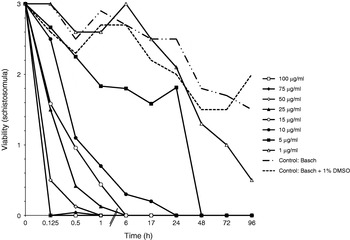
The effect of incubation with different concentrations of MQ on the viability of schistosomula for up to 96 h is depicted in Fig. 1. In the absence of the drug, schistosomula showed normal viability without any morphological change for up to 24 h. Slight morphological changes such as granularity, a shorter body length with a falcate shape and reduced activity, were observed after 48 h of incubation. Schistosomula were viable for at least 96 h.
Effect of different concentrations of MQ on the viability of Schistosoma mansoni schistosomula. Mean values of viability using a viability score. Numbers derived from a minimum of 6 experiments (500–1000 schistosomula per experiment were scored at each concentration). Open squares: 100 μg/ml; filled diamonds: 75 μg/ml; open diamonds: 50 μg/ml; filled triangles: 25 μg/ml; open circles: 15 μg/ml; filled circles: 10 μg/ml; filled squares: 5 μg/ml; open triangles: 1 μg/ml; dashed and dotted line: Control (Basch medium); dashed line: Control (Basch medium+1% DMSO).

Following incubation with the 2 highest concentrations of MQ (100 and 75 μg/ml) schistosomula died rapidly; no movement was observed 3–4 min after incubation. At this time-point, schistosomula incubated with 1–10 μg/ml of MQ showed normal movements, while schistosomula incubated with 15–50 μg/ml of MQ showed slowed activity (Fig. 1).
At 1 h after incubation with 25 and 50 μg/ml of MQ all worms had died. Schistosomula incubated with 5, 10 and 15 μg/ml showed only minimal activity, including convulsions and contractions. Severe morphological changes, such as dark granular and crescent-shaped bodies associated with a reduction in body lengths were seen. No effect was observed at the lowest concentration of MQ (1 μg/ml) at the 1 h time-point. Hence, the MLC and MEC of MQ on schistosomula determined after 1 h were 25 and 5 μg/ml, respectively. In the presence of 1 μg/ml of MQ schistosomula revealed first morphological changes and a decreased motility 48 h post-incubation and 96 h after incubation most of the schistosomula were dead.
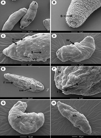
The structure and appearance of S. mansoni schistosomula, incubated for 1 h in the absence of MQ were similar to those described in the literature (Crabtree and Wilson, Reference Crabtree and Wilson1980; Basch and Basch, Reference Basch and Basch1982). In brief, schistosomula had a round shape, clearly defined head and tail regions and an incompletely developed ventral (acetubulum) and oral sucker (Fig. 2A). The tegument was covered by tegumental spines, which were directed towards the posterior end (Fig. 2B).
SEM observations of Schistosoma mansoni schistosomula after in vitro incubation. (A) Control, showing the ventral sucker (VS) (=acetabulum), the anterior end (A) and the posterior end (P). (B) Control. Posterior region with clearly defined tegumental spines (S). (C) Thirty min after incubation with MQ (100 μg/ml). Shortened spines (S) and blebbing (b) near the ventral sucker (VS) visible. (D) Thirty min after incubation with MQ (10 μg/ml). Contraction and formation of wrinkles (W) visible. (E) Six h after incubation with MQ (10 μg/ml). A shrunken body with swelling (sw) and many furrows (f) observed. (F) Six h after incubation with MQ (10 μg/ml). The tegument shows deep furrows (f) and flattened spines (fs). (G) Twenty-four h after incubation with of MQ (1 μg/ml). Shrunken (sh) tegument and bending of the body. (H) Seventy-two h after incubation with MQ (1 μg/ml). A shrunken (sh) tegument visible.

Thirty min after incubation with 10 and 100 μg/ml of MQ, all schistosomula examined showed a change in shape and extensive tegumental alterations. Worms incubated with 100 μg/ml of MQ showed intensive blebbing and shortened spines on the entire tegumental surface (Fig. 2C). The incubation of schistosomula with 10 μg/ml of MQ showed contraction and bending of the worm body, resulting in the formation of wrinkles and a reduction of body length by ~50%. In addition, spines were shortened (Fig. 2D). Schistosomula exposed to 1 μg/ml of MQ showed no morphological changes 30 min after incubation, and their structure and appearance was similar to the controls.
At 6 h after incubation with 10 μg/ml MQ the tegument of schistosomula showed many deep furrows, swelling and flattened spines (Fig. 2E and F). At this same time-point, the lowest concentration of MQ induced no tegumental alterations on schistosomula.
Slight tegumental alterations were observed on worms incubated with 1 μg/ml of MQ after 72 h; schistosomula were bending backwards (Fig. 2G) and showed a shrunken tegument (Fig. 2G and H).
In vitro studies of adult S. mansoni – dissecting microscopic and SEM evaluations
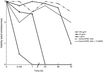
The dose-response relationships of MQ on adult schistosomes for up to 72 h are given in Fig. 3. Control female and male schistosomes showed normal viability for up to 72 h. Egg production was observed starting 24 h post-incubation. After 3–4 min of incubation with 100 μg/ml of MQ, schistosomes displayed only minimal activities, including slow convulsions and contractions. At 1 h after incubation, adult worms exposed to this concentration were dead (Fig. 3). Worms incubated with 10 and 1 μg/ml of MQ showed normal activity at the 1 h time-point. After 24 h, all schistosomes incubated with 10 μg/ml of MQ were dead. In the presence of 1 μg/ml of MQ, there was a reduction in the viability of female worms after 72 h compared with untreated schistosomes. MQ suppressed egg production in female worms incubated with all 3 concentrations used.
Effect of different concentrations of MQ on the viability of adult worms of Schistosoma mansoni. Mean values of viability using a viability score. Numbers derived from a minimum of 6 experiments (in total 24 worms were scored at each concentration). Open squares: 100 μg/ml; filled circles: 10 μg/ml; open triangles: 1 μg/ml; dashed and dotted line: Control (RPMI 1640); dashed line: Control (RPMI 1640+1% DMSO).

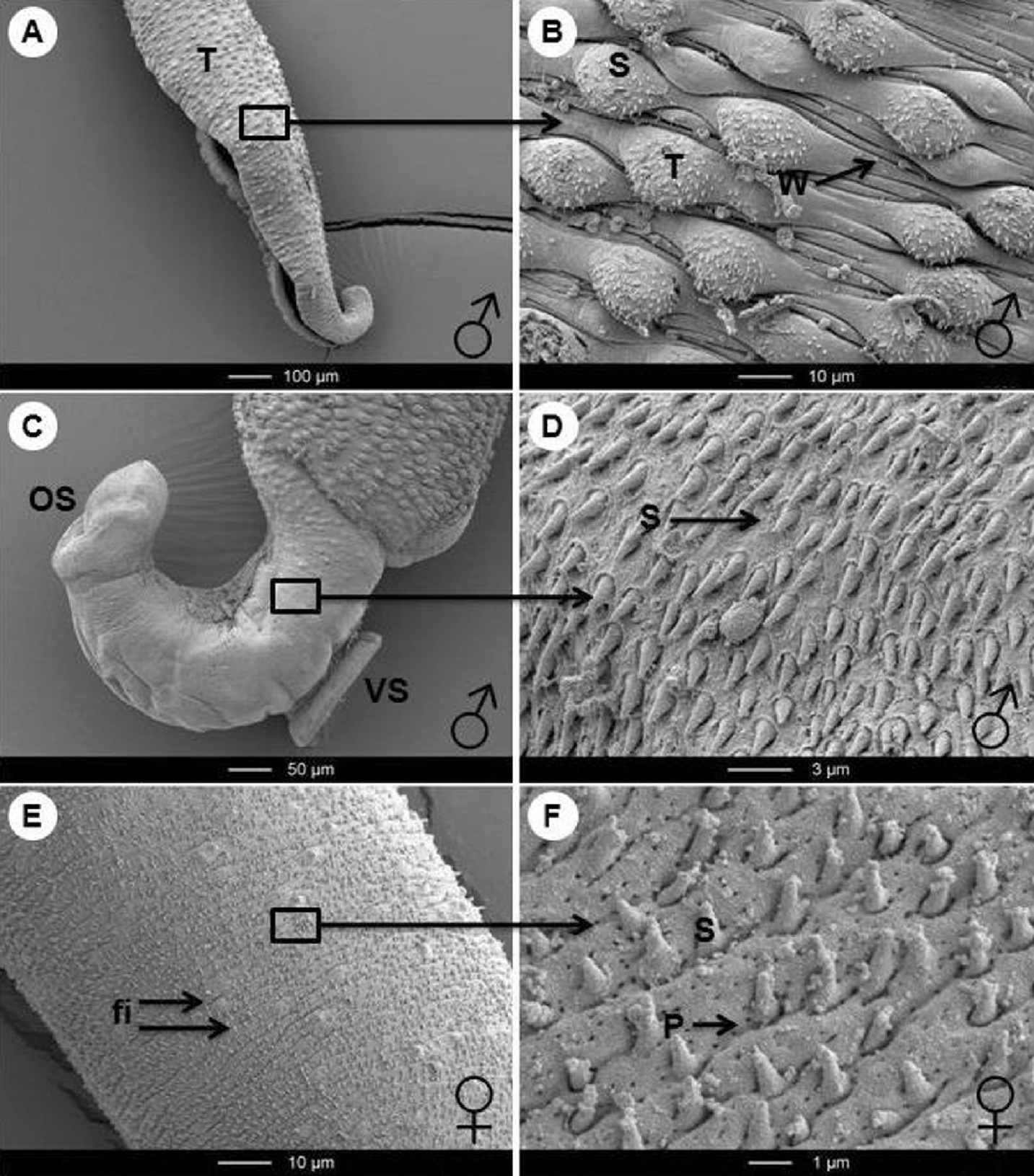
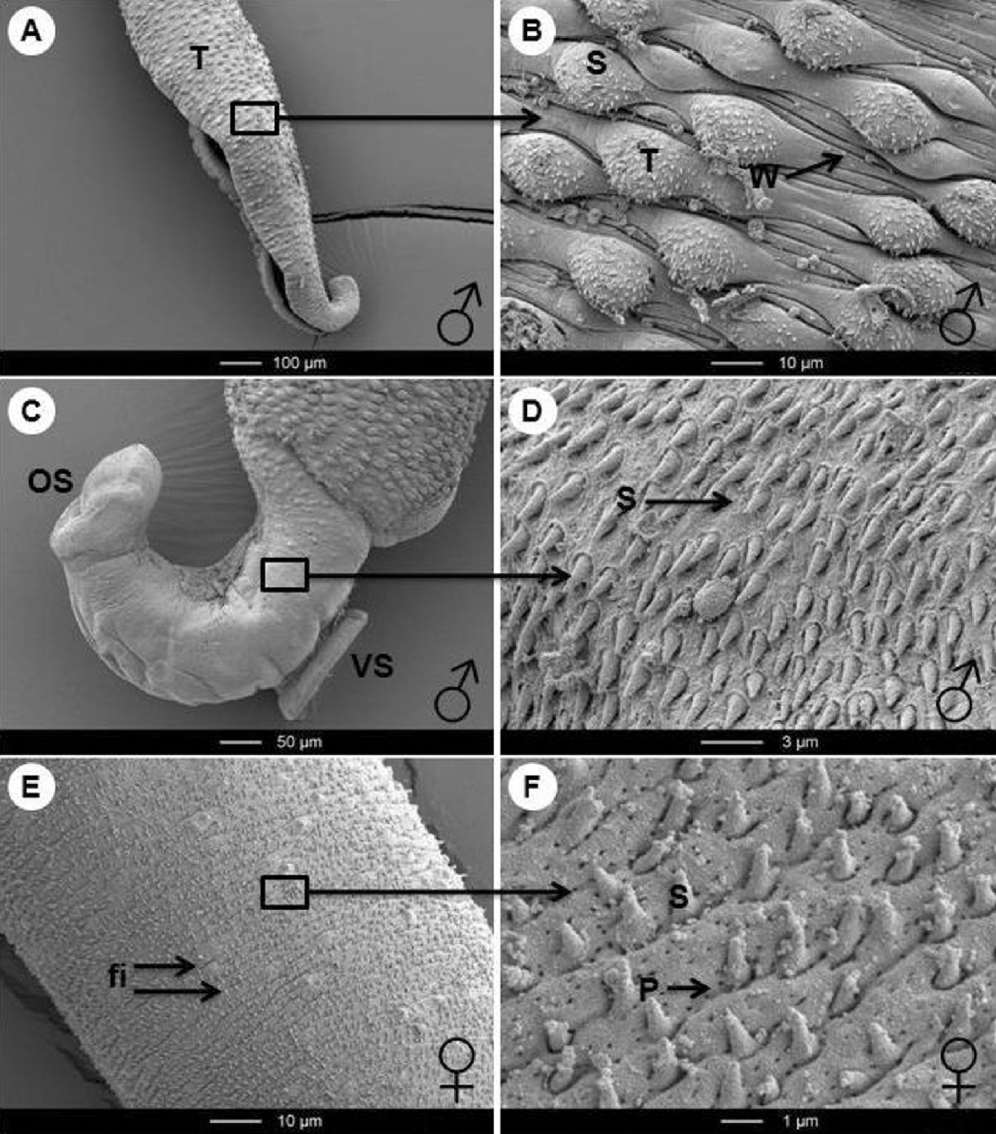
The tegumental surface of adult S. mansoni has been described in a number of SEM studies (Miller et al. Reference Miller, Tulloch and Kuntz1972; Hockley, Reference Hockley1973; Hockley and McLaren, Reference Hockley and McLaren1973). Briefly, the surface of female schistosomes is simpler and more uniform when compared with the male worm (Basch and Basch, Reference Basch and Basch1982; Gupta and Basch, Reference Gupta and Basch1988). The latter sex showed many tubercles on the dorsal body surface of the tail region (Fig. 4A). At a higher magnification, spines on the top of the tubercles and parallel-arranged wrinkles on the tegument were visible (Fig. 4B). The head region of both sexes of S. mansoni is characterized by an oral and a ventral sucker (Fig. 4C) and a smooth tegument with numerous tegumental spines (Fig. 4D). The mid-body region of female adults had parallel arranged fissures (Fig. 4E) and, at a higher magnification, tegumental spines and pores were visible (Fig. 4F).
SEM observations of untreated controls of adult Schistosoma mansoni recovered from a mouse 49 days post-infection by portal perfusion. (A) Tail region of male worm, showing tubercles (T). (B) Higher magnification of (A): tegument with tubercles (T), spines (S) and parallel-arranged wrinkles (W). (C) Head region of a male worm, showing the ventral (VS) and the oral sucker (OS). (D) Higher magnification of (C): tegument of the head region with spines (S) visible. (E) Female tegument of the mid-body region with parallel arranged fissures (fi). (F) Higher magnification of (E). Spines (S) and pores (P) visible.
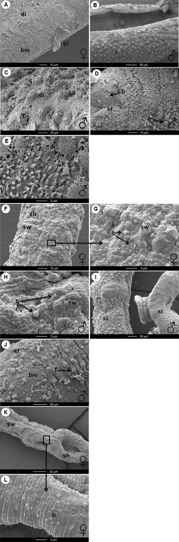
By 30 min after incubation with 100 μg/ml of MQ extensive destruction was apparent on the entire tegument of all adult schistosomes examined. For example, on the mid-body region of female worms, extensive sloughing was visible, leaving the basal membrane exposed. In addition, the tegument showed a roughened appearance and started to disintegrate (Fig. 5A). Figure 5B depicts a roughened surface of a male specimen; at a higher magnification, disintegration of the tegument, resulting in a fibrous appearance, loss of tubercles, spines and the parallel-arranged wrinkles, was visible (Fig. 5C). The whole tegumental layer was destroyed.
SEM observations of adult Schistosoma mansoni after in vitro incubation with MQ. (A) Thirty min after incubation with MQ (100 μg/ml). Tegument of the mid-body region of a female worm. Extensive sloughing (sl) exposing to view the basal membrane (bm). Tegument shows roughening and disintegration (di). (B) Thirty min after incubation with MQ (100 μg/ml). Lateral and dorsal tegument around the gynecophoral canal of a male worm depicting a roughened (r) surface. (C) Higher magnification of (B). Tubercles (T) and the lateral tegument disintegrate (di) resulting in disappearance of the knobs, spines and the parallel-arranged wrinkles. (D) One h after incubation with MQ (10 μg/ml). Blebbing (b) and sloughing (sl) on the inner surface of the oral sucker of a male specimen is visible. (E) Higher magnification of (D) reveals disintegration (di) of the tegument. Spines (S) are still apparent, but not as clear and regular as in the control. (F) Twenty-four h after incubation with MQ (10 μg/ml). The tegument of the ventral mid-body region of a female worm is shrunken (sh) and swollen (sw). (G) Higher magnification of (F) shows many blebs (b) and swelling (sw) of the tegument. (H) Twenty-four h after incubation with MQ (10 μg/ml). Swollen (sw) male tegument of the ventral mid-body region with many blebs (b) seen. (I) Forty-eight h after incubation with MQ (10 μg/ml). Sloughing (sl) on the anterior region and at the ventral sucker of a male worm visible. (J) Forty-eight h after incubation with MQ (10 μg/ml). Mid-body region of a male worm: tegument with tubercles (T). The tegument shows extensive sloughing (sl). In some parts it lifted away leaving the basal membrane (bm) exposed. (K) Forty-eight h after incubation with MQ (1 μg/ml). Female with a shrunken (sh) and swollen (sw) mid-body tegument. (L) At higher magnification of this body region numerous blebs (b) were seen.

After 1 h of incubation with 10 μg/ml of MQ, tegumental alterations were observed on the oral suckers of male worms and on the anterior and mid-body regions of both males and females. For example, blebbing and sloughing were observed on the head region of a male worm. Even the inner surface of the oral sucker of this worm was affected (Fig. 5D). Higher magnification revealed a disintegration of the interspinal tegument of the oral sucker, resulting in a roughened and fibrous appearance, with a loss of regularly arranged spines (Fig. 5E). A roughening of the tegument was also observed on the ventral suckers of male worms (not shown). While part of the female mid-body regions showed no damage, other mid-body regions revealed heavy sloughing, leaving the underlying membrane exposed.
After incubation for 24 h with 10 μg/ml of MQ, massive shrinking and swelling of the tegument was observed on all female worms examined (Fig. 5F). At a higher magnification of the ventral mid-body region, numerous blebs and swelling of the tegument, associated with a loss of spines, fissures and pores were seen (Fig. 5G). Similar observations were made when the tegument of the male flukes was examined. The male tegument was swollen and showed some blebs (Fig. 5H).
By 48 h after incubation with 10 μg/ml of MQ, sloughing affected the whole bodies of male and female worms. The ventral suckers were also affected (Fig. 5I). In many parts, the tegument lifted away and left the basal membrane exposed. In addition, tubercles had lost their spines (Fig. 5J). Additionally, many blebs of different sizes were visible on the basal membrane. At the same time-point, worms incubated with the lowest concentration of MQ (1 μg/ml) also revealed tegumental alterations with a more severe tegumental response observed on female worms. The female body was shrunken and formed many wrinkles and deep furrows. In addition, the tegument was swollen in some parts and blebbing was observed (Fig. 5K and L). On the other hand, the teguments of male worms were only slightly roughened.
In vivo studies of adult S. mansoni – hepatic translocation
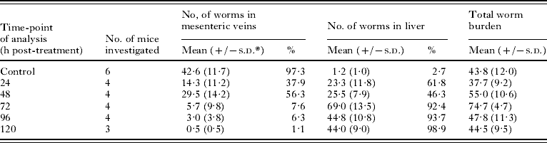
The distribution of adult worms in the mesenteric veins and the liver of S. mansoni infected mice 24, 48, 72, 96 and 120 h post-treatment with MQ (400 mg/kg) is summarized in Table 1. A hepatic shift was already observed 24 h post-treatment with MQ, with 61·8% of S. mansoni harboured in the liver. At 72 h and 120 h post-treatment >92% and >98%, respectively, of all worms had shifted to the liver.
Hepatic shift test following a single 400 mg/kg oral dose of MQ administered to mice infected with Schistosoma mansoni
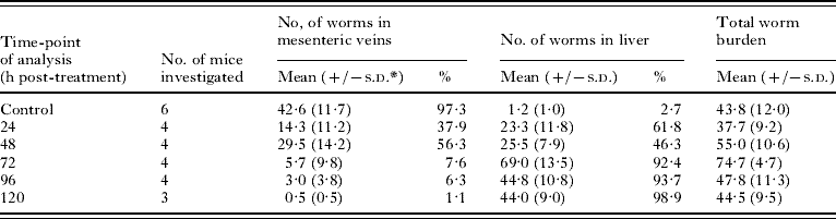
* s.d., standard deviation.
In vivo studies of adult S. mansoni – dissecting microscope and SEM evaluations
All adult flukes recovered from the mesenteric veins of mice at 24, 48, 72, 96 and 120 h after MQ treatment were alive and viable. Worms which had shifted to the liver could not be examined.
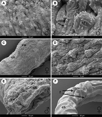
Twenty-four h after S. mansoni-infected mice had received a single oral dose of MQ (400 mg/kg) localized blebbing on the tegument was the main response to drug action observed on worms recovered from the mesenteric veins (Fig. 6A). By 48 h post-treatment, blebbing increased, affecting the entire tegumental surface of the worms (Fig. 6B).
SEM observations of adult Schistosoma mansoni recovered from mice treated with a single oral dose of MQ (400 mg/kg). (A) Twenty-four h after treatment. Blebbing (b) on the dorsal mid-body tegument of a male worm is visible. (B) Forty-eight h after treatment. Male dorsal tegument showing blebs (b). (C) Seventy-two h after treatment. Tegument of the mid-body region of a female has a shrunken (sh) and swollen (sw) appearance. (D) Seventy-two h after treatment. Dorsal mid-body male tegument reveals many blebs (b). (E) Seventy-two h after treatment. Oral sucker (OS) of a male specimen shows a shrunken (sh) and swollen (sw) tegument. (F) Ninety-six h after treatment. The shrunken (sh) mid-body region of a female S. mansoni with many furrows (f) is visible.

At 72 h after treatment, tegumental damage was more severe and several worms, in particular the females, were now affected. For example, the mid-body tegument of a female S. mansoni became shrunken (Fig. 6C). Again, many blebs were visible on the teguments as depicted in Fig. 6D. The oral sucker of a male specimen had a shrunken appearance resulting in formation of deep furrows. Moreover, some parts of the oral sucker around the pharyngeal opening were swollen. Small spines, which cover the inner surface and the outer margin of the oral sucker were not affected (Fig. 6E).
At 96 h after treatment a slightly increased tegumental response to drug treatment was observed on female worms when compared to previous time-points. For example, the mid-body region of a female worm examined showed a shrunken appearance with deep furrows (Fig. 6F).
DISCUSSION
Recently, promising anti-schistosomal properties of MQ against both adult and juvenile schistosomes harboured by mice were found (Keiser et al. Reference Keiser, Chollet, Xiao, Mei, Jiao, Utzinger and Tanner2009) and proof-of-concept trials have been launched with MQ in S. mansoni or S. haematobium-infected children in geographical regions in which malaria and schistosomiasis are co-endemic. To further deepen our understanding of the activity of this anti-malarial drug against schistosomes, we investigated morphological effects and tegumental alterations of MQ on S. mansoni juveniles (schistosomula) and adults in vitro and in vivo. Dissecting microscopic investigations demonstrated the temporal effect of MQ on viability and morphology as well as dose-response relationships on schistosomula and adult S. mansoni. In addition, SEM examinations revealed the progressive stages of damage to and disruption of the tegumantal surface of the worm.
MQ had a very fast onset of action on schistosomula in vitro. In the presence of 100 and 75 μg/ml worms died immediately. Concentrations of ⩾25 μg/ml killed all schistosomula after 1 h of incubation. Interestingly, a slightly slower onset of action was observed when adult worms were incubated with MQ in vitro. For example, while 10 μg/ml of MQ killed schistosomula already after 6 h, the adults did not die until 24 h of exposure in vitro. A much slower onset of action was observed on adult worms collected following in vivo treatment with MQ. Worms collected from the mesenteric veins were still alive up to 96 h following treatment, and only moderate tegumental disruption, such as blebbing and a shrunken body, was observed. However, severely damaged worms might have already been expelled and worms that had translocated to the liver could not be examined. Hence, the evidence presented might have underestimated the amount of damage inflicted by the drug to the worm population as a whole. Nonetheless, the discrepancy between the effect of the drug and the onset of action of MQ in vitro and in vivo might be related to lower MQ concentrations present in the livers and mesenteric veins in mice compared with in vitro results. Pharmacokinetic (PK) studies, measuring drug concentrations in the body and the target organs, might aid elucidation of these differences observed in vitro and in vivo. In addition, the effect as well as onset of action of MQ on different stages of juvenile worms (e.g. 21-day-old worms) in vitro and in vivo remains to be elucidated.
Interestingly, worms incubated with MQ in vitro died without the presence of haemin or red blood cells. In contrast, no apparent effect was observed when schistosomes were incubated with the anti-malarial artemether in vitro in the absence of haemin, suggesting that artemether interacts with haemin to exert a toxic effect on the worms (Xiao et al. Reference Xiao, Collet, Utzinger, Matile, Mei and Tanner2001). Though the exact mechanism of action of MQ on malaria parasites has not yet been entirely elucidated, a disturbance of the haemoglobin metabolism due to inhibition of the haematin polymerization and formation of an insoluble polymer, termed haemozoin, has been suggested (Dorn et al. Reference Dorn, Vippagunta, Matile, Jaquet, Vennerstrom and Ridley1998). A recent morphological study of S. japonicum in mice, showed a pronounced dilatation of the gut of adult worms, accompanied by focal or extensive peeling of gut epithelial cells, or even focal collapse of the gut wall (Zhang et al. Reference Zhang, Xiao, Utzinger, Chollet, Keiser and Tanner2009). Erythrocytes with accumulated MQ entering the worm gut might inhibit haem detoxification, resulting in gut damage (Zhang et al. Reference Zhang, Xiao, Utzinger, Chollet, Keiser and Tanner2009). Hence, while an interference of MQ with the haemoglobin digestion of schistosomes might play a role in vivo, MQ seems to attack additional drug targets in vitro. Further studies are necessary to elucidate the multiple mechanisms of action of MQ, which seems to be involved in the killing of schistosomes. It might also be useful to investigate whether MQ acts synergistically with the host immune response, similar to the chemotherapeutic effect of PZQ, which has been shown to be dependent on the host antibody response (Brindley and Sher, Reference Brindley and Sher1987).
SEM is often used for documenting the efficacy of anti-schistosomal drugs. The 10–20 μm thick tegument (Neves et al. Reference Neves, Machado-Silva, Pelajo-Machado, Oliveira, Coutinho, Lenzi and Gomes2001) that covers the entire surface of schistosomes represents an important drug target. It consists of a double outer membrane (2 lipid bilayers), has no glycocalyx (Hockley, Reference Hockley1973; Wilson and Barnes, Reference Wilson and Barnes1974; McLaren and Hockley, Reference McLaren and Hockley1977), is responsible for absorptive activities (Rogers et al. Reference Rogers, Shannon and Reeder1983) and is a protection barrier between the parasite and the host (Hoffmann and Strand, Reference Hoffmann and Strand1996). In addition, it contains numerous ciliated or unciliated bulb-shaped tubercles, which are sensory receptors and associated with ring-shaped actin filaments (Zhou and Podesta, Reference Zhou and Podesta1992). Also, the spines of the tubercles contain bundles of highly packed actin filaments (Zhou and Podesta, Reference Zhou and Podesta1992).
We observed extensive tegumental damage on the larvae and adult developmental stages of S. mansoni, in particular following in vitro incubation, which intensified progressively as the incubation period and the concentration of MQ increased. Blebbing was visible on the tegument of both treated juvenile and adult flukes. Blebbing is an indicator for stress and has been observed in previous SEM studies evaluating anti-schistosomal drugs (Jiraungkoorskul et al. Reference Jiraungkoorskul, Sahaphong, Sobhon, Riengrojpitak and Kangwanrangsan2005). Nonetheless, vesicle formation induced by MQ might also occur due to the focal lysis of muscles (Zhang et al. Reference Zhang, Xiao, Utzinger, Chollet, Keiser and Tanner2009). Interestingly, while in the present study blebbing was observed on the tegument of both male and female adult S. mansoni, vesiculation was absent from female S. japonicum (Zhang et al. Reference Zhang, Xiao, Utzinger, Chollet, Keiser and Tanner2009). Flattened spines were observed on the tegument of schistosomula and roughening, sloughing and disintegration of the tegument were other typical features observed on the tegument of adult worms in the present investigation. In addition, extensive swelling and furrowing were observed on both development stages.
The effect of PZQ on the tegument of S. mansoni shows distinct differences, when compared to MQ, though related tegumental alterations such as sloughing, swelling, and loss or shortening of spines have also been documented for PZQ (Xiao et al. Reference Xiao, Binggui, Chollet and Tanner2000). First, in contrast to small- and also medium-sized blebs observed in the present study a typical feature seen on the tegument of S. mansoni treated with PZQ was the presence of large-sized vesicles protruding from the tegumental surface, most likely due to swelling of the cytoplasm (Xiao et al. Reference Xiao, Yang, Shu and Wang1981). Second, in the current study, the tegument of female worms was slightly more affected and females died more rapidly when compared to male worms, in particular following in vitro exposure to MQ, male worms had a higher sensitivity to PZQ than female worms in vitro (Pica-Mattoccia and Cioli, Reference Pica-Mattoccia and Cioli2004) and exhibited more extensive tegumental damage than female worms following PZQ treatment (Shaw, Reference Shaw1990). The greater MQ susceptibility of females is consistent with results from a previous in vivo study (Keiser et al. Reference Keiser, Chollet, Xiao, Mei, Jiao, Utzinger and Tanner2009), in which greater reductions in the female worm burden were observed compared with total worm burden reductions. We speculated that either a sex-specific interference of the drug with the target might occur or that there are different targets for MQ in females compared with males (Keiser et al. Reference Keiser, Chollet, Xiao, Mei, Jiao, Utzinger and Tanner2009). Alterations induced by artemether on the tegument of S. mansoni have also been well studied. Briefly, severe tegumental damage was observed on juvenile schistosomes, correlating with the high efficacy of artemether in killing 21-day-old schistosomes (Xiao et al. Reference Xiao, Binggui, Chollet and Tanner2000). Focal swelling and fusion of tegumental ridges and occasionally sloughing was observed on adult S. mansoni recovered from mice. Similar to the present results obtained using MQ, female worms were more affected following treatment with artemether (Xiao et al. Reference Xiao, Binggui, Chollet and Tanner2000).
We documented distinct morphological characteristics of schistosomes incubated with MQ. Schistosomula treated with MQ had a crescent-shaped body and showed contraction resulting in a reduction of the body length by approximately half. In addition, a dark granular body was seen. Adult worms showed convulsions after incubation with MQ in vitro. In contrast, rapid spasmodic contractions of the worm body are the most prominent morphological alterations induced by PZQ (Xiao et al. Reference Xiao, Friedman, Catto and Webster1984). Finally, in vivo studies showed that extensive structural changes to worms occurred within 15 min of treatment, and both female and male adult worms died within 24 h following treatment with PZQ (Shaw, Reference Shaw and Erasmus1983; Xiao et al. Reference Xiao, Yang, Yang, Guo and Shao1983). In contrast, in our study worms were still alive several days post-treatment of mice with MQ and nearly all worms had shifted to the liver after 72 h. Interestingly, a more rapid hepatic translocation was observed with MQ on S. japonicum, all worms had moved to the liver 48 h after treatment (Zhang et al. Reference Zhang, Xiao, Utzinger, Chollet, Keiser and Tanner2009).
In conclusion, we demonstrated that MQ induces extensive morphological and tegumental alterations on both S. mansoni schistosomula and adults in vitro and in vivo. Importantly, given the distinct morphological and tegumental changes caused by MQ compared with PZQ, the only other effective anti-schistosomal drug available, we speculate that both drugs attack different targets; hence, it is suggested that no cross-resistance will occur. Thus, the present findings provide a sound basis for further in-depth studies of the anti-schistosomal properties of MQ, both in the laboratory and in the field.
We thank Yvette Endriss for maintenance of the S. mansoni life cycle at the Swiss Tropical Institute. We thank Mr Daniel Mathys, Mrs Evi Bieler and Mr Marcel Düggelin, at the Centre for Microscopy at the University of Basel, for expert help with the SEM studies. This investigation received financial support from the Swiss National Science Foundation to J.K. and T.M. (project no. PPOOA-114941).