Introduction
Dairy cattle are remarkable amongst domestic animals as bacterial contamination of the uterine lumen is ubiquitous after parturition (Elliott et al., Reference Elliott, McMahon, Gier and Marion1968; Griffin et al., Reference Griffin, Hartigan and Nunn1974; Sheldon et al., Reference Sheldon, Noakes, Rycroft, Pfeiffer and Dobson2002). As the cervix dilates to allow passage of the calf at parturition, the anatomical barrier to bacterial contamination is breached, allowing micro organisms from the cow’s skin, faeces and surrounding environment to enter the uterus (Sheldon and Dobson, Reference Sheldon and Dobson1999). In one study, 93% of uteri obtained within 2 weeks of calving were contaminated with bacteria and others have shown the presence of bacteria in more than 60% of animals 3 weeks after parturition (Elliott et al., Reference Elliott, McMahon, Gier and Marion1968; Griffin et al., Reference Griffin, Hartigan and Nunn1974; Sheldon et al., Reference Sheldon, Noakes, Rycroft, Pfeiffer and Dobson2002). Many cows spontaneously eliminate this contamination; however, at least 20% of cows fail to clear the bacteria and in 10% to 15% of animals infection persists within the uterus, causing endometritis (Griffin et al., Reference Griffin, Hartigan and Nunn1974; Borsberry and Dobson, Reference Borsberry and Dobson1989; Dhaliwal et al., Reference Dhaliwal, Murray and Woldehiwet2001). In addition, there is emerging evidence that other microbes such as Bovine herpesvirus 4 may contribute to the disease process (Donofrio et al., Reference Donofrio, Herath, Sartori, Cavirani, Flammini and Sheldon2007).
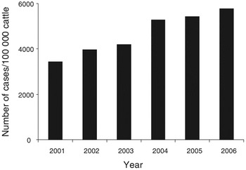
Uterine infection and the associated inflammatory and immune responses compromise animal welfare, causing subfertility and infertility (Borsberry and Dobson, Reference Borsberry and Dobson1989; Sheldon and Dobson, Reference Sheldon and Dobson2004). Endometritis is associated with lower conception rates, increased intervals from calving to first service or conception, and more culls for failure to conceive (Borsberry and Dobson, Reference Borsberry and Dobson1989). Furthermore, the subfertility associated with endometritis persists even after clinical resolution of the disease. In typical studies of cows treated successfully for clinical endometritis, conception rates are approximately 20% lower than in unaffected animals and an extra 3% of animals remain infertile (Borsberry and Dobson, Reference Borsberry and Dobson1989, LeBlanc et al., Reference LeBlanc, Duffield, Leslie, Bateman, Keefe, Walton and Johnson2002a and Reference LeBlanc, Duffield, Leslie, Bateman, Keefe, Walton and Johnson2002b). The financial losses associated with uterine infection depend on the direct cost of treatment, reduced milk yields and subfertility. Despite great effort to control uterine disease in cattle, the number of animals treated annually for endometritis reported by the National Animal Disease Information Service (NADIS, the UK Veterinary Sentinel Practice Network, http://www.nadis.org.uk) has steadily increased since 2001 (Figure 1; NADIS); in 2006, almost 6% of animals required direct intervention by a veterinarian. These data, however, only reflect a proportion of animals with clinical disease and do not quantify the numbers of animals with subclinical disease. An increased incidence of uterine disease and the associated costs will have major implications for the economic sustainability of both the individual animal and the national herd. Thus, an important challenge facing reproductive biologists is to understand the mechanisms underlying uterine disease in cattle.
Cases of uterine disease requiring veterinary treatment between 2001 and 2006, as reported by 36 attending veterinarians. Data generously supplied by the National Animal Disease Information Service (NADIS).

Post partum uterine infection
The flora cultured in vitro from the early post partum uterus represents a wide spectrum of environmental contaminants and some anaerobic species, and bacteria isolated from the uterine lumen have been categorised according to their pathogenicity within the uterus (Ruder et al., Reference Ruder, Sasser, Williams, Ely, Bull and Butler1981; Olson et al., Reference Olson, Ball, Mortimer, Farin, Adney and Huffman1984; Farin et al., Reference Farin, Ball, Olson, Mortimer, Jones, Admey and McChesney1989; Noakes et al., Reference Noakes, Till and Smith1989; Bonnett et al., Reference Bonnett, Martin, Gannon, Miller and Etherington1991; Laven et al., Reference Laven, Biggadike, Proven, Halfacre and Tickle2000). Bacteria are classed as ‘recognised uterine pathogens’ associated with uterine endometrial lesions; ‘potential pathogens’ frequently isolated from the bovine uterine lumen and cases of endometritis, but not commonly associated with uterine lesions; and, ‘opportunist contaminants’ transiently isolated from the uterine lumen but not associated with endometritis (Sheldon et al., Reference Sheldon, Noakes, Rycroft, Pfeiffer and Dobson2002; Williams et al., Reference Williams, Fischer, Pfeiffer, England, Noakes, Dobson and Sheldon2005). The recognised uterine pathogens are E. coli, Arcanobacterium pyogenes, Fusobacterium necrophorum, Prevotella melaninogenica and Proteus species, and these bacteria are associated with greater endometrial inflammation and more severe signs of clinical uterine disease (Farin et al., Reference Farin, Ball, Olson, Mortimer, Jones, Admey and McChesney1989; Bonnett et al., Reference Bonnett, Martin, Gannon, Miller and Etherington1991; Sheldon et al., Reference Sheldon, Noakes, Rycroft, Pfeiffer and Dobson2002; Williams et al., Reference Williams, Fischer, Pfeiffer, England, Noakes, Dobson and Sheldon2005). We have recently shown that potential uterine pathogens or opportunistic contaminant bacteria in the uterine lumen do not have this same relationship (Williams et al., Reference Williams, Fischer, Pfeiffer, England, Noakes, Dobson and Sheldon2005).
The innate immune system in the uterus is responsible for host recognition of pathogens. Pattern recognition receptors (PRRs) recognise a wide host of pathogens via their pathogen-associated molecular patterns (PAMPs). A key group of receptors that recognise PAMPs are the Toll-like receptors (TLRs), which were first identified on immune cells such as macrophages, but have since been identified on other cell types (Akira and Takeda, Reference Akira and Takeda2004; Akira et al., Reference Akira, Uematsu and Takeuchi2006; Beutler, Reference Beutler2003 and Reference Beutler2004). Engagement of these receptors initiates a signalling cascade stimulating cytokine release. Production of immune mediators such as tumour necrosis factor α (TNFα) and nitric oxide (NO), orchestrate the resultant immune response to clear bacterial infection. Cytokines including TNFα are potent stimulants of acute phase proteins production by liver hepatocyte cells (Baumann and Gauldie, Reference Baumann and Gauldie1994). Acute phase proteins, such as α1-acid glycoprotein (AGP), act at the site of infection to limit tissue damage and promote tissue repair (Baumann and Gauldie, Reference Baumann and Gauldie1994). The severity of uterine bacterial contamination, as determined by the bacterial growth density, is correlated with the peripheral circulating concentrations of the acute phase protein AGP (Sheldon et al., Reference Sheldon, Noakes, Rycroft and Dobson2001). Furthermore, concentrations of AGP are particularly increased in those animals from which E. coli are isolated from the uterine lumen, but not other bacteria.
The bacterium E. coli is the most commonly isolated pathogen from the post partum uterus and, in the first few days after calving, dominates the uterine flora (Hussain et al., Reference Hussain, Daniel and O’Boyle1990; Huszenicza et al., Reference Huszenicza, Fodor, Gacs, Kulcsar, Dohmen and Vamos1999). Of greater importance is the infection with E. coli, which appears to increase the susceptibility of the endometrium to subsequent infection with A. pyogenes (Williams et al., Reference Williams, Fischer, Noakes, England, Rycroft, Dobson and Sheldon2007); and A. pyogenes is associated with inflammation of the endometrium both in vivo and in vitro (Bonnett et al., Reference Bonnett, Martin, Gannon, Miller and Etherington1991; Miller et al., Reference Miller, Williams, Sibley, Herath, Lane, Fishwick, Nash, Rycroft, Dobson, Bryant and Sheldon2007). These observations highlight the substantial impact E. coli has on post partum uterine health.
Effects of E. coli in the whole animal
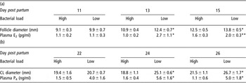
Uterine bacterial infection or bacterial products suppress pituitary luteinising hormone (LH) secretion, and are associated with inhibition of folliculogenesis, decreased ovarian steroidogenesis and abnormal luteal phases (Peter et al., Reference Peter, Bosu and DeDecker1989; Huszenicza et al., Reference Huszenicza, Fodor, Gacs, Kulcsar, Dohmen and Vamos1999; Opsomer et al., Reference Opsomer, Grohn, Hertl, Coryn, Deluyker and de Kruif2000; Mateus et al., Reference Mateus, Lopes da Costa, Bernardo and Silva2002 and Reference Mateus, Lopes da Costa, Diniz and Ziecik2003; Sheldon et al., Reference Sheldon, Noakes, Rycroft, Pfeiffer and Dobson2002; Williams et al., Reference Williams, Fischer, Noakes, England, Rycroft, Dobson and Sheldon2007). In the field, infections are associated with changes in luteal phase length and more cystic ovarian disease (Peter et al., Reference Peter, Bosu and DeDecker1989; Opsomer et al., Reference Opsomer, Grohn, Hertl, Coryn, Deluyker and de Kruif2000); and the first post partum ovarian follicle is smaller and produces less oestradiol in animals with higher numbers of bacteria in the post partum uterus (Sheldon et al., Reference Sheldon, Noakes, Rycroft, Pfeiffer and Dobson2002). More recently, we have shown that animals with high numbers of the recognised uterine pathogens including E. coli have retarded ovarian follicle growth, lower peripheral plasma concentrations of oestradiol and an inability to form a competent corpus luteum (CL) (Table 1; Williams et al., Reference Williams, Fischer, Noakes, England, Rycroft, Dobson and Sheldon2007).
Mean ± s.e. (a) follicle diameter and plasma oestradiol concentrations on days 11, 13 and 15 post partum and (b) corpus luteum (CL) diameters and plasma progesterone concentrations on days 22, 24 and 26 post partum in animals with high v. low uterine pathogen load

Values differ between groups within day *P < 0.05, **P < 0.01.
Lipopolysaccharide (LPS), the main pathogenic moiety of E. coli, is detectable in the uterus, peripheral plasma and ovarian follicular fluid of cows with post partum uterine infection (Peter et al., Reference Peter, Bosu and DeDecker1989; Mateus et al., Reference Mateus, Lopes da Costa, Diniz and Ziecik2003; Herath et al., Reference Herath, Williams, Lilly, Gilbert, Dobson, Bryant and Sheldon2007; Williams et al., Reference Williams, Fischer, Noakes, England, Rycroft, Dobson and Sheldon2007). Administration of intravenous LPS disrupts neuroendocrine activity and results in interference with the oestrous cycle. In heifers infused intrauterine with LPS, the pre-ovulatory LH surge was blocked, resulting in the formation of cystic follicles (Peter et al., Reference Peter, Bosu and DeDecker1989). In sheep, hypothalamic GnRH secretion is suppressed, there is a reduction in pituitary responsiveness to GnRH and pulsatile LH secretion from the pituitary is inhibited, following the intravenous infusion of LPS (Williams et al., Reference Williams, Harris, Battaglia, Viquie and Karsch2001). However, uterine infection does not affect peripheral plasma FSH concentrations or the consequent emergence of a wave of growing follicles (Sheldon et al., Reference Sheldon, Noakes, Rycroft, Pfeiffer and Dobson2002; Williams et al., Reference Williams, Fischer, Noakes, England, Rycroft, Dobson and Sheldon2007).
There are also localised effects of LPS in the ovary. Following administration of LPS, an inhibition in peripheral plasma oestradiol concentrations has been observed despite normal plasma LH concentrations (Xiao et al., Reference Xiao, Xia-Zhang, Barth, Zhu and Ferrin1998; Battaglia et al., Reference Battaglia, Krasa, Padmanabhan, Viguie and Karsch2000); and in heifers, LPS delays ovulation by interrupting the preovulatory oestradiol rise, thus delaying the LH surge (Suzuki et al., Reference Suzuki, Yoshioka, Iwamura and Hirose2001). These observations are likely to be biologically relevant as we have recently demonstrated the presence of LPS at concentrations of up to 0.8 μg/ml in the follicular fluid of cows with clinical uterine disease. Animals with subclinical disease had mid-range concentrations, while in normal animals, LPS was not detectable in follicular fluid (Herath et al., Reference Herath, Williams, Lilly, Gilbert, Dobson, Bryant and Sheldon2007).
Cellular mechanisms
Besides being responsible for prostaglandins E2 and F2α (PGE, PGF) production, the uterine mucosa has a role in immunity. The endometrium provides a barrier against infection and an opportunity to detect bacteria by innate immune receptors recognising PAMPs (Beutler, Reference Beutler2004). In the human endometrium, nine TLRs are expressed at the mRNA level including TLR4 (Pioli et al., Reference Pioli, Amiel, Schaefer, Connolly, Wira and Guyre2004; Young et al., Reference Young, Lyddon, Jorgenson and Misfeldt2004; Hirata et al., Reference Hirata, Osuga, Hirota, Koga, Yoshino, Harada, Morimoto, Yano, Nishii, Tsutsumi and Taketani2005; Schaefer et al., Reference Schaefer, Fahey, Wright and Wira2005). It is the role of TLR4 to detect LPS, although signalling through TLR4 also requires accessory molecules such as CD14, LBP and MD2 (Beutler, Reference Beutler2003 and Reference Beutler2004). Bovine uterine endometrial and stromal cells express TLR4 and the accessory molecules required for LPS recognition (Herath et al., Reference Herath, Fischer, Werling, Williams, Lilly, Dobson, Bryant and Sheldon2006). Following treatment with LPS, epithelial and stromal cells respond to E. coli or LPS treatment by secreting PGE or PGF, respectively, and this effect is abrogated in the presence of Polymixin B, a compound that binds to LPS, thus reducing the amount available to bind to MD2/TLR4. Thus, it appears that besides fulfilling an endocrine role, endometrial cells have an immune role in detecting and responding to bacterial pathogens (Herath et al., Reference Herath, Fischer, Werling, Williams, Lilly, Dobson, Bryant and Sheldon2006). Uterine tissue explants secrete more PGE than PGF in response to LPS (Herath et al., Reference Herath, Fischer, Werling, Williams, Lilly, Dobson, Bryant and Sheldon2006). It is well known that secretion of PGF, in response to endometrial oxytocin receptor activation, induces luteolysis and initiates the follicular phase of the ovarian cycle (Poyser, Reference Poyser1995). On the other hand, PGE has a luteotrophic role to help maintain the CL, particularly during pregnancy. Infection of the uterus with bacteria disrupts luteolysis and, between 1960 and 1980, short luteal phases or an ‘inadequate CL’ after calving were a substantial area of research interest. Post partum uterine infection and the release of PGF was thought to reduce luteal phase length. Although our recent data support the hypothesis that infection results in the formation of an inefficient CL (Williams et al., Reference Williams, Fischer, Noakes, England, Rycroft, Dobson and Sheldon2007), it is perhaps now accepted that uterine disease is mainly associated with extended luteal phases. The altered prostaglandin secretion following E. coli LPS treatment of uterine cells provides further evidence that E. coli may play a role in perturbed ovarian cycles.
In addition to the likely effects on luteal function associated with changes in endometrial cell prostaglandin production, LPS can also directly affect ovarian cell function. Bovine granulosa cells isolated from selected and dominant follicles express the transcripts for TLR4 and the accessory molecules MD2 and CD14 required for LPS recognition (Herath et al., Reference Herath, Williams, Lilly, Gilbert, Dobson, Bryant and Sheldon2007). When treated with LPS, oestradiol production by these cells is inhibited and mRNA for the aromatase enzyme, which catalyses the conversion of androstenedione to oestradiol, is down-regulated. Furthermore, the effect of LPS on oestradiol biosynthesis was not due to the inability of the cells to respond to oestradiol as transcripts for ERα were unchanged by LPS treatment and ERβ was not detected in control or treated granulosa cells, appearing to be down-regulated by culture. Neither was it due to the bioavailability of androstenedione substrate as the LPS treatment of ovarian theca cells did not affect androstenedione production (Herath et al., Reference Herath, Williams, Lilly, Gilbert, Dobson, Bryant and Sheldon2007).
Summary and conclusions
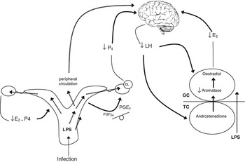
The bacterium E. coli is the most common uterine pathogen early post partum, paving the way for A. pyogenes infection and clinical disease. Uterine infections with E. coli affect fertility at multiple levels, including the neuroendocrine axis and ovaries as well as in the uterus. At all of these sites, LPS from E. coli perturbs the delicate balance of hormone regulation with detrimental consequences for the reproductive function of the animal. Notably, the key endocrine cells of the reproductive tract express the receptor complex for recognition of LPS, and LPS modulates their endocrine function (Figure 2). Thus, treatment regimes tailored to disrupting the actions of E. coli in the early post partum period may diminish the detrimental effects of this pathogen. This would lead to improved animal welfare and an increase in the economic viability of dairy farming.
Summary of the effects of E. coli lipopolysaccharide (LPS) on the reproductive axis of the cow. E. coli ascend the reproductive tract and LPS is recognised by uterine endometrial and stromal cells. The prostaglandin production by these cells is modulated in response to LPS and this in turn may affect luteal phase length. At the ovary, theca cells produce androstenedione, which is aromatised to oestradiol by granulose cells (GC). GC can recognise LPS following which expression of aromatase enzyme is down-regulated and less oestradiol is produced. The corpus luteum produces less progesterone in animals with E. coli infection. The disruption of the hormonal milieu will have effects on the hypothalamus and pituitary, and also at the uterus. The presence of LPS in the peripheral circulation also disrupts LH production and release, which may have additional effects on ovarian theca and GC. Together, these observations may explain the infertility in cattle associated with E. coli infection of the uterus.

Acknowledgements
The authors would like to thank Dave Williams at the National Animal Disease Information Service for kindly providing data. We also thank The Wellcome Trust for funding part of the work. S Herath is supported by the BBSRC (F005129) and IM Sheldon is a BBSRC Research Development Fellow (BB/D02028X/1).