Introduction
Echinostoma caproni Richard, 1964 (Trematoda: Echinostomatidae) has been used extensively as an experimental model to study host–parasite relationships in intestinal helminth infections, as its life cycle can be maintained easily under laboratory conditions (Toledo et al., Reference Toledo, Espert, Carpena, Trelis, Muñoz-Antoli and Esteban2004a, b; Toledo & Fried, Reference Toledo and Fried2005). It has been established that Wistar rats, Rattus norvegicus, show low compatibility with E. caproni, developing acute infections in which the adult worms are rapidly expelled and the pathological effects in the intestine are apparently minor (Hansen et al., Reference Hansen, Nielsen, Hindsbo and Christensen1991; Toledo et al., Reference Toledo, Espert, Carpena, Muñoz-Antoli, Fried and Esteban2004b, Reference Toledo, Carpena, Espert, Sotillo, Muñoz-Antoli and Esteban2006). Several host-related factors have been involved in the expulsion of the worms in echinostome infections and, in this case, immunological responses against E. caproni appear to be implicated (Toledo et al., Reference Toledo, Esteban and Fried2009).
The differential leucocyte count is a useful tool to determine the immunological status of the definitive host and its capacity to overcome the infection (Parvathi & Karemungikar, 2011). Variations in the levels of lymphocytes, eosinophils, basophils and monocytes are indicative of variations in the mechanisms of resistance developed by the definitive host to control the infections. However, the haematological profile has been scarcely studied in echinostome infections: E. revolutum infections in domestic chickens and golden hamsters; E. caproni (referred to as E. liei) infections in golden hamsters; and E. paraensei infections in Wistar rats (Fried & Wilson, Reference Fried and Wilson1981; Huffman et al., Reference Huffman, Michos and Fried1986, Reference Huffman, Iglesias and Fried1988a, Reference Huffman, Alcaide and Friedb; García et al., Reference García, Pinheiro, Hooper, Simões, Ferraz and Maldonado2012).
In the present study, we use the model E. caproni–Wistar rat to analyse the consequences of an intestinal helminth infection on the haematological parameters of the host. Considering that the Wistar rat is a host of low compatibility with E. caproni, the data obtained may be of interest when determining the haematological elements involved in worm expulsion.
Materials and methods
Parasitological procedures
To analyse the haematological alterations induced by E. caproni infection, a total of 24 male Wistar rats weighing 80–100 g were used. Twelve rats were individually exposed to 100 E. caproni metacercariae as described previously (Sotillo et al., Reference Sotillo, Trelis, Fried, Marcilla, Esteban and Toledo2012). Blood samples of 300 μl were obtained by saphenic puncture, collected in heparinized vials, 1, 2 and 3 weeks post-exposure, while at week 4 post-exposure blood was extracted at necropsy. The success of the infection (egg-positive or negative rats) was established weekly with the discovery of E. caproni eggs by the Kato–Katz coprological technique, and confirmed by finding adult worms after dissection (worm-positive or negative rats). A total of 12 uninfected rats were used as controls and blood samples were obtained as described for E. caproni-exposed rats.
Data analysis
Blood samples were analysed to determine total red blood cells (RBC), total white blood cells (WBC), platelets (PLT), haematocrit (HCT), haemoglobin (HGB), mean corpuscular volume (MCV), mean corpuscular haemoglobin (MCH) and the mean corpuscular haemoglobin concentration (MCHC). Haematological values were measured using the haemocytometer Sysmex XS-21 (MYCO-Instrumentation Inc., Seattle, Washington, USA). Blood smears stained with Giemsa were used to determine the differential leuococyte counts (lymphocytes, neutrophils, eosinophils, basophils and monocytes).
One-way analysis of variance analysis (one-way ANOVA) was used to evaluate the differences among worm-positive, egg-positive, negative and control rats at each time point, and the Bonferroni t-test of the difference between means was used as a post-hoc analysis. Previously, data were log transformed to achieve normality. P< 0.05 was considered significant.
Results and discussion
A total of six rats experimentally exposed to metacercariae of E. caproni developed infection, as determined coprologically. However, adult worms were only found in three of them after dissection and they were considered as ‘worm-positive rats’. The mean number of worms recovered from rats was 22.0 with a range of 9–32 worms, with a mean recovery rate of 7.3%. The remaining three rats that became infected but from which no adult worms were recovered at 4 weeks post-exposure were considered as ‘egg-positive rats’. Those rats that did not become infected were considered as ‘negative rats’.
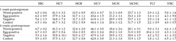
Haematological parameters such as HGB, MCV, MCH and MCHC did not show differences among the different rat groups during the entire experiment (table 1). However, at 2 weeks post-exposure mean levels of RBC showed significant differences between worm-positive and control rats (P= 0.023). Similarly, mean levels of WBC from worm-positive rats were found to be significantly different to those from control (P= 0.002) and negative rats (P= 0.037). Moreover, those values from egg-positive rats showed significant differences when compared to control rats (P= 0.001) and negative rats (P= 0.026). At week 4 post-exposure, significant differences in mean levels of MCHC were found (P= 0.013) between worm-positive and control rats. Finally, mean values of PLT showed significant differences between worm-positive rats compared to control (P= 0.023) and negative rats (P= 0.014) at 2 weeks post-exposure, and between egg-positive and control rats at 4 weeks post-exposure (P= 0.048).
Haematological changes including counts (±SD) of red blood cells (RBC), haematocrit (HCT), haemoglobin (HGB), mean corpuscular volume (MCV), mean corpuscular haemoglobin (MCH), mean corpuscular haemoglobin concentration (MCHC), platelets (PLT) and white blood cells (WBC) in rats infected with Echinostoma caproni at weeks 1 and 4 post-exposure.

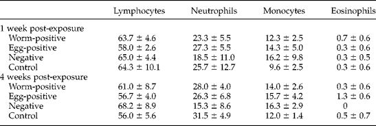
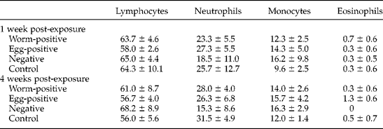
Differential leucocyte counts obtained from E. caproni-infected rats showed similar values of lymphocytes, neutrophils, monocytes and eosinophils in worm-positive, egg-positive, negative and control rats during the entire experiment (table 2). Although levels of eosinophils in worm-positive, egg-positive, negative and control rats were similar, the values were higher in egg-positive rats than in worm-positive rats at 4 weeks post-exposure. Finally, mean levels of basophils were negligible in worm-positive rats, but significant differences with respect to egg-positive or negative rats were not found.
Mean percentages of leucocyte counts (±SD) in blood samples of rats infected with Echinostoma caproni at weeks 1 and 4 post-exposure.
Variations in haematological parameters are commonly used in the diagnosis of physiological alterations (Valdéz-Cruz et al., Reference Valdéz-Cruz, Hernández-Gil, Galindo-Rodríguez and Alonso-Díaz2013). For example, low RBC values may indicate pathological processes such as anaemia, dehydration, haemorrhage, infections or parasitism, among others (Barros Monteiro et al., Reference Barros Monteiro, Coutinho, de Araujo, Barros Monteiro, de Castro, da Silva, Benigno and Vicente2009). In relation to intestinal helminths, it has been shown that they may induce significant reductions in RBC, HCT and HGB levels (Qadir et al., Reference Qadir, Dixit, Dixit and Sharma2011). In echinostome infections, rats infected with E. paraensei showed lower RBC, HCT and HGB levels than those observed in control animals, indicating the development of normocytic and normochromic anaemia with an anisocytic alteration (Garcia et al., Reference García, Pinheiro, Hooper, Simões, Ferraz and Maldonado2012). In hamsters infected with E. revolutum or E. caproni an increase in HGB levels was observed (Huffman et al., Reference Huffman, Iglesias and Fried1988a, b). However, our results are in agreement with those of Karanja et al. (Reference Karanja, Ngatia and Wandera1994), who did not find associations between intestinal nematode infections and those haematological values in equids.
In the present work, a significant reduction of MCHC levels at week 4 post-exposure was observed, indicating that the infection could induce normocytic hypochromic anaemia, microcytic hypochromic or macrocytic hypochromic anaemia (Martins et al., Reference Martins, Tavares-Dias, Fujimoto, Onaka and Nomura2004; Preet & Prakash, Reference Preet and Prakash2011; Qadir et al., Reference Qadir, Dixit, Dixit and Sharma2011). MCHC represents the quotient between HGB and HCT. Nevertheless, we did not find significant changes in these parameters. As HCT indicates the fraction composed by RBC in the total blood volume, the observed decrease in MCHC, together with an increase in the total RBC count and the lack of changes in HGB levels, appears to indicate that the main haematological alteration induced by E. caproni infection in rats is not only related to the number of RBC but also to their functionality. In this sense, it should be noted that the effect of the intestinal helminth infections on haematological parameters is normally related to elevated parasite loads (Soulsby, Reference Soulsby1987; Lassen & Swardson, Reference Lassen and Swardson1995; Shah et al., Reference Shah, Parveen, Mir, Sarwar and Yousuf2009; Valdéz-Cruz et al., Reference Valdéz-Cruz, Hernández-Gil, Galindo-Rodríguez and Alonso-Díaz2013). The low parasite burden of E. caproni in rats could be the cause of the slight alterations observed in the haematological values.
In the present study, we have found that E. caproni infection in rats has a slight effect on leucocyte counts and significant differences were not detected. This could be due to the fact that infiltration of immunological cells at the local site is scarce in hosts of low compatibility, such as rats, whereas elevated levels of inflammation are described exclusively in mice and other hosts of high compatibility with the parasite (Toledo et al., Reference Toledo, Carpena, Espert, Sotillo, Muñoz-Antoli and Esteban2006; Muñoz-Antoli et al., Reference Muñoz-Antoli, Sotillo, Monteagudo, Fried, Marcilla and Toledo2007; Sotillo et al., Reference Sotillo, Muñoz-Antoli, Marcilla, Fried, Esteban and Toledo2007; Trelis et al., Reference Trelis, Sotillo, Monteagudo, Fried, Marcilla, Esteban and Toledo2011). In this work, we observed that WBC values in worm-positive rats reflected the course of E. caproni infection. A sudden WBC increase in worm-positive rats was observed at 2 weeks post-infection and, thereafter, the values progressively decreased. As mentioned above, the rat is a host of low compatibility with E. caproni and those rats that become infected start to expel the adult worms rapidly (Toledo et al., Reference Toledo, Espert, Carpena, Muñoz-Antoli, Fried and Esteban2004b). This is in contrast with the data presented by Garcia et al. (Reference García, Pinheiro, Hooper, Simões, Ferraz and Maldonado2012) in E. paraensei infections in rats, whose WBC values remained elevated until the end of their experiment at 5 weeks post-infection.
Although helminth infections are often related to eosinophilia (Parvathi & Kerenungikar, 2011), in the present study the level of eosinophils in peripheral blood was very low, as described previously in other echinostome infections (Huffman et al., Reference Huffman, Michos and Fried1986, Reference Huffman, Alcaide and Fried1988b; García et al., Reference García, Pinheiro, Hooper, Simões, Ferraz and Maldonado2012). This is probably due to the low level of systemic immune response generated by E. caproni in the rat, together with the increased number of eosinophils at the site of infection (Hansen et al., Reference Hansen, Nielsen, Hindsbo and Christensen1991; Toledo et al., Reference Toledo, Espert, Carpena, Muñoz-Antoli, Fried and Esteban2004b, Reference Toledo, Carpena, Espert, Sotillo, Muñoz-Antoli and Esteban2006; Toledo & Fried, Reference Toledo and Fried2005). Moreover, it should be considered that elevated eosinophil levels are commonly correlated with the parasite load (Barros Monteiro et al., Reference Barros Monteiro, Coutinho, de Araujo, Barros Monteiro, de Castro, da Silva, Benigno and Vicente2009). A striking feature in our study is the fact that several parameters in those rats that were infected but in which adult worms did not remain established (egg-positive rats) were markedly different from those observed in the other groups. This suggests that egg-positive rats were affected by exposure to metacercariae, resulting in a higher increase of eosinophil levels than observed in worm-positive rats. Although this fact is difficult to explain, it may be related to delayed responses to parasite molecules absorbed through the intestinal wall or, even, allergic responses. Conversely, the WBC values in egg-positive rats decreased at 3 and 4 weeks post-exposure with respect to those in worm-positive rats, which could be caused by the expulsion of juvenile worms, confirming that leucocyte counts may serve as a marker of healing and worm expulsion.
In conclusion, E. caproni infection does not appear to alter the blood parameters of rats, RBC functionality being the main alteration induced by the infection. Consequently, there is no evidence of an indirect effect of E. caproni infection on the haematological condition of the host of low compatibility.
Acknowledgements
We are grateful to Dr Miguel Asensi Miralles, Department of Physiology, University of Valencia, for his help in haematological analysis; to Servei Central de Suport a la Investigació Experimental - Universitat de València Estudi General for providing the blood samples and for maintenance of experimental animals.
Financial support
This work was supported by Projects: INV-AE13-136845 (University of Valencia, Valencia, Spain); PROMETEO/2009/081 of Conselleria d'Educació, Generalitat Valenciana (Valencia, Spain); and n°RD12/0018/0013 of RICET, RETICS/FEDER of Ministerio Sanidad y Consumo (Madrid, Spain). This work has been carried out while the second author (A.C.) was a recipient of a pre-doctoral fellowship from the Vicerectorat d'Investigació i Política Científica de la Universitat de València (Valencia, Spain).
Conflict of interest
None.
Ethical standards
The authors assert that all procedures contributing to this work comply with the ethical standards of the relevant national guides on the care and use of laboratory animals and has been approved by the Comisió d'Etica en Investigació Experimental de la Universitat de València.