Introduction
The invasive eucalypt galling insect Leptocybe invasa (Hymenoptera: Eulophidae) was described in 2004 and subsequently identified as two cryptic lineages: a Western lineage (lineage A) and a Chinese lineage (lineage B) (Nugnes et al., Reference Nugnes, Gebiola, Monti, Gualtieri, Giorgini, Wang and Bernardo2015; Dittrich-Schröder et al., Reference Dittrich-Schröder, Hoareau, Hurley, Wingfield, Lawson, Nahrung and Slippers2018). These wasps induce swellings in midribs and leaf petioles of young eucalypts causing abnormal growth, and in severe cases, death in seedlings of several economically important species (Mendel et al., Reference Mendel, Protasov, Fisher and La Salle2004; FAO, 2012; Branco et al., Reference Branco, Battisti, Mendel, Paine and Lieutier2016). Some Australian parasitic eulophid wasps (Hymenoptera: Eulophidae), Selitrichodes kryceri, Quadrastichus mendeli (Kim et al., Reference Kim, Mendel, Protasov, Blumberg and La Salle2008) and S. neseri (Kelly et al., Reference Kelly, La Salle, Harney, Dittrich-Schröder and Hurley2012), are used as Leptocybe spp. biocontrol agents. These have been reared, released, monitored and, to different extents, reported to occur adventively in numerous countries (Mendel et al., Reference Mendel, Protasov, La Salle, Blumberg, Brand and Branco2017; Huang et al., Reference Huang, Li, Lu, Zheng and Yang2018; Le et al., Reference Le, Nahrung, Griffiths and Lawson2018).
The genus Megastigmus (Hymenoptera: Megastigmidae) (Janšta et al., Reference Janšta, Cruaud, Delvare, Genson, Heraty, Křížková and Rasplus2018) also contains potential biocontrol agents of Leptocybe spp. (Protasov et al., Reference Protasov, Doĝanlar, La Salle and Mendel2008; Doğanlar and Hassan, Reference Doğanlar and Hassan2010; Le et al., Reference Le, Nahrung, Griffiths and Lawson2018). Currently, 145 valid Megastigmus species are described, of which about one-third are recorded only in Australia, and four shared between Australia and other countries (Noyes, Reference Noyes2020). Nineteen Megastigmus species are reported as Leptocybe associates: seven endemic Australian species and twelve non-Australian ‘local’ species thought to represent host-switches in Leptocybe's invasive range (Huang et al., Reference Huang, Li, Lu, Zheng and Yang2018; Le et al., Reference Le, Nahrung, Griffiths and Lawson2018). Two Australian species, M. zvimendeli and M. lawsoni, were released in Israel where they contribute to the control of L. invasa populations (Mendel et al., Reference Mendel, Protasov, La Salle, Blumberg, Brand and Branco2017). Among the non-Australian species, M. leptocybus was identified as a potential L. invasa biocontrol agent in the Mediterranean (Viggiani et al., Reference Viggiani, Laudonia and Bernardo2002; Mendel et al., Reference Mendel, Protasov, Fisher and La Salle2004; Le et al., Reference Le, Nahrung, Griffiths and Lawson2018), while M. dharwadicus in India and M. thitipornae in Thailand were described and trialled for Leptocybe spp. biocontrol (Narendran et al., Reference Narendran, Girish Kumar and Vastrad2010; Ramanagouda et al., Reference Ramanagouda, Vastrad, Narendran, Basavanagoud and Viraktamath2011; Sangtongpraow and Charernsom, Reference Sangtongpraow and Charernsom2013). Megastigmus zebrinus, of Australian origin (Grissell, Reference Grissell2006), is thought to associate with Leptocybe in all infested continents except Europe (Grissell, Reference Grissell2006; Doğanlar, Reference Doğanlar2015; Hernández et al., Reference Hernández, Aquino, Cuello, Andorno and Botto2015).
In the pre-Leptocybe period, almost all entomophagous Megastigmus records were Australian, with only three of twenty-four entomophagous species found outside Australia, none of which associated with Hymenoptera or eucalypts (Grissell, Reference Grissell1999). In contrast, non-Australian Megastigmus were mainly phytophagous (Milliron, Reference Milliron1949; Grissell, Reference Grissell1999; Roques and Skrzypczyńska, Reference Roques and Skrzypczyńska2003). The recent associations of local Megastigmus with invasive Leptocybe thus suggests the occurrence of a previously unknown non-Australian entomophagous group. Considering the Australian origin of eucalypts and Leptocybe spp., this could be attributed to rapid host-shift of previously undescribed Megastigmus species outside Australia, and/or invasion of undescribed Australian Megastigmus species with or following the introduction of host Leptocybe. Understanding the drivers behind these novels Megastigmus-Leptocybe association is important and requires species confirmation as a prerequisite.
Progress in identifying Megastigmus species was made by Doğanlar (Doğanlar and Hassan, Reference Doğanlar and Hassan2010; Doğanlar, Reference Doğanlar2015), who described and redescribed eucalypt-associated Megastigmus species in Australia and in many parts of the world, including M. zvimendeli as the most extensively used Megastigmus species in biocontrol programs worldwide (Mendel et al., Reference Mendel, Protasov, La Salle, Blumberg, Brand and Branco2017; Le et al., Reference Le, Nahrung, Griffiths and Lawson2018). Although taxonomical keys are available, significant challenges remain in species identification. Separation at several dichotomous couplets relies mainly on the size and ratio of body parts, however, variation in sizes and shapes of parasitoid Megastigmus is not well understood, resulting in taxonomical uncertainty. Additionally, molecular markers have not been used despite their increasing importance in taxonomy (DeSalle et al., Reference DeSalle, Egan and Siddall2005; Hajibabaei et al., Reference Hajibabaei, Singer, Hebert and Hickey2007) and in Megastigmus studies (Auger-Rozenberg et al., Reference Auger-Rozenberg, Kerdelhué, Magnoux, Turgeon, Rasplus and Roques2006; Roques et al., Reference Roques, Copeland, Soldati, Denux and Auger-Rozenberg2016).
To address these challenges, we analyse morphology and species delimitation of Australian parasitic Megastigmus using M. zvimendeli as a case study, against its sibling M. manonae, sp. nov., described herein. Specimens of these two species are measured and analysed using multivariate ratio analysis (MRA) (Baur and Leuenberger, Reference Baur and Leuenberger2011) and DNA-based species delimitation. Our study provides robust insights into Megastigmus morphology, which plays a major role in resolving their identities for biocontrol studies.
Materials and methods
Materials
Specimens were collected in roadside surveys between February 2015 and January 2019 in eastern Australia. Gall-bearing young shoots and leaves of eucalypts were collected and assigned codes linked to a database recording collection date, coordinates, gall type and plant details. Galls were placed in zip-lock bags, stored in a cooled insulated box and transferred to the laboratory within 7 days of collection. In the laboratory, galls were placed in separate plastic emergence vials containing moistened tissue paper. Vials were kept in a controlled temperature cabinet maintained at 25 ± 2°C, 50–70% RH for approximately 30 days until no further insect emergence was recorded. Emerging Megastigmus wasps were placed in small glass vials containing 100% ethanol (volumetric ratio of insect: ethanol <1:10) and stored at −20°C.
Specimens of the target species (Megastigmus zvimendeli and a commonly collected, previously undescribed sibling, described herein as M. manonae sp. nov.) were used for morphometric and DNA analyses. All collected females were used in analyses, except those that were physically damaged, and with the exception of collection sites at Miva, QLD and Nanango, QLD, for which 41 and 25 specimens were analysed, respectively. Specimens from each locality used in morphometric analyses and representative specimens from morphometric measurement were subsequently used for molecular analyses. Additional DNA extractions were performed on non-Australian specimens supplied by colleagues in South Africa, China, Israel and Kenya; and Australian specimens from locations where specimen scarcity precluded morphometric examination.
Details of specimens extracted for DNA are provided in Supplementary Document 1.
Morphometric measurement
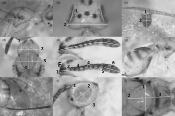
Measurements were taken for 39 body characters, detailed in table 1 and fig. 1. Terminology follows Baur et al. (Reference Baur, Kranz-Baltensperger, Cruaud, Rasplus, Timokhov and Gokhman2014), Bouček (Reference Bouček1988), Gibson et al. (Reference Gibson, Read and Fairchild1998), Graham (Reference Graham1969), and Roques and Skrzypczyńska (Reference Roques and Skrzypczyńska2003). In total, 97 specimens (58 M. zvimendeli, 39 M. manonae) were examined.
Measurement of body characters. (a) mss.ll; (b1-2): eye.d, head.b; (c1-2): stg.l, stg.b; (d1-2-3-4): pnc.l, pnc.b, msc.b, msc.l; (e) pdl.flg; F1-2-3-4-5-6: ped.l, ped.b, fu3.l, fu4.b (drawn as example, similarly measured for other funicle segments), clv.l, clv.b; (g) gst.ll; (h) ovi.l; (i1-2-3): eye.b, eye.h, hea.hl; (j1-2-3): sct.b, sct.l, ppd.l.

Body characters of female Megastigmus measured for multivariate ratio analysis

Microscopic observations and photographs were taken under a binocular microscope (NIKON SMZ800N) with an attached digital camera (TUCSEN H500), resolution 2584 × 1936 pixels. For measurement, body parts were placed so that the entire length was oriented on an imaginary plane perpendicular to the viewing (photographing) angle. Sizes were measured in pixels by the software Image-Pro® (Media Cybernetics) and ImageJ 1.52a (National Institute of Health, USA) and converted to mm using an object micrometre (Carl Zeiss 5 + 100/100 mm) for species description. If curved, ovipositor and antenna lengths comprised the total length of straight segments approximating the curvature of these parts.
One commonly used trait, scape length, was not measured due to difficulties in taking measurable photos without breaking apart the insect body. Similarly, ppd.l and pnc.l were removed from analysis due to failure in taking reliable measurements.
Measurement data are provided in Supplementary Document 2.
Multivariate ratio analysis
Body ratios were examined by the tool MRA, which analyses body proportions of morphologically similar arthropod species (Baur and Leuenberger, Reference Baur and Leuenberger2011). Body ratios with best discriminant power were determined using the algorithm linear discriminant analysis (LDA) ratio extractor. Structure of variation of all specimens was analysed by the algorithm PCA in shape space, identifying the principal components accounting for variation in shape space, and principal component analysis (PCA) ratio spectrum which visualizes the contribution of each character (Baur and Leuenberger, Reference Baur and Leuenberger2011). Computation was conducted in R statistical software R studio version 1.2.5019 (RStudioTeam, 2019). Codes for analysis were obtained from Baur and Leuenberger (Reference Baur and Leuenberger2011) and the package MASS (Ripley et al., Reference Ripley, Venables, Bates, Hornik, Gebhardt and Firth2018).
DNA extraction, polymerase chain reaction (PCR) and sequencing
DNA was extracted from entire insects using ISOLATE II Genomic DNA Kit (Bioline, Eveleigh NSW, AUS), or a prepGEM® Insect kit (ZyGEM, Hamilton, Aotearoa, NZ), eluting into 40 μl extraction volume. Undiluted genomic DNA was used in PCR amplification using MyTaq™ HS Red DNA Polymerase (Bioline, Eveleigh NSW, AUS). Total reaction volumes were 10 μl including DNA template (1 μl), primer (1 μl each, at 10 μM concentration), premixed 5 × buffer (2 μl), HSTaq DNA polymerase (0.1 μl), and H2O (4.9 μl). PCR thermo-cycling in a Bio-Rad T100 (Greenslopes, QLD, AUS): 95°C, 1 min + 35 cycles of (95°C, 1 min + 55°C, 1 min + 72°C, 1 min) + 72°C, 5 min final extension then holding at 10°C. When primer 1775-COI-F was used, the annealing temperature was reduced to 50°C.
DNA primers (table 2) followed previous work on Megastigmus (Scheffer and Grissell, Reference Scheffer and Grissell2003; Boivin et al., Reference Boivin, Henri, Vavre, Gidoin, Veber, Candau, Magnoux, Roques and Auger-Rozenberg2014; Roques et al., Reference Roques, Copeland, Soldati, Denux and Auger-Rozenberg2016) targeting the inner region of the mitochondrial gene cytochrome oxidase 1 (COI mtDNA) and nuclear fragment coding 28S rDNA, from the D1 to D3 region. Amplification was first attempted with primers 1775-COI-F/2773-COI-R (amplicon size 1040 bp) and 28S-D1F/28S-D3R (amplicon size c.a. 1090 bp) (Boivin et al., Reference Boivin, Henri, Vavre, Gidoin, Veber, Candau, Magnoux, Roques and Auger-Rozenberg2014; Roques et al., Reference Roques, Copeland, Soldati, Denux and Auger-Rozenberg2016). The alternative combination 28S-D1F/28S-1059R (amplicon size c.a. 1080 bp) was used to amplify the 28S fragment when necessary. For COI, because the primer 1775-COI-F co-amplified a pseudogene (nuclear copy of mitochondrial DNA, numt) in M. zvimendeli, the upstream forward primer LCO1490 (Folmer et al., Reference Folmer, Black, Hoeh, Lutz and Vrijenhoek1994) was alternatively used to amplify an extended fragment (amplicon size 1304 bp). Even in difficult cases, the reverse sequences 2773-COI-R successfully provided clean reading results covering the 1040 bp target region. For part of the dataset, the internal reverse primer 2399-COI-R resulted in a fully matched fragment of 904 bp extending from the middle of the target region to the start of the barcoding fragment, allowing BLAST searches for matching partial barcode and subsequent analysis based on the barcoding region.
Names, sequences and reference sources of primers used for DNA extractions

PCR products with a single band at the desired fragment size, visualized by electrophoresis on 1 × TBE and agarose gel with GelRed® (Biotium, California, USA), were sent to Macrogen Inc. (Seoul, ROK) for purification and Sanger sequencing. For smaller batches (<10 samples), PCR products were purified on-site using ExoSAP-IT (Thermo Fisher Scientific, MA, USA). Sequencing reactions were conducted on-site using BigDye Terminator v3.1 Cycle Sequencing Kit (Thermo Fisher Scientific, MA, USA), and products were sent to the Australian Genome Research Facility (QLD, Australia) for sequencing.
Localities of analyzed M. zvimendeli and M. manonae in Australia, including the number of morphometric specimens, are illustrated in fig. 2.
Distribution of Australian specimens of Megastigmus zvimendeli (triangles) and M. manonae (circles) used in the study. Numbers indicate morphometric samples in each location. Species identity was confirmed by DNA sequences of representative samples from all localities.

DNA sequence analysis
Forward and reverse sequences were aligned and edited using Geneious 11.0.2 (Biomatters, Auckland, NZ). Primer sequences were removed, and alignments were trimmed to equal lengths (867 bp for 28S rDNA and 885 bp for COI mtDNA). Mitochondrial DNA was verified by translating the genetic code to amino acids using Geneious 11.0.2 to check for stop codons which suggested the presence of pseudogenes. Sequences from two specimens of M. manonae contained ambiguous sites, illustrated by double peaks nested within regions of clear, unambiguous signal, likely representing within-individual mitochondrial copy differences, so the sites were labelled with a degenerative base following the IUPAC ambiguity code.
Genetic distances and base compositions were calculated using MEGA X software (Kumar et al., Reference Kumar, Stecher, Li, Knyaz and Tamura2018). Trees were constructed using Maximum Likelihood (ML) algorithm with the PHYML plugin (Guindon et al., Reference Guindon, Dufayard, Lefort, Anisimova, Hordijk and Gascuel2010) in Geneious 11.0.2 (Biomatters, Auckland, NZ). For single-gene ML analysis, the optimal model of DNA evolution was determined using the Bayesian Information Criterion in the program Jmodeltest 2.1.0 (Darriba et al., Reference Darriba, Taboada, Doallo and Posada2012). Support values were calculated by bootstrap resampling 1000 times. The concatenated dataset was partitioned into four blocks (28S + CO1 separated into 1st, 2nd and 3rd codon position) in PartitionFinder 2.1.1 (Lanfear et al., Reference Lanfear, Frandsen, Wright, Senfeld and Calcott2016) and analysed to determine the best GTR model (Tavaré and Miura, Reference Tavaré and Miura1986) for use in constructing a ML tree in RAxML 8.2.11 (Stamatakis, Reference Stamatakis2014). Support values were calculated using the RAxML's Boostraping algorithm with 1000 replicates.
DNA-based species delimitation was performed using the web version of the Automatic Barcode Gap Discovery (ABGD) tool (Puillandre et al., Reference Puillandre, Lambert, Brouillet and Achaz2012). ABGD was applied to the trimmed, aligned 885 bp COI region (from 1798 to 2688 in reference to Drosphila yakuba mtDNA). Genetic distance was based on the KP80 model, a common parameter in mtDNA-based species delimitation (Boykin et al., Reference Boykin, Armstrong, Kubatko and De Barro2012; Collins et al., Reference Collins, Boykin, Cruickshank and Armstrong2012; Evans and Paulay, Reference Evans, Paulay and Walker2012).
Genbank searches for matching sequences were performed using the Geneious BLAST tool. Queries were representative sequences of M. manonae and M. zvimendeli corresponding to the barcode region and the Clyde-Bonnie region of the COI gene. The BLAST search returned two sequences (KF938926.1, JN559766.1) of 654 and 686 bp at the barcoding region and one sequence (KU984684.1, 417 bp) from the downstream end, that matched >99% with M. zvimendeli query sequences, with all other entries <93.2% (Supplementary Document 3). Trees containing Genbank-obtained sequences were built separately by alignment with the sequences generated in this study then trimmed to the reduced length of the Genbank sequences. The approximate fragment locations are presented in Supplementary Document 4.
Scanning electron microscopy
Representative specimens were dehydrated with hexamethyldisilazane (HMDS) and gold-coated using a Dynavac SC150 Sputter Coater (Dynavac High Vacuum Pty. Ltd., Victoria, Australia). Scanning electron microscope images were taken using a Hitachi Tabletop Microscope model TM-1000 (Hitachi High-Technologies Corporation, Tokyo, Japan).
Results
Multivariate ratio analysis of variation in body size and shape
Application of the function PCA ratio spectrum to all specimens as a single group identified principal components contributing to morphometric variations without prior species-determinant input. In shape space, PC1 and PC2 accounted for 50% of the variation of the entire sampled population. The first principal component is in congruent with the separation of species, although a clear cut between two clusters could not be established (fig. 3).
Scatterplot of isometric size against PC1 in shape space. Specimens confirmed by COI DNA sequence were marked with arrows. Confidence ellipses assuming multivariate normal distribution.

The PCA ratio spectrum (fig. 4) identified ool.l at the extreme high end, eye.h, eye.b and breadth of funicle segment fu2 to fu6 at the extreme low end. These characters, except fu2.b to fu6.b, were found to contribute to species discrimination. LDA ratio extractor identified eye.b/ool.l, fu1.l/stg.l and eye.h /fu2.l as the three best discriminating ratios. Combining the best two ratios successfully separated the studied species in a scatterplot (fig. 5). The calculated ratios for LDA-suggested characters (M. zvimendeli vs M. manonae, range 5th percentile–95th percentile) are: eye.b/ool.l (2.9–3.9) vs (4.3–6.4); stg.l/fu1.l (2.4–3.0) vs (3.0–3.7); eye.h/fu2.l (5.9–8.0) vs (7.9–10.0). This suggests that the two species can be separated when judgement is based on a series of individuals. Further, the calculated D.shape are much higher than D.size in all of the three best discriminative ratios, indicating that species are mostly separated by differences in the shape of characters (Supplementary Document 5).
PCA Ratio Spectrum of the first principal component in shape space. Horizontal bars show 68% bootstrapping confidence intervals. Characters at two extreme ends explain most of variation in the first principal component.

Scatterplots illustrating discrimination of M. manonae and M. zvimendeli with two best discriminating ratios selected by LDA Ratio Extractor (eye.b/ool.l vs fu1.l/stg.l). Dots pointed with arrows denote specimens confirmed by COI DNA sequence. Confidence ellipses were based on an assumed multivariate normal distribution.

Molecular species delimitation
Sequences of nuclear DNA coding partial 28S rRNA were obtained for M. manonae (13 specimens) M. zvimendeli (19 specimens) and M. pretorianensis (4 specimens), all trimmed to 867 bp. Guanine and cytosine accounted for 57.5% of the total bases on average, illustrating a slight GC bias. Within the studied group, nuclear 28S DNA sequences were highly conserved: intraspecific pairwise differences were 0% in all three species, maximum interspecific divergence was 1% (8 bases).
Partial mitochondrial DNA coding COI were sequenced and trimmed to 885 bp for 18, 13 and 4 specimens of M. zvimendeli, M. manonae and M. pretorianensis, respectively. The sequences contained 74.6% adenine and thymine, demonstrating a strong AT bias as in previous hymenopteran mtDNA studies (Castro et al., Reference Castro, Austin and Dowton2002; Rokas et al., Reference Rokas, Nylander, Ronquist and Stone2002). Thymine alone accounted for 46.1–46.8% of the total number of bases in the coding strands. COI sequences of M. manonae (maximum 2.3% intraspecific difference) were more divergent than those of M. zvimendeli (maximum 0.3% intraspecific difference).
The generated sequences, including those of the outgroup (M. zebrinus) have been submitted to Genbank (Accession number MN165877 to MN165951).
Application of the ABGD tool to the aligned 885 bp COI sequences (Supplementary Document 6) revealed a clear barcode gap between KP80 distance of 0.02 and 0.06, which respectively represented the maximum intraspecific and minimum interspecific distances. DNA sequences were firmly assigned to four groups (M. zvimendeli, M. manonae, M. pretorianensis and the outgroup M. zebrinus). Specimens identified as M. zvimendeli from Kenya and Israel, and a specimen identified as M. sichuanensis, were also placed within the M. zvimendeli group.
The program PartitionFinder 2.1.1 recommended the models GTR + G, GTR + I + G, GTR and GTR + G for the 28S and the 1st, 2nd and 3rd codon position, respectively. The model GTR + G represented most of the variation (28S and the 3rd codon position of COI) in the partitioned concatenated dataset and was therefore selected for phylogeny reconstruction in RAxML (Stamatakis, Reference Stamatakis2014). The reconstructed phylogeny was congruent with the ABGD species assignment, with bootstrap support from 90 to 100% (fig. 6a). The ABGD and ML analyses assigned a specimen from NSW-Australia with M. pretorianensis from South Africa, which was then verified using diagnostic characters (Doğanlar, Reference Doğanlar2015), confirming the presence of M. pretorianensis in Australia. The Australian M. pretorianensis specimens were from a local gall-inducing Leptocybe sp. that differs from the two invasive Leptocybe spp. in COI sequence (Le et al., unpublished).
ML trees inferred from different DNA markers. (a) Phylogeny based on the concatenated COI + 28S sequences (867 + 885 bp), model of evolution (GTR + G), data partitioned into four blocks (28S + three COI codon positions) using RAxML. (b) Placement of the Genbank entry KU984684.1 (M. icipeensis, Kenya) based on COI downstream 417 bp sequences, model of evolution (TPM1uf + I), using PHYML. (c) Placement of the Genbank entry JN559766.1 (M. viggianii, India) and KF938926.1 (Megastigmus sp., India) based on 613 bp sequences at COI barcoding region, model of evolution (TIM3 + G), using PHYML. (b) and (c) are from non-overlapping regions. (GTR: Generalized time-reversible; TPM: 3-parameter model; TIM: Transitional model; uf: unequal base frequency; G: with gamma-distributed among-site rate variation; I: with a proportion of invariable sites). Outgroup specimen was M. zebrinus for all trees. Star-marked nodes are from non-Leptocybe galls. Numbers adjacent to nodes indicate bootstrap support (n = 1000; only values ≥80 are shown). Number of substitution rate categories = 4 for all PHYML analysis. One representative was selected for identical sequences from the same locality. Identical sequences from different localities and non-identical sequences from the same locality were retained in ML analysis.

Before analysis of Genbank sequences, 13 and 21 bases were respectively removed from the downstream ends of KF938926.1 and JN559766.1 due to overlap with primer region, and a strong indication of technical misread commonly found in Sanger sequencing, illustrated in a disproportionately high ratio of mismatch of this region to published congeneric sequences. Sequences were subsequently aligned to available data and trimmed to the same length for tree construction and distance calculation. KP80 distance (≤0.8%) and ML inference (fig. 6b, c) confidently confirmed that the Genbank sequences and Australian M. zvimendeli are the same species.
Species description
Megastigmus manonae sp. nov.
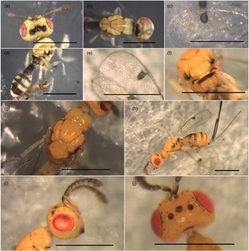
(figs 7 and 8)
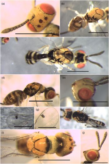
Megastigmus manonae (scale bars = 0.5 mm). Female: (a). pedicel and flagellum; (b, d): light form and dark form, dorsolateral view; (e) face showing toruli position; (g) forewing stigma; (i) mesoscutal midlobe, pronotal collar and occiput; (j) head and antenna from lateral view. Male: (c, h): light and dark form, dorsal view; (f) forewing stigma.

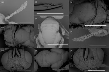
SEM photographs of Megastigmus manonae. Female: (a) pedicel and flagellum; (b) scutellum and propodeum; (c) toruli and scrobal depression; (d) drilling tip of ovipositor stylet; (e) pronotum and mesoscutum. Male: (f) pronotum and mesoscutum; (g) scutellum and propodeum; (h) pedicel and flagellum; (i) toruli and scrobal depression. Scale bars: a, b, c, f, g, h, i = 0.2 mm; d = 0.1 mm; e = 0.3 mm.

Type material
Holotype: [-26.653896, 151.956021 / D'Anguilar Hwy, near Parsons Rd / Nanango Queensland 4615] // [HOLOTYPE ♀ / Megastigmus manonae] Queensland Museum (QM) South Brisbane, QLD, Australia. Reg. No. T245894, 1♀; card mounted; emerged from Leptocybe spp. gall on roadside Eucalyptus tereticornis; collected 16.i.2018.
Paratypes: Same data as holotype: QM 7♀, 4♂; Australian National Insect Collection, Black Mountain (ANIC), Black Mountain, ACT, Australia 7♀, 3♂ (ANIC 32-141414 to 23); Queensland Department of Primary Industries Insect Collection (QDPC), Dutton Park, QLD, Australia: 7♀, 2♂ (QDPC-0 −176208 to 176216).
[-27.487666, 152.662933 / Fairneyview – Fernvale Rd, / Fairney View QLD 4306], Australia; QM 3♀, 3♂; ANIC 2♀, 2♂ (ANIC 32-141424 to 141427); QDPC: 3♀, 1♂ (QDPC-0 −176208 to 176216); card mounted; emerged from Leptocybe galls on roadside saplings of Eucalyptus tereticornis; collected 17.xi.2017.
[-28.818824, 153.080054 / Manifold Rd, N. Casino NSW 2470], Australia; QM, 1♀; card mounted; emerged from Leptocybe galls on roadside saplings of Eucalyptus sp.; collected 14.xii.2018.
Same collection data as holotype: QM: 3♂, 3♀; HMDS treated, sputter coated, mounted on one metal stub for SEM.
Etymology
The specific epithet manonae (a noun in the genitive case) is named after Dr Manon Griffiths, an entomologist and NHL's supervisor who sadly passed away during the preparation of this manuscript.
Description
Diagnosis: 1. small, gst.ll + mss.ll <1.4 mm, with 2 pairs of scutellar setae, the anterior pair inserts at the approximate level of the posteriormost point of axilla and much longer than the posterior pair; 2. body darkly pigmented, most conspicuous in male: lightest male form having dark black colour on vertex, propodeum, anterior edge of mesoscutal midlobe and anterior half of tergites; darkest male form with scutellum and mesoscutal midlobe entirely black; 3. female eyes large from a lateral view, making up 80% length from vertex to mouth margin, fu3 not shorter than fu2.
Female
Colour and colour variation: Body ranging from dominantly yellow to almost black to unassisted eyes. Colour variation most readily observed in mesosoma; the lightest form having notauli, scutoscutellar suture and anterior edge of mesoscutal midlobe black; the darkest form with scattered black pigments, making mesoscutum superficially black. Face and vertex from yellowish orange to whitish-yellow. Eye reddish. Occiput, mandibles and eye margin black. Pedicel and flagellum dark brown from dorsal view to yellowish-orange from ventral view; scape and pedicel much darker dorsally than ventrally. The lateral panel of pronotum, mesopleuron, prepectal shelves and prostenum dark yellow to dominantly black. Pronotal collar from concolourous to strongly paler than mesoscutum. Anterior of abdominal sclerites each with an anteriorly darkened band, grey to black, becoming orange or yellow posteriorly except the median of first and second tergites. Ovipositor sheaths black. Ovipositor stylets copper yellow.
Head. Head shape from lateral view cuneate (fig. 7j). Eye large, from lateral view making 80% dorsoventral length from vertex to mouth margin (eye.h/hea.hl 0.72–0.86 in morphometric specimens), surrounded by a ring of setae along the margin. Head breadth at posterior eye margin 0.30–0.45 mm. Ratio head.b/pol.l 2.8–3.7, head.b/eye.d 1.5–1.8, pol.l/ool.l 2.5–3.9. Lower face with striae convex towards mandibles. Antenna clavate; funicle and clava segments with one row of sensilla. Scape reaching the level of ocelli. Ratio pdc.flg/head.b 1.1–1.3. Pedicel significantly longer and wider than fu1 (ped.l/fu1.l 1.6–2.4). Funicles subequal in length (fu1.l > fu3.l > fu2.l with few exceptions), widened from fu1 to fu7 (fu1.b/fu7.b 0.5–0.7), hence segments appear transverse except first and second funicle nearly cylindrical. Club length varied (clv.l/fu7.l 2.7 −3.7).
Mesosoma: approximate length of gaster (mss.ll/gst.ll 0.8–1.2), mss.ll/msc.b 1.6–2.0, msc.b/head.b 0.8–1.1. Pronotum posteriorly overlaps but not completely concealing the concave anterior edge of mesoscutal midlobe. From dorsal view, pronotal collar sub-rectangular, twice as broad as length (pnc.l/pnc.b 0.4–0.6). Pronotal collar setae from dorsal view: two longitudinal rows near lateral edges, sparse long setae nearly forming two submedian longitudinal rows, a transverse row at posterior edge. Mesoscutal midlobe and scutellum with fine transverse, sometimes inconspicuous sculpture. Frenal groove absent, frenum indistinct. Dorsellum conspicuous, paler than two lateral panels of metanotum, the lateral panels frequently with a brighter posterior region forming a transverse, near-oval shape at each panel. Propodeum slightly reticulated, more densely and hence appears punctate between two plicae. Propodeal callus with a patch of hairs paler than body setae. Legs white except tarsal claws dark and hind coxa brownish, darker basally. Forewing (fig. 7g) hyaline, stigma darkened brown. Basal setal line incomplete. Cubital fold conspicuous. Submarginal vein (smv) with 6–7 setae, narrower than parastigmal vein (pv) and post marginal vein (pmv). Smv length approximate (pv + pmv). Pmv forms an acute angle with and approximates the length of stigmal vein. Upper part of stigma vein (stigma petiole) short, slightly longer than uncus, stigma knob length approximately 1.5 × breadth (stg.l/stg.b 1.4–1.7).
Metasoma: inconspicuous petiole, hence gaster rather sessile. Tergites from dorsal view with a ring of black setae at posterior edges. Ratio ovi.l/gst.ll 1.4–2.0.
Male
Body smaller than females (paratypes, mss.ll + gst.l 1.0–1.3 mm); body colour (fig. 7c and h) as in diagnosis character, usually darker than females. Head: head and antennal shape similar to females, a lower face from lateral and frontal view paler than mesoscutum, vertex with a black butterfly-shaped region covering ocelli; ool.l short. Eye reddish with black margin surrounded by a ring of black setae like in females. Antennal segments paler ventrally than from dorsal view. Size (paratypes, mm): head.b 0.33–0.42, eye.d 0.2–0.25. ratio: pol.l/ool.l 3.4–4.4, pol.l/lol.l 2.2–2.3, head.b/eye.d 1.6–1.7, eye.d/pol.l 1.7–2.0, ped.l/fu1.l 1.6–2.2. Mesosoma: shape of mesosoma, mesoscutum and scutellum like in females. The transverse sculpture on mesoscutal midlobe usually conspicuous, most noticeable on darker specimens. Scutellum sometimes appears punctate in lighter forms. Scutellum with 1 additional pair of scutellar setae in addition to the characteristic two pairs. Pronotum shape like in females, paler than mesonotum form dorsal view, concolourous to lower face. Legs generally whitish to yellow, darker at tarsal claws. Stigma knob with a clear margin like in females but less elongated. Length and breadth of the uncus and stigma petiole vary. Size: mss.ll 0.5–0.7, msc.b 0.3–0.4; Ratio: stg.l/stg.b 1.2–1.4, mss.ll/msc.b 1.7–2.0, msc.b/head.b 0.9. Metasoma usually much smaller than mesosoma, gst.ll 0.5–0.6 mm with the first tergite elongated, petiolate.
Biology
Megastigmus manonae emerged from Leptocybe spp. galls on Eucalyptus tereticornis seedlings and from a small blister, non Leptocybe spp. – induced galls on leaves of young Eucalyptus sp. Females that emerged from field-collected Leptocybe spp. galls oviposited on laboratory-induced Leptocybe spp. galls and reproduced successfully. In laboratory condition, development time (from oviposition to emergence of the new generation) varied from 19 to 22 days and a maximum number of offspring was nine (NHL, pers. obs., four parental females).
Taxonomical Remarks
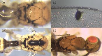
Megastigmus manonae grouped to M. zvimendeli and M. pretorianensis in a phylogenetic tree of eucalypt-associated Megastigmus with minute size, short and clavate antennae, ovipositor similar in relative length to mesosoma, similar forewing and stigma knobs shape, and two pairs of scutellar setae (Le et al., unpublished). In Australia, emergence from the same collected material, collection site and eucalypt plant can include any of the combination of M. manonae, M. zvimendeli, M. lawsoni and an unidentified species (Megastigmus sp. 1). Small individuals of Megastigmus sp. 1 and M. zvimendeli are similar in size and body colour but males and females of Megastigmus sp. 1 can be distinguished from M. zvimendeli by their three pairs of black scutellar setae (fig. 9a). Males of M. lawsoni have a black patch on the mesonotum but the coloration is always confined to the median part of the transscutal articulation, never reaching the anterior edge of mesonotum (fig. 9c). This coloration delimits these species: the black patch is lacking in M. zvimendeli; while in M. manonae the black pigments are unconfined (fig. 7c and h). Females of M. lawsoni and M. zvimendeli have similar colour and shapes; rare cases of M. lawsoni females have four scutellar setae instead of the typical single pair as in Doğanlar and Hassan (Reference Doğanlar and Hassan2010). These two species can be misidentified without the presence of males, but M. lawsoni can be identified by longer stigma knobs and different, somewhat irregular placement of scutellar setae (fig. 9b and d).
Possible misleading forms of Australian species that can be misidentified for Megastigmus manonae and M. zvimendeli. (a) scutellar setae, female Megastigmus sp. 1; (b) female stigma, M. lawsoni; (c) mesonotum colouration, male M. lawsoni; (d) scutellar setae, female M. lawsoni.

Within the group of M. zvimendeli, M. manonae and M. pretorianensis, the first species can be identified by the dominantly yellowish-orange female colour, rarely with darkened notauli or scutoscutellar suture and never with black colour on the anterior edge of mesoscutum. M. manonae and M. pretorianensis both have darkly pigmented bodies, but M. pretorianensis appears closer to M. zvimendeli than to M. manonae in terms of body shapes: they can be separated by the same two best ratios that worked for M. manonae and M. zvimendeli (Supplementary Document 7). The ratio that could be most conveniently interpreted into body proportion is eye.h/hea.hl, representing the ‘smaller eye’ when observed from a lateral view. This ratio varied from 0.68 to 0.74 in 11 out of 12 measured M. pretorianensis, well within the 5th −95th percentile range of M. zvimendeli (0.64–0.76) and not exceeding that of M. manonae (0.74–0.85). In the study, M. manonae specimens with less conspicuously large eyes were further identified by having fu3.l > fu2.l. The fu3 was significantly shorter for the Australian M. pretorianensis specimen (1♀ available for study), and the examined South African M. pretorianensis having fu2 longer or at least subequal to fu3. For this reason, the character ‘large eye’ and fu3.l ≥ fu2.l were mentioned in the diagnosis of M. manonae. Additionally, M. manonae females usually have longer pronotal and mesoscutal setae which are much more conspicuous than that of M. pretorianensis.
Megastigmus pretorianensis was described with ‘antennae inserted slightly above lower ocular line’ (Doğanlar, Reference Doğanlar2015). We found this character highly subjective to observational error due to the generally low position of toruli in the studied group and the absence of an actual lower ocular line. This character was therefore disregarded for delimitation of M. manonae and M. pretorianensis. In addition, the breadth of funicle segments (fu2.b to fu6.b) were not selected by LDA Ratio Extractor despite being placed at the lower end of PCA spectrum. This indicates that the interspecific variation of funicle breadths for the two studied species were not strong enough to outweigh the intraspecific variation under the selected measurement technique. Removal of funicle breadths in fact improved the separation of two species (Supplementary Document 8). Funicle breadths together represent the thinness of flagellum and consequently the differently perceived length of the antenna (Supplementary Document 9), which should be considered if species identification is based on few specimens.
Megastigmus zvimendeli Doğanlar & Hassan 2013
(figs 10 and 11)
Description and diagnostic characters of M. zvimendeli were provided by Doğanlar (Reference Doğanlar2015), Doğanlar and Hassan (Reference Doğanlar and Hassan2010), and Roques et al. (2016, as M. icipeensis) (Roques et al., Reference Roques, Copeland, Soldati, Denux and Auger-Rozenberg2016). We provide additional data on sizes and diagnostic characters of female specimens, and comments on synonymies and possible misidentification.
Megastigmus zvimendeli (scale bars = 0.5 mm). Male: (a) vertex and ocelli; (b) mesoscutal midlobe and pronotum; (c) forewing stigma; (d) abdomen. Female: (e) forewing stigma; (f) scutellum and propodeum; (g) mesoscutal midlobe and pronotum; (h) dorsolateral habitus; (i) head from lateral view; (j) vertex and ocelli.

SEM photographs of Megastigmus zvimendeli. Male: (a) mesoscutal midlobe; (b) mesoscutum from dorsal view; (c) face, scape and toruli; (f) flagellum; (j) scutellum and propodeum. Female: (d) drilling tip of ovipositor stylet; (e) scape and flagellum; (g) face, scape and toruli; (h) scutellum and propodeum; (i) mesoscutal midlobe. Scale bars: a, b, g = 0.3 mm; c, e, f, i = 0.2 mm; h, j = 0.1 mm; d = 0.05 mm.

Synonyms
Megastigmus icipeensis Roques & Copeland, 2016. Syn. n.
Megastigmus sichuanensis Doğanlar & Zheng 2017. Syn. n.
Megastigmus judikingae Doğanlar & Hassan Reference Doğanlar and Hassan2010. Syn. n.
Sizes (Australian morphometric specimens, mm): head.b 0.3–0.4; hea.hl 0.2–0.3; eye.h 0.15–0.21; eye.b 0.12–0.15; pdl.fgl 0.4–0.6; eye.d 0.21–0.26; mss.ll 0.5–0.7; msc.b 0.3–0.4; gst.ll + mss.ll 0.9–1.5; ovi.l 0.8–1.3; Ratio: eye.b/ool.l 2.6–4.0; pol.l/ool.l 2.0–3.1; ped.l/fu1.l 1.5–2.3; fu1.b/fu7.b 0.5–0.7; eye.h/fu2.l 5.7–8.0; stg.l/fu1.l 2.3–3.2; fu1.l/fu7.l 0.8–1.2; msc.b /head.b 0.8–1.0; mss.ll/msc.b 1.5–2.1; stg.l/stg.b 1.3–1.7; ovi.l/gst.ll 1.4–2.1; (gas.ll + mss.ll)/ovi.l 1.0–1.3.
Diagnosis (females)
1. Scutellum with two pairs of setae, the anterior pair longer, inserting at the approximate level of the posteriormost point of axilla; 2. body almost concolourous orange; both male and female with mesoscutum and scutellum entirely concolourous yellowish-orange from dorsal view; darkest male body dominantly yellowish-orange mesonotum with narrow black scutoscutellar suture; pilosity of thorax mostly pale; 3. female eyes small, making up 65–75% of the head height from lateral view.
Taxonomical Remarks
Megastigmus judikingae and M. zvimendeli were distinguished by size and ratio of body characters in the original description (Doğanlar and Hassan, Reference Doğanlar and Hassan2010; Doğanlar, Reference Doğanlar2015). Synonymy of M. zvimendeli and M. judikingae is based on the observations below:
• Remeasurement of paratypes of M. judikingae (2♀, ANIC 111465, ANIC111466) based on characters delimiting M. judikingae and M. zvimendeli in the original paper (Doğanlar and Hassan, Reference Doğanlar and Hassan2010) (ped.l/fu1.l 1.9–2.0, fu1.b/fu7.b 0.6–0.7; combined length of funicle 5, 6, 7/clv.l 1.1; ovi.l/gst.ll 1.6 to 1.8) varied within intra-species limit of M. zvimendeli. PCA clustering and ratio-based scatterplot comfortably placed the two paratype specimens into M. zvimendeli group (Supplementary Document 10).
• Labels on abovementioned paratypes record E. tereticornis as the host plant, rather than Corymbia tessellaris as in the original publication. E. tereticornis is a common host of Leptocybe-associated M. zvimendeli, whereas C. tessellaris does not support Leptocybe development (Phạm et al., Reference Phạm, Dell and Isobel Burgess2009).
• These specimens and M. zvimendeli shared key characters of body size, body colour, scutellar setae position and colour, and placement of toruli.
Because the name M. zvimendeli has been cited in publications regarding the species' use in biocontrol (Dittrich-Schröder et al., Reference Dittrich-Schröder, Harney, Neser, Joffe, Bush, Hurley, Wingfield and Slippers2014; Bush et al., Reference Bush, Dittrich-Schröder, Neser, Gevers, Baffoe, Slippers and Hurley2017; Mendel et al., Reference Mendel, Protasov, La Salle, Blumberg, Brand and Branco2017), precedence was given to M. zvimendeli Doğanlar and Hassan 2013 (the Principle of the First Reviser, Article 24.2, ICZN Code). Megastigmus judikingae Doğanlar and Hassan Reference Doğanlar and Hassan2010 becomes a junior synonym.
Megastigmus icipeensis Roques & Copeland was described in 2016 (Roques et al., Reference Roques, Copeland, Soldati, Denux and Auger-Rozenberg2016) from Malaise trapped Kenyan specimens. The available COI DNA sequence from these specimens fully grouped with Australian M. zvimendeli and with Leptocybe invasa-associated Megastigmus specimens sent from Kenya by our colleagues and subsequently identified as M. zvimendeli (fig. 6). Characters including the minute size, distinctive orange body colour and pale bristles fully matched with M. zvimendeli. We, therefore, treat M. icipeensis Roques & Copeland 2016 as a junior synonym (Article 23. Principle of Priority, ICZN Code).
Megastigmus sichuanensis Doğanlar & Zheng was described in 2017 (Doğanlar et al., Reference Doğanlar, Huang, Guo, Lu, Yang, Yang and Zheng2017; Huang et al., Reference Huang, Liu, Zhang, Guo, Yang, Lu and Zheng2017). Nuclear 28S DNA and mitochondrial COI DNA sequences obtained from specimens provided by our colleagues in China fully matched M. zvimendeli (fig. 6). As in M. judikingae, the sizes (ovi.l 1.2 mm), body ratios (mesosoma length/msc.b 2.1, clv.l/clv.b 1.85, stg.l/stg.b 1.4) fell within the variation range of M. zvimendeli in our study. The number of scutellar setae, body and pilosity colour matched diagnostic characters of M. zvimendeli. We, therefore, synonymize M. sichuanensis and M. zvimendeli, with the latter name having precedence (Article 23. Principle of Priority, ICZN Code).
Megastigmus viggianii Narendran & Sureshan was described from specimens collected from the Indian Ukshi plant Calycopteris floribunda (Narendran and Sureshan, Reference Narendran and Sureshan1988) and linked with Leptocybe in subsequent studies (see Huang et al., Reference Huang, Li, Lu, Zheng and Yang2018; Le et al., Reference Le, Nahrung, Griffiths and Lawson2018). KP80 distance and phylogenetic analysis firmly placed two Genbank entries, JN559766.1 (recorded as M. viggianii) and KF938926.1 (unidentified Megastigmus, India), into the M. zvimendeli group. M. viggianii is described as of significantly larger body size (1.67–2.13 mm), distinct malar sulcus, all funicles elongated, and smooth frenum (Narendran and Sureshan, Reference Narendran and Sureshan1988). The examined holotype (Reg. No. T57893, donated to QM by Narendran in 1999 (Wright, 2019, pers. comm.) bears three pairs of scutellar setae, elongated funicles, and toruli at higher than the lower eye margin. These characters are not observed in M. zvimendeli; therefore, we believe JN559766.1 was misidentified. Megastigmus viggianii and M. zvimendeli remain two valid species, but with no association between M. viggianii and Leptocybe or eucalypts.
Discussion
Within Megastigmus, Bouček (1988) suggested the divergence of a distinctive entomophagous group with small size, low antennal sockets, convex lower face, short antennae and similarities in male and female antennal form. Megastigmus associated with Leptocybe galls in our study also differs from phytophagous species in minute sizes and distinctly clavate antenna in males and females. While phylogenetic relationships among them and their phytophagous congeners await elucidation, their size and morphological variation have confused researchers attempting morphological classification.
Our study successfully utilized morphometric and genetic data to discriminate two closely related species: M. manonae, sp. nov., and M. zvimendeli. Key morphological characters contributing to variation among all specimens (ool.l, eye.h, eye.b, fu1.l, fu2.l and stg.l) were revealed by MRA. Moreover, the two species could not be separated by ratios involving funicle breadths and several body characters which are relied on in current taxonomic keys to parasitic Megastigmus, e.g. pdc.fgl, clv.l, ovi.l. This reiterates the need for exhaustive examination (Bouček, Reference Bouček1988; Auger-Rozenberg et al., Reference Auger-Rozenberg, Kerdelhué, Magnoux, Turgeon, Rasplus and Roques2006; Protasov et al., Reference Protasov, Doĝanlar, La Salle and Mendel2008). In addition, phylogenetic analyses revealed greater genetic diversity within M. manonae than M. zvimendeli.
The generated knowledge of morphological variation and complementary DNA data resulted in the synonymies of M. zvimendeli with M. judikingae, M. sichuanensis and M. icipeensis, and the disassociation of M. viggiannii as a Leptocybe-eucalypt gall associate. In turn, this illustrates the successful spread and establishment of M. zvimendeli, an Australian endemic parasitoid, in the invasive range of Leptocybe spp., unveiling the previously under-recognized success of this species in biocontrol. The findings also emphasize the need for coordinated examination and possible revision of Megastigmus spp. from Leptocybe galls worldwide. Gall-inducing insects are highly specialized (Price, Reference Price2005) and arose from evolutionary pathways starting from plant tissue miners or sedentary plant feeders (Price et al., Reference Price, Fernandes and Waring1987). Considering the Australian origin of eucalypts, the long co-evolutionary history of insect-plant systems and the paucity of non-Australian entomophagous Megastigmus, a significant proportion of the purportedly fortuitous local Megastigmus spp. associated with Leptocybe spp. in their invasive range could have come from unintentional introductions from Australia. Megastigmus pretorianensis, which we confirmed present in Australia and South Africa, is a tentative example requiring further specimens for an origin-tracing study.
Attention should be drawn to the co-occurrence of Megastigmus species in Leptocybe spp. galls. Megastigmus zvimendeli and M. manonae have been found in galls collected from the same location in Queensland (fig. 6a). Co-occurrence of M. zvimendeli with other Megastigmus species, even within the same gall, was observed frequently in collections (Le et al., unpublished data). This could possibly be due to the multi-chambered, large Leptocybe galls being exploited at the same time by multiple parasitoids. Co-occurrence can confound identification and cause the unintended release of mixed species in biocontrol programs.
The conserved COI DNA sequences in M. zvimendeli precluded further population analysis of its worldwide movement, although its geographic origin is partly known through data on its discovery, collection and release. This high conservation is at odds with previous observations in which the rate of mutation in parasitic Hymenoptera is higher than that of nonparasitic insects (Castro et al., Reference Castro, Austin and Dowton2002). Nevertheless, like Auger-Rozenberg et al. (Reference Auger-Rozenberg, Kerdelhué, Magnoux, Turgeon, Rasplus and Roques2006) and Roques et al. (Reference Roques, Copeland, Soldati, Denux and Auger-Rozenberg2016), we found DNA markers highly successful for Megastigmus species discrimination. Continued use of these markers will facilitate overall understanding of the genus and relationships between species that do and do not associate with eucalypts, from Australia and elsewhere, and phytophagous vs entomophagous species.
Like Borowiec et al. (Reference Borowiec, La Salle, Brancaccio, Thaon, Warot, Branco, Ris, Malausa and Burks2019), we have demonstrated the utility of morphological characters and molecular methods to discriminate species in cryptic eucalypt-gall associated wasps. While this successful combination of morphological and DNA tools will be applied to a broader range of endemic Australian Megastigmus species (Le et al., in prep), we recommend that non-Australian species unavailable for our study (e.g. M. dharwadicus, M. thailandiensis, M. thithipornae, M. brasiliensis, M. leptocybus) be examined in light of our findings. Understanding the status of Megastigmus species associated with Leptocybe will assist with identifying new biocontrol candidates and the potential host-switching and/or invasion processes that may underpin the diversity and distribution of eucalypt-associated Megastigmus worldwide.
Supplementary material
The supplementary material for this article can be found at https://doi.org/10.1017/S000748532000022X.
Acknowledgements
The authors thank colleagues who supplied specimens, supplementary information and advice: Professor Zvi Mendel, Department of Entomology, The Volcani Center, Israel; Dr Beryn A. Otieno, Kenya Forestry Research Institute, Kenya; Dr Gudrun Dittrich-Schröder, FABI, University of Pretoria, South Africa; Dr Zheng Xialin, College of Agriculture, Guangxi University, China; and Dr Arakalagud Nanjundaiah Shylesha, Indian Council of Agricultural Research, India. They are very grateful to colleagues who assisted with work on museum specimens: Susan Wright, QM; Juanita Rodriguez Arrieta, ANIC; Justin Bartlett, QDPC. Owen Seeman (QM) assisted with SEM specimen preparation and proof-reading. The authors are particularly in debt to Dr Petr Janšta, Department of Zoology, Charles University, Prague, Czech Republic, for the comments and assistance that significantly improved the manuscript.
NHL received an Endeavour Award Post Graduate Scholarship from the Australian Government Department of Education and Training. Additional funding was provided through The Australian Centre for International Agricultural Research (ACIAR) project FST/2012/091 with support from the Department of Agriculture and Fisheries, Queensland, and the University of the Sunshine Coast.
The authors sincerely thank the anonymous reviewers for their constructive feedback and suggestions.