Introduction
Interest in the synthesis and biologic application of semiconductor nanoparticles (NPs) also known as quantum dots (QDs) has become an ever-increasing research area.[Reference Clift and Stone1–Reference Lim, Kim, Nakayama, Stott, Bawendi and Frangioni3] This is due to their excellent optical properties compared to organic dyes such as broad and continuous absorption spectrum, high fluorescence and long-term stability, narrow and symmetric emission spectrum, and good photo- and chemical stability. These properties have also led to their applications in many areas ranging from optical where they can be used for the fabrication of devices such as solar cells, light emitting diodes, etc. to catalysis, sensor, water purification, and biological applications where they can be used for imaging, labeling, diagnostics, and therapeutic purposes. As a result of these diverse areas of applications, several methods have been reported for the synthesis of semiconductor NPs which can be broadly divided into three approaches namely organic, two-phase (organic to aqueous and vice visa), and aqueous approaches.[Reference Chandan, Jessica, Schiffman and Geetha Balakrishna4–Reference Oh, Liu, Nel, Gemill, Bilal, Cohen and Medintz6] Among these approaches, aqueous synthesis is the most suitable for biologic applications. Though this approach has some challenges in the past regarding the quality of the material produced, improvements on this approach over the years have been able to overcome some of these challenges. Hence, more efforts are now focused on how to make this method of synthesis more environmentally benign and cost-effective especially for their biologic applications.[Reference Oluwatobi, Daramola and Ncapayi7–Reference Jin, Hu, Gu, Liu and Wu9] Among the II–IV semiconductor NPs reported to date via aqueous synthesis, CdTe QDs have shown to be one of those with equal and better quality in comparison with those synthesized via organic approach.[Reference Mohan and Oluwafemi10–Reference Yong, Law, Roy, Jing, Huang, Swihart and Prasad13] Hence, it is important to investigate its cell viability against many cell lines before its biologic application for imaging or therapeutic purposes. In our previous work, we have reported the cellular toxicity of CdTe-based QDs on KM-Luc/GFP cell line at different concentrations. KM-Luc/GFP cell line is a mouse malignant fibroblast histiocytoma cells stably expressing the firefly luciferase gene.[Reference Ncapayi, Oluwafemi, Songca and Kodama14–Reference Ncapayi, Parani, Songca, Kodama and Oluwafemi16] In this work, we report the synthesis of CdTe/CdSe QDs in an ambient environment and investigated its cytotoxicity against the LM-8 cell line. LM8 cell line is a murine osteosarcoma cell line associated with bone tumor and is commonly found in young adult and children.[Reference Hosono, Nishida, Knudson, Knudson, Naruse, Suzuki and Ishiguro17, Reference Asai, Ueda, Itoh, Yoshioka, Aoki, Mori and Yoshikawa18] These osteosarcoma cells were derived from Dunn osteosarcoma and have been identified as wild-type in terms of local tumorigenicity and a high metastatic potential to the lung when injected internally by consistently forming local tumor masses. LM-8 nuclear factor-κB makes this cell high resistant to radiation and limit its treatment to chemotherapy and surgery.[Reference Hosono, Nishida, Knudson, Knudson, Naruse, Suzuki and Ishiguro17–Reference Sugiyasu, Nanno, Tamai, Hashimoto, Kishida, Yoshikawa and Myoui19] It is hoped that the as-synthesized CdTe/CdSe QDs will be able to impact some cytotoxicity on this resistant cell for improved therapeutic action when conjugated to an anticancer drug. This will open up an avenue for a new theranostic agent that can be used for the treatment of this tumor via minimally invasive techniques than the conventional chemotherapy and surgery which is usually associated with a lot of side effects. The as-synthesized QDs were characterized using ultraviolet–visible (UV-vis) absorption and photoluminescence (PL) spectroscopy, transmission electron microscopy (TEM), high-resolution transmission electron microscopy (HRTEM), and energy dispersive spectroscopy (EDS). The cell viability study shows that the as-synthesized QDs posed good cellular toxicity against osteosarcoma cell in a concentration- and time-dependent manner.
Experimental
Cadmium acetate (Cd(Ac), ACS, 99+%), potassium tellurite (K2TeO3, 99.99%), 3-mercaptopropanoic acid (MPA, 98%), selenium powder, and sodium borohydride were purchased from Sigma Aldrich; Dulbecco's modified Eagle medium (DMEM) , bovine serum albumin, and cloned murine osteosarcoma cell LM 8 derived from parental Dunn cells were provided by Graduate School of Biomedical Engineering Tohoku University, Japan. The QDs were prepared according to our previous report with slight modification.[Reference Ncapayi, Parani, Songca, Kodama and Oluwafemi15] The synthesis involved the dissolution of cadmium precursor in 100 mL of water followed by the addition of tellurite precursor and mercaptopropanoic acid. The reaction temperature was increased to 97 °C followed by refluxing for 1 h to produce CdTe core. After this, selenium precursor (sodium selenosulfate) was added to the reaction solution to produce the CdTe/CdSe core–shell QDs. The entire solution was refluxed at 97 °C for 7 h and aliquots were taken at different intervals to monitor the growth of the QDs. The stability of the as-synthesized CdTe/CdSe QDs was monitored by optical characterization for 6 months after preparation before evaluating its cytotoxicity. The cytotoxicity of the as-synthesized QDs was performed by using the 3-(4,5-dimethylthiazol-2-yl)-2,5-diphenyltetrazolium bromide (MTT) assay. Typically, LM-8, the murine osteosarcoma cell line was cultured in DMEM containing 10% fetal bovine serum and a 1% penicillin/streptomycin mixture in an air incubator with 5% CO2 at 37 °C and incubated for 24 h. The cells were seeded at a density of 1 × 10⁴ cells per well in 96-well plates and incubated at 5% CO2 in an incubator. After 24 h, the cell medium was discarded and the cells were incubated with fresh medium containing a different concentration of MPA-capped QDs for additional 24 h. A 20 µL portion of MTT solution was added to the plates to replace the culture medium followed by incubation for an additional 4 h. After 4 h, 200 µL of dimethyl sulfoxide was added to each well to dissolve the purple formazan crystals. Finally, the absorbance of each well was measured in a monochromator-based multifunction microplate reader with background subtraction. The mean of the cell with each concentration/mean of the cell with control × 100% was used to calculate the cell viability
Results and discussion
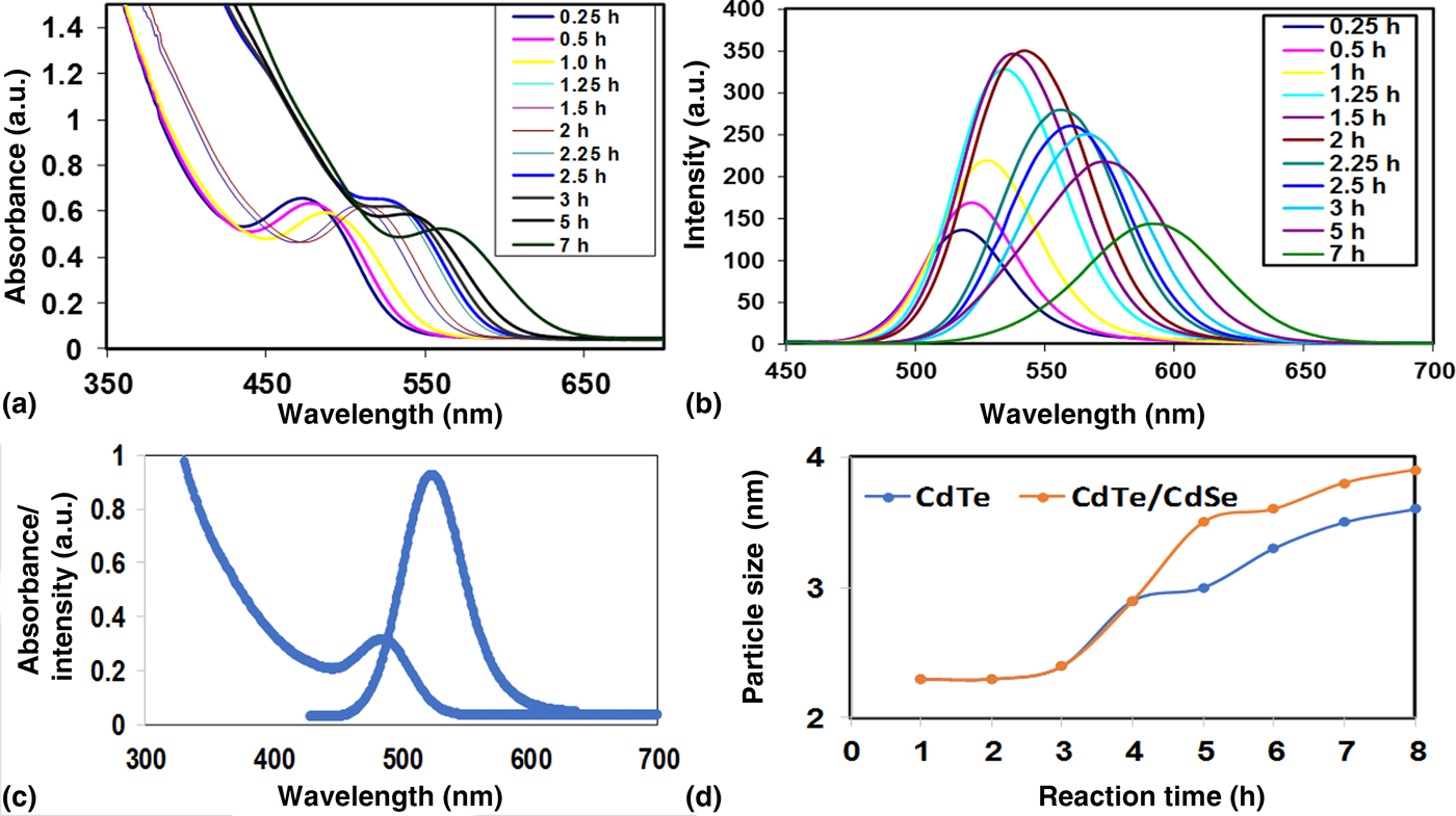
Figure 1 shows the absorption and emission spectra of the as-synthesized CdTe/CdSe QDs. The spectra show a steady increase in absorption and emission wavelength position as the reaction time increased. However, after the addition of the selenium precursor, a remarkable increase was observed indicating the formation of a core–shell structure. The average particle size as calculated using the Yu et al. method,[Reference Yu, Qu, Guo and Peng20] ranges from 2.1 to 3.5 nm. The emission spectra [Fig. 1(b)] show that the remarkable increased in the maximum emission position was accompanied by an enhancement in the fluorescence intensity (about threefold increases compare to the core QDs) after the shell formation. The fluorescence intensity reached maximum intensity at 2 h. The narrow emission spectra and sharp excitonic peaks indicate the formation of materials with focused size distribution and the growth of the crystallites with few electronic defect sites. The decrease in intensity after the 2 h shell formation is due to the formation of larger particles with a wider size distribution (increased emission width). The as-synthesized QDs exhibited band edge luminescence for excitation at 400 nm with the absence of deep or surface trap emission [Fig. 1(c)]. This is attributed to the effective passivation of the surface defects (which are usually associated with the core QDs) by the shell formation. Figure 1(d) shows an increase in particle size as the reaction time increased (for the core) and after the shell formation. The increase in particle size (lower band gap) with time could be attributed to quantum confinement while the increase in particle size after selenium addition is attributed to the shell formation.[Reference Ncapayi, Oluwafemi, Songca and Kodama14, Reference Ncapayi, Parani, Songca, Kodama and Oluwafemi15]
The absorption (a), PL, (b) overlay of absorption and emission spectra at 3 h (c), and the effect of shell on the particle size (d) of the as-synthesized CdTe/CdSe QDs as a function of time.

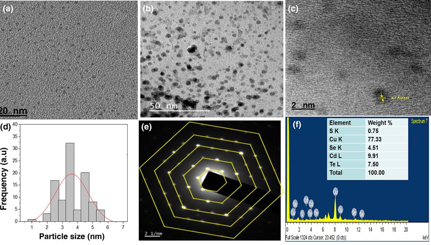
Figure 2 shows the TEM micrographs of the as-synthesized QDs at different reaction times. The micrographs show that the as-synthesized QDs [Figs. 2(a) and 2(b)] are small, spherical in shape, and highly monodispersed. The existence of a lattice fringe on the representative HRTEM image [Fig. 2(c)] confirms the crystallinity of the as-synthesized core–shell QDs. The measured lattice spacing of 3.7 corresponds to the 111 planes of the wurtzite structure. The particle size distributions [Fig. 2(d)] show an average particle size of 3.5 ± 0.60 nm for CdTe/CdSe QDs. The selected area electron diffraction (SAED) pattern of CdTe/CdSe core/shell QDs [Fig. 2(e)] consists of a clear singular reciprocal point arranged in a distinct hexagonal pattern. This further confirms the crystallinity of the material and their existence in the wurtzite phase. The EDS spectrum in Fig. 2(f) shows the presence of the corresponding elements present in the core–shell.
TEM micrograms (a) 0.25 h, (b) 3 h, (c) representative HRTEM, (d) representative particle size distribution, (e) SAED, and (f) EDS of the as-synthesized CdTe/CdSe NPs.

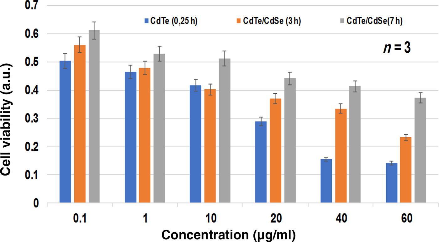
The cytotoxicity of the as-synthesized CdTe/CdSe QDs against LM-8 osteosarcoma cell as presented in Fig. 3 increased with increase in concentration. The cell viability ranges from 50% at a lower concentration of 0.1 µg/mL to 16% at the highest concentration of 60 µg/mL for the core CdTe QDs produced at 0.25 h reaction time [Fig. 3(a)]. However the shell formation (3 and 7 h) shows an increase in cell viability range from 56% to 61% and the 23% to 37% as the concentration increased from 0.1 to 60 µg/mL, respectively. These results show that the as-synthesized QDs exhibited toxicity in a concentration-dependent manner. In addition, the core QDs were more toxic to the cell compared to the core–shell. This decrease in cell viability is attributed to the proper passivation of the core QDs by the shell and the capping agent. This prevents fast dissolution of Cd ion into the cell solution, aggregation of the QDs and instability of the QDs. Nevertheless, the core–shell QDs still exert toxicity on the osteosarcoma cell. This indicates that despite the passivation and stability of the core–shell QDs, the osteosarcoma cell was still more vulnerable to the QDs and hence it can be used as a therapeutic agent against this tumor. The as-synthesized QDs were stable for several months and the cytotoxicity assay was carried out 6 months after the synthesis. This result is different from our previous work on KM-Luc cell line which gave 90% and 60% cell viability at lower (0.1 µg/mL) and higher (60 µg/mL) concentrations respectively after the shell formation. This indicates that the as-synthesized QDs can be used to enhance the therapeutic effect of a photosensitizer used in minimally invasive cancer therapy such as photodynamic therapy (PDT).
The MTT assay of LM8 cell line as a function of the concentration of the core (0.25 h) and core–shell (3 and 7 h).

Conclusion
In conclusion, CdTe/CdSe QDs were synthesized via a simple and reproducible environmentally benign method, and the cytotoxicity action was tested against LM-8 cell line, a murine osteosarcoma cell line associated with bone tumor. The optical analyses show that the as-synthesized QDs are highly fluorescent with band edge emission and focused size distribution. The structural analyses revealed that the as-synthesized QDs are small, spherical in shape, and highly crystalline with wurtzite structure. The cell viability study shows that the as-synthesized QDs are cytotoxic to the LM- 8 cell line. This indicates that these QDs can be used to enhance the therapeutic effect of a photosensitizer used in minimally invasive cancer therapy such as PDT.
Acknowledgments
The authors thank the National Research Foundation (NRF), South Africa under the Nanotechnology Flagship Programme (Grant no. 97983), Competitive Programme for Rated Researchers (Grant no. 106060), and the University of Johannesburg, Faculty of Science Research Committee, and University research Committee for financial support.