Introduction
The different components of physical examination can be viewed as diagnostic tools used by the clinicians at the bedside. An astute clinician should be aware of the diagnostic accuracy of the various physical examination findings he or she uses at the bedside. The apical impulse is one such physical finding, which is used in cardiovascular examination.
The apical impulse that represents the outermost and lowermost point of apical pulsation can give us an idea of the cardiac size. As a non-invasive and readily accessible bedside clinical sign, the apical impulse provides valuable information about the size, contractile function of the ventricles, and a clue to underlying structural heart diseases.Reference Basta and Bettinger1,Reference Perloff2 The importance of cardiac examination has seemingly declined over the years due to availability of diagnostic modalities like echocardiography.Reference Basta and Bettinger1,Reference Alawani, Raj, Sudhakar and Kumar3,Reference Oliver, Hunter, Ikeda and Galletly4
The importance of cardiac physical examination becomes more pronounced in settings where echocardiography is not available. Examination of the location, size, and duration of the apical impulse not only provides valuable clues to the examiner regarding the possibility of heart disease, but it may also point to the type of the underlying cardiac ailment.
Studies done in adults have shown conflicting results regarding the sensitivity and specificity of a displaced apical impulse in detecting ventricular enlargement. A major limitation of these studies has been related to the presence of palpable apical impulse, wherein the proportion of impalpable apical impulse has varied from 5% to 50% in different studies.Reference Eilen, Crawford and O’Rourke5–Reference Mulkerrin, Saran, Dewar, Harding, Bayer and Finucane10
There have been a limited number of research publications on this topic in the paediatric population.Reference Antia, Maxwell, Gough and Ayeni11–Reference Suriyanarayanan, Rajkumar, Muthuswamy and Naveena13 No study has previously been done to evaluate the diagnostic accuracy of a displaced apical impulse in detecting ventricular enlargement in infants. The present study was done to fill this lacuna in the current literature on this topic.
Materials and methods
This cross-sectional observational study was conducted at the Division of Pediatric Cardiology, of a tertiary care hospital in North India. The study was carried out over a 15-month period from April 2023 to June 2024. Infants between 1 month and 1 year of age, who were referred for trans-thoracic echocardiography for various clinical indications, were assessed for eligibility for enrollment in the study. The inclusion and exclusion criteria were as given below
Inclusion Criteria
-
• Infants between 1 month to 1 year of age who are undergoing echocardiography.
Exclusion Criteria
-
• Structural deformities of the chest wall or spine.
-
• History of cardiothoracic surgery.
-
• Presence of a chest tube or significant pleural/pericardial disease.
-
• Cardiac malposition, congenital diaphragmatic hernia, or congenital lobar emphysema.
-
• Infants unable to lie supine or apex not palpable (e.g., meningomyelocele, infection, burn injuries).
-
• Haemodynamically unstable infants precluding adequate physical examination.
-
• Indeterminate ventricular morphology or unbalanced ventricles requiring single-ventricle palliation, precluding assessment of ventricular enlargement by echocardiography.
Sample size was calculated using the sensitivity of displaced apical impulse (59%) from prior literature, a 30% prevalence of ventricular enlargement, 10% precision, and a 95% confidence level.Reference O’Neill, Barry, Smith and Graham6 The estimated minimum sample size was 309 infants.
After satisfying the enrollment criteria informed consent was taken from the parents or guardians. Enrolled children underwent physical examination to localise the apical impulse followed by echocardiography for the assessment of cardiac chamber enlargement.
Localisation of apical impulse
All infants were examined in a quiet, well-lit room in supine position. The measurement of the apical impulse was done as shown in Figure 1.
Showing the method measurement of the horizontal distance of the apical impulse from the mid-sternal line.

The apical impulse was first localised using the palm and then precisely defined using the pulp of the index and middle fingers. Its location was marked and measured in reference to two anatomical landmarks:
-
• Vertical axis: Intercostal space, counted downward from the second intercostal space at the angle of Louis.
-
• Horizontal axis: Distance from the mid-sternal line, measured using a flexible measuring tape.
The line joining the midpoint of the suprasternal notch and the xiphisternal junction defined the midsternal line. A displaced apical impulse was defined as one located below the 4th intercostal space and more than 3 cm lateral to the mid-sternal line.
For inter-observer variability assessment, a subset of 50 infants was independently examined by two examiners; one with over five years of experience and the other with one month of focused training in cardiac palpation. Discordant findings were jointly re-evaluated to reach a consensus; if unresolved, the experienced examiner’s findings were taken as final.
Those infants in whom the apical impulse had been localised were sent for echocardiography.
Echocardiography
Echocardiography was performed by a single experienced paediatric cardiologist using a Philips EPIQ 7 ultrasound system equipped with S12/S8 phased array probes. Standardised paediatric protocols were followed.Reference Lopez, Colan and Frommelt14
Dimensions of the following cardiac chamber were obtained:
-
• Left ventricle: Left ventricle end-diastolic volume, measured using the area-length method in apical four-chamber view.
-
• Left atrium: Left atrium volume, measured in apical 4 chamber and 2 chamber view.
-
• Right ventricle: Minor axis and long-axis dimension: These measurements were done in diastole in right ventricle focused view. The minor axis was measured in the basal region and the long axis was measured from the midpoint of a line connecting the tricuspid annulus hinge points in end diastole to the right ventricle apex.
Z-scores were computed based on body surface area using established normative data.Reference Colan, Lai, Cohen, Geva and Mertens15–Reference Cantinotti, Scalese and Murzi17 A z-score > 2 was considered indicative of chamber enlargement.
Sensitivity, specificity, positive predictive value, negative predictive value, and overall accuracy were calculated for the clinical finding of a displaced apical impulse, using echocardiography as the gold standard. Data were entered and analysed using SPSS version 23.0. Continuous variables were expressed as mean ± standard deviation or median with interquartile range, as appropriate. Categorical variables were presented as frequencies and percentages. Diagnostic test characteristics were calculated with 95% confidence intervals. Correlation between apical impulse displacement and left ventricular end diastolic volume was assessed using Spearman’s rank correlation coefficient. A p-value < 0.05 was considered statistically significant.
Results
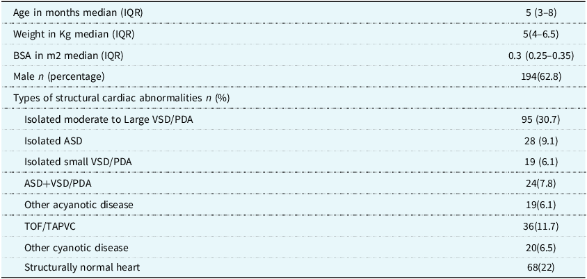
The demographic and echocardiography data of the patients who were enrolled in the study are as shown in Table 1. Flow of patient enrollment in the study was as shown in Figure 2. In 5.9 % of the infants who were evaluated for enrollment, the apex beat was not palpable. They were excluded from the study in accordance with the exclusion criteria. Finally, a total of 309 infants between 1 and 12 months of age were enrolled in the study. Among those who were enrolled, 241(78%) had heart disease on echocardiography. Majority of them had acyanotic heart disease 185 (76.8%), while 68 (22%) of the study participants had structurally normal hearts.
Demography and echocardiography data of the study subjects

IQR = inter-quartile range; BSA = body surface area; ASD = atrial septal defect; VSD = ventricular septal defect; PDA = patent ductus arteriosus.
Showing the flow of patient enrollment in the study.

There was a moderate positive linear correlation between distance of apical impulse from the mid-sternal line and left ventricle end diastolic volume as well as left atrium volume on echocardiography (r = 0.42. p = < 0.001, r = 0.54, p < 0.001, respectively). The correlation between the horizontal distance of apical impulse from the mid-sternal line and right ventricle dimensions was assessed. Mild positive correlation was noted between the horizontal distance of apical impulse from the mid-sternal line with the right ventricle minor axis dimension (r = 0.26, p < 0.001) and moderate correlation with right ventricle long-axis dimension (r = 0.43, p < 0.001).
Based on the data of a previous study by Anita et al., an apical impulse located >3 cm from mid sternal line and >4th intercostal space was defined as a displaced apical impulse.Reference Antia, Maxwell, Gough and Ayeni11 The diagnostic accuracy of a displaced apical impulse using this definition in detecting cardiac chamber enlargement was assessed. The diagnostic accuracy was 71.5% and 64.9% for left ventricle and left atrial enlargement, respectively. For the right ventricle, the results were discordant with the diagnostic accuracy of right ventricle minor axis and long-axis measurements being 49.5% and 68.7%, respectively.
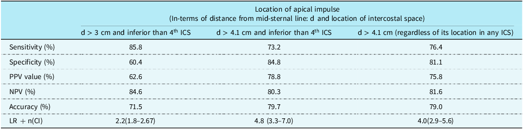
Receiver operating characteristic curve analysis identified that apical impulse located ≥4.1 cm from the mid-sternal line was the most accurate cut-off for detecting left ventricle enlargement. The area under the curve for this cut-off was 0.83 (0.78–0.88).
Therefore, we also analysed the accuracy of a displaced apical impulse using the above cut-off, wherein a displaced apical impulse was defined as being located >4.1 cm from the mid-sternal line and below the >4th intercostal space. Using this new cut-off value to define a displaced apical impulse increased the specificity and diagnostic accuracy while the sensitivity decreased marginally. The diagnostic accuracy of a displaced apical impulse in detecting left ventricle enlargement using different cut-offs are shown in Table 2.
Diagnostic accuracy of various apical impulse displacement criteria for detecting left ventricular enlargement (LVEDV z-score >2)

ICS = intercostal space; PPV = positive predictive value; NPV = negative predictive value; LR + positive likelihood ratio.
Inter-observer variations
For the continuous variable, distance (cm), we calculated the intra-class correlation coefficient to assess agreement, resulting in an inter-correlation coefficient of 0.91. This indicates a very strong positive correlation between the measurements taken by the two observers, showing a high level of consistency in their assessment of the apical impulse distance from the mid-sternal line. For the categorical variable intercostal space, Cohen’s Kappa was used to evaluate agreement, yielding a value of 0.80. This also reflects a strong level of agreement between the observers in categorising the intercostal space of the apical impulse.
Discussion
This study evaluated the diagnostic accuracy of a displaced apical impulse to detect ventricular enlargement in infants between 1 to 12 months of age, using two-dimensional echocardiography as the reference standard. There are no previous studies on this subject in the paediatric population.
The findings demonstrate that a displaced apical impulse, when defined as being located beyond the 4th intercostal space and more than 3 cm from the mid-sternal line, detects an enlarged left ventricle with a diagnostic accuracy of 71.5 %. This cut-off to define a displaced apical impulse was based on a previous study in normal infants.Reference Antia, Maxwell, Gough and Ayeni11
Based on our data, a receiver operating characteristic curve was generated to identify the best cut-off to predict left ventricular enlargement. It showed that the most accurate parameter in detecting left ventricle enlargement was the location of apical impulse >4.1 cm from the mid-sternal line. Using this information, we used an alternative definition of a displaced apical impulse (greater than 4 cm form mid sternal line and inferior to 4th intercostal space) and assessed its diagnostic accuracy. Using this alternate definition, the diagnostic accuracy improved to 79.7 %. When a displaced apical impulse was defined simply as one that was located more than 4 cm from the mid sternal line, regardless of its location in any intercostal space, the diagnostic accuracy in detecting left ventricular enlargement remained high (79%).
Based on the findings of this study, identifying left ventricular enlargement can be done with fairly good accuracy in infants by just measuring the horizontal distance of the apical impulse from the mid-sternal line regardless of its vertical location in terms of the intercostal space. Localising the apical impulse in-terms of its vertical location in a particular intercostal space might not be necessary. This is important because identification of the manubrium-sternal joint is crucial to identifying the correct intercostal space and in infants it is time consuming and sometimes very difficult to identify this landmark.
In this study, 5.9 % of infants who were examined had an impalpable apical impulse and had to be excluded from the study. This is less than compared to adults where different studies have suggested that 10–50% of subjects had an impalpable apical impulse. Reference Eilen, Crawford and O’Rourke5–Reference Mulkerrin, Saran, Dewar, Harding, Bayer and Finucane10
Since diseases involving the right as well as the left ventricle can cause displacement of the apical impulse, both right and left ventricular enlargement were assessed. In this study, right ventricle enlargement was assessed using linear dimension in minor and long axis of the right ventricle. However, there were discrepant findings, with the accuracy of detecting right ventricle enlargement using right ventricle minor axis dimension and right ventricle long-axis dimension being 49.5 % and 68.7 %, respectively. Linear right ventricle dimension using echo-cardiography has significant limitation in estimating the true right ventricular size.Reference Lai, Gauvreau, Rivera, Saleeb, Powell and Geva18,Reference Kim, Srinivasan and Seoane19 Therefore, it was difficult to come to a conclusion regarding the value of a displaced apical impulse in detecting right ventricular enlargement.
This study helps to shed light on the diagnostic accuracy of a commonly elicited physical sign in the course of cardiovascular examination in children. The result suggests that identification of a displaced apical impulse has fairly good accuracy in detecting left ventricular enlargement.
The strengths of this study are its prospective design, standardised methods for precordial palpation, assessment of inter-observer variation, and adequate sample size for the study. These findings hold particular relevance in low-resource settings, where there might be limited access to echocardiography. In such contexts, a carefully assessed displaced apical impulse can serve as an effective triage indicator, guiding early referral for imaging and paediatric cardiology consultation.
Limitations
Several limitations of this study must be acknowledged. First, the patients in this study were a selected group of patients visiting a tertiary care hospital for echocardiography on clinical suspicion of heart disease. The generalisability of the findings to infants in the community setting has to be assessed. Second, the interpretation of right ventricular enlargement on echocardiography was limited by the discrepant findings seen in right ventricle minor and long-axis dimensions making its interpretation difficult. Subjectivity in defining the right ventricle focused view, fore-shortening of view, multiple trabeculations in right ventricle apex may have introduced errors in the measurement of the right ventricle. Finally, the z scores, on the basis of which the cardiac enlargement was defined, were based on a western population and might not be applicable in our population.
Conclusion
This study demonstrates that a displaced apical impulse—defined as being located below the 4th intercostal space and more than 3 cm from the mid-sternal line—is an accurate marker of left ventricular enlargement in infants. Horizontal displacement of the apical impulse 4 cm or more from the mid-sternal line (regardless of the level of intercostal space) is a simpler and more accurate method to identify left ventricular enlargement in this population.
Acknowledgements
The authors thank Mr. Khushal Sagar and Mr. Naveen Kumar.
Financial support
This research received no specific grant from any funding agency, commercial, or not-for-profit sectors.
Competing interests
None.
Ethical standard
The authors assert that all procedures contributing to this work comply with the ethical standards of the relevant national guidelines on human experimentation (India) and with the Helsinki Declaration of 1975, as revised in 2008, and has been approved by the IRB committee of ABVIMS and Dr RML Hospital.