

Introduction
Sauropod dinosaurs achieved the largest body sizes (Alexander Reference Alexander1989) and the longest necks of any terrestrial vertebrate (Taylor and Wedel Reference Taylor and Wedel2013). The long necks and tails are hypothesized to have been held aloft (Coombs Reference Coombs1975) because trackways lack tail drag marks (e.g., Bird Reference Bird1941). Sauropod necks and tails were loaded like cantilevers, beams supported at only one end (i.e., the body) and free at the other end. As a result, the forces of weight support were likely experienced as compression of the vertebral centra and tension of the muscles, tendons, and ligaments attaching to the neural arches, as in mammals (Slijper Reference Slijper1946) and extant reptiles (Hoffstetter and Gasc Reference Hoffstetter and Gasc.1969). Unlike a rigid cantilevered beam, however, necks and tails are segmented, with joints between vertebrae held together in life by muscles, tendons, ligaments, and cartilage. Intervertebral joints permit mobility but are also potential sites of catastrophic dislocation. In order for sauropods to achieve their characteristic large body sizes and long necks, the intervertebral joints required stabilization against increasingly large stresses without compromising the extent of mobility required for survival. One way in which sauropod intervertebral joints may have been stabilized without compromising flexibility is by the evolution of concavo-convex centra (e.g., opisthocoely, procoely) in the neck and, in some lineages, in the tail (J. A. Fronimos and J. A. Wilson unpublished). Previous research on concavo-convex intervertebral joint function in crocodylians has suggested they resist dislocation by shear due to the nesting of one centrum in another and the increased surface area of contact relative to planar joints (Salisbury and Frey Reference Salisbury and Frey2001; J. A. Fronimos and J. A. Wilson unpublished).
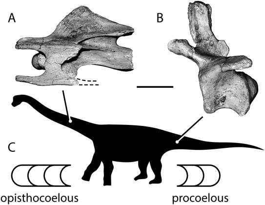
The concavo-convex joints of the sauropod neck and tail show strong, conservative patterns in the polarization of the articular surfaces. Most vertebrae can be classified into two polarities, depending upon which end of the centrum bears the convex articulation (i.e., the condyle) and which the concave articulation (i.e., the cotyle). Opisthocoelous centra (Fig. 1A) have the condyle at the anterior end and the cotyle at the posterior end. Procoelous centra (Fig. 1B) have the reverse anatomical orientation, with the cotyle facing forward and the condyle facing backward. In sauropods, opisthocoely is characteristic of the presacral spine (Fig. 1C). Only the most basally diverging sauropods (i.e., Antetonitrus, Lessemsaurus, Gongxianosaurus) retain the ancestral condition of amphiplatyan (flat-ended) cervical centra, and these taxa are not known from complete cervical series. In all other sauropods, all postaxial cervical centra are invariantly opisthocoelous, as are at least some centra in the pectoral region (e.g., Tazoudasaurus [Allain et al. Reference Allain, Aquesbi, Dejax, Meyer, Monbaron, Montenat, Richir, Rochdy, Russell and Taquet.2004]; Shunosaurus [Zhang Reference Zhang1988]). This pattern extends to the sacrum in macronarians (Wilson and Sereno Reference Wilson and Sereno1998).
Sauropod centrum articular morphology and the anatomical distribution of centrum types. A, Opisthocoelous cervical vertebra of the titanosaur Trigonosaurus. B, Procoelous caudal vertebra of the titanosaur Trigonosaurus. The serial distribution of opisthocoely and procoely is depicted on a sauropod silhouette (C) with schematic representations of the two concavo-convex joint polarities. In both the neck and the tail, the concave articular surfaces of the centra face toward the body. Vertebrae are shown in left lateral view. Dashed lines indicate missing regions of vertebrae. Scale bar, 10 cm (A) and 6 cm (B). Silhouette modified from Wilson and Sereno (Reference Wilson and Sereno1998).

Procoelous anterior caudal vertebrae (Fig. 1C) evolved independently three or four times in sauropods according to our current understanding of their interrelationships: (1) in the Late Jurassic basal eusauropod Mamenchisaurus (Young Reference Young1954; Xing et al. Reference Xing, Miyashita, Zhang, Li, Ye, Sekiya, Wang and Currie2015); (2) within the diplodocoids (Calvo and Salgado Reference Calvo and Salgado.1995); (3) within the titanosaurs (McIntosh Reference McIntosh1990; Wilson and Sereno Reference Wilson and Sereno1998; Upchurch et al. Reference Upchurch, Barrett and Dodson2004); and (4) in a tail of uncertain taxonomic position from the Late Jurassic of Tanzania (Bonaparte et al. Reference Bonaparte, Heinrich and Wild.2000). In some Late Cretaceous titanosaurs, procoely extends to middle and distal caudal vertebrae (McIntosh Reference McIntosh1990; Wilson Reference Wilson2002; Upchurch et al. Reference Upchurch, Barrett and Dodson2004). Opisthocoelous anterior caudal vertebrae are known in only one sauropod, the titanosaur Opisthocoelicaudia (Borsuk-Bialynicka Reference Borsuk-Bialynicka1977). Other patterns occurring in the first caudal vertebra and the distal caudal vertebrae are summarized in J. A. Fronimos and J. A. Wilson (unpublished).
Opisthocoelous cervical centra also occur in some theropod and ornithopod dinosaurs and many ungulates, and procoelous caudal vertebrae occur in crocodylians, squamates, and a few other reptiles. Cervical opisthocoely and caudal procoely occur together only in sauropods (see Nopcsa Reference Nopcsa1930: p. 19; J. A. Fronimos and J. A. Wilson unpublished: Table 1) and trionychid turtles (Williams Reference Williams1950; Hoffstetter and Gasc Reference Hoffstetter and Gasc.1969). Sauropods are distinct from the other taxa listed, in that both the neck and tail are long, heavy cantilevers.
Although cervical opisthocoely and caudal procoely have the opposite anatomical orientation, they are mechanically equivalent; that is, in each case the concave articulation faces toward the body and the convex articulation faces away (Fig. 1C). In the intervertebral joints of the sauropod neck and tail, the forces of weight support are transferred proximally from cotyle to condyle, not from condyle to cotyle (with the exception of Opisthocoelicaudia mentioned above).
Previous Functional Studies of Concavo-Convex Joints
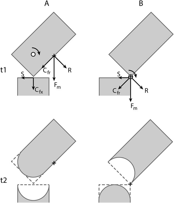
The consistent mechanical polarity of concavo-convex joints in the sauropod neck and tail suggests there is a functional advantage for centra that are concave toward the body compared with the opposite polarity. Several previous studies sought to identify the factors that determine concavo-convex joint polarity. Early proposals by L. Fick (Reference Fick1845) and Henke and Reyher (Reference Henke and Reyher.1874) assumed that muscles play an active role in developing joint shape during ontogeny, leading those authors to the conclusion that the location of muscle insertion sites on the more mobile bone (i.e., the one farther from the body) determines whether its articular surface is convex or concave. This was expanded upon by R. Fick (Reference Fick1890), who, like his predecessors, focused predominately on the concavo-convex joints of the human appendicular skeleton. Summarizing Henke and Reyher (Reference Henke and Reyher.1874), Fick (Reference Fick1890: p. 392) wrote that, “the one articular surface where the muscles insert near the joint will always be ground concave, the joint end with distant muscle attachments, in contrast, will be convex” (translated from the German by J.A.F. and J. Fahlke). In other words, when muscle insertion sites on the free element are located close to the joint, the free element will develop a cotyle, whereas when insertion sites are distally located, it will develop a condyle instead (Fig. 2). The fixed element of the joint will develop a corresponding condyle or cotyle, respectively. Although Fick (Reference Fick1890) described the shaping of the joint surface as a process of abrasion, Fick (Reference Fick1904: pp. 40–44; following Roux Reference Roux1891) later clarified that the same outcome would result from bone growth being inhibited by pressure at the contact points between joint surfaces.
Schematic depiction of the forces interpreted by Fick (Reference Fick1890) to act on two articulated vertebrae when the muscle insertion site on the free element is located far from the joint surface (A) and close to the joint surface (B). Fick (Reference Fick1890) proposed that the location of the muscle insertion site on a moving bone determines which joint surface is convex and which is concave. The applied muscular force (F m) has a rotational component (R) and a compressional component (C fr). At the joint surface, the compressional component can be further decomposed into compression acting on the fixed element (C fx) and shear acting along the joint (S). When the muscle insertion site (black cross) is located far from the joint, the rotational and shear components are widely separated, and the center of rotation (white circle) lies between them in the free element. When the distal end of the free element rotates laterally, the proximal end rotates medially (curved arrow). According to Fick (Reference Fick1890), over time the edges of the proximal end would be ground down, producing a condyle. When the muscle insertion site is located near the joint, the rotational component of force directly counteracts the shear component. As a result, the center of rotation is at the joint surface, and the entire free element rotates laterally. In this case, Fick (Reference Fick1890) believed the proximal end would be ground into a cotyle over time. Adapted from Fick (Reference Fick1890: Fig. 2, reproduced in Supplementary Fig. 1). t1, starting time; t2, time later in ontogeny.
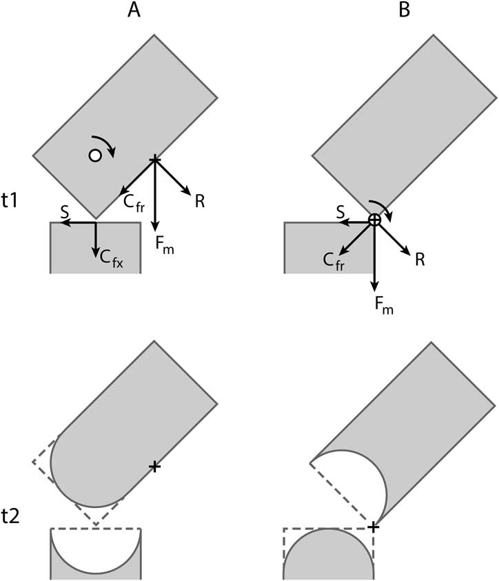
Fick (Reference Fick1890) predicted the relationship between muscle insertion site and joint polarity based on his reconstruction of the forces acting on the free element in each scenario (Fig. 2). He decomposed the applied muscular force (F m) into a rotational component (R) that moves the free element and a compressional component parallel to the free element (C fr). At the joint surface, the compressional force acts oblique to the fixed element and so can be further decomposed into compression acting on the fixed element (C fx) and a shear component (S) acting along the joint surface. When the site of force application is far from the joint (Fig. 2A, t1), the opposing rotational and shear components are widely spaced, and there is a center of rotation in between them within the free element; as the distal end rotates laterally, the proximal end rotates medially. Although not clearly stated by Fick (Reference Fick1890), if the muscle originates from the fixed element, its pull will have a greater medial component the farther distally it inserts on the free element, which will also contribute to the described movement. Given the assumption that muscular action determines joint shape, Fick (Reference Fick1890) concluded that the pressure of this movement would abrade or inhibit bone growth at the corners of the free element, eventually rounding its end into a condyle (Fig. 2A, t2); the corresponding pressure on the central part of the fixed element would generate the cotyle. In contrast, when muscles insert on the free element near the joint, the rotational component of the applied force acts close to the articular surface (Fig. 2B, t1). With the rotational force more directly opposed to the shear component, the center of rotation is located at the joint surface, and the proximal end of the free element must rotate laterally, along with the rest of the bone. The pulling muscle may also have a lesser medial component and a shorter moment arm, reducing the magnitude of the shear. As the free element is pulled laterally over the edge of the fixed element, the corners of the fixed element would be rounded off into a condyle, and the free element would bear the cotyle (Fig. 2B, t2). To test his hypothesis, Fick created flat-ended elements in plaster and attached strings to the free element at insertion sites near to or far from the joint. When the strings were repeatedly pulled side to side by a motor, the articular surfaces were ground into a condyle and cotyle in accordance with the predictions above (see Fick Reference Fick1890: Fig. 5, reproduced in Supplementary Fig. 1).
Although the mechanics described by Fick (Reference Fick1890) are plausible, the hypothesis presented does not explain the distribution of opisthocoely and procoely in the sauropod spine. Sauropod cervical vertebrae are hyperelongated, displacing the ligament and muscle insertions of the neural spine, transverse process, and cervical rib distally. According to the hypothesis, this should result in the formation of proximal condyles, that is, procoelous cervical vertebrae, which is opposite the observed pattern. By the same logic, the shorter caudal vertebrae would be more likely to be opisthocoelous. In the only case of opisthocoelous anterior caudal vertebrae among sauropods, Opisthocoelicaudia, the neural arch morphology closely resembles that of other titanosaurs, showing no evidence for distal displacement of insertion sites (Borsuk-Bialynicka Reference Borsuk-Bialynicka1977). In light of this, we conclude that intervertebral joint polarity in sauropods is not mediated by the position of muscle insertions.
Although the hypothesis that muscle insertion location determines joint polarity does not explain the patterns observed in sauropods, the observation that a free element with distal muscle insertion sites will experience medial rotation of the proximal end is significant. Medial rotation is potentially destabilizing because the proximal end of the vertebra rotates out and away from the joint. As discussed above, any tendency toward dislocation of an intervertebral joint is disadvantageous. We hypothesize that the polarity observed in sauropods is opposite to that predicted by the hypothesis of Fick (Reference Fick1890) to counteract medial rotation and thereby enhance joint stability.
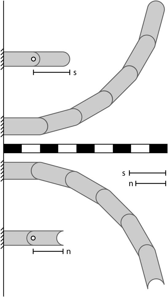
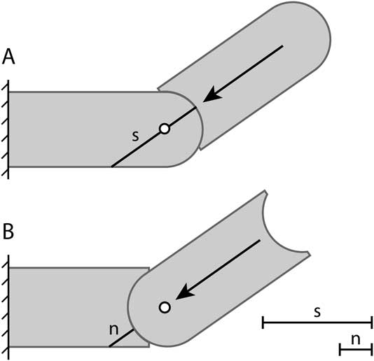
Later investigations into the significance of concavo-convex joint polarity that specifically addressed opisthocoely and procoely were conducted by Troxell (Reference Troxell1925) and Nopcsa (Reference Nopcsa1930). Troxell (Reference Troxell1925) sought to explain the presence of procoelous vertebrae in crocodylians using the observation that the center of rotation (COR) of a concavo-convex joint is invariably found in the condyle. Troxell (Reference Troxell1925) hypothesized two advantages for proximally concave vertebrae over proximally convex ones. The first advantage is that the proximal displacement of the COR in proximally concave vertebrae would increase the displacement of the vertebral column per unit of rotation. According to Troxell (Reference Troxell1925), under these conditions the free element has a greater functional length because it rotates about a point outside itself (Fig. 3, s). As a result, during rotation the distal end of the vertebra is displaced farther from the midline for any given angle of rotation compared with the opposite polarity. In contrast, proximally convex vertebrae rotate about an internal COR, so the functional length is less than the length of the centrum (Fig. 3, n) and displacement per unit of rotation is reduced. Troxell (Reference Troxell1925) assumed that the increase in functional length of crocodylian procoelous caudal vertebrae was additive serially. This means that a swimming crocodylian would generate significantly more displacement, and therefore more thrust, with its tail for a given energy input than would a hypothetical crocodylian with an opisthocoelous tail. This interpretation cannot be correct because it overlooks the effect of successive vertebrae on the functional length of one another. The functional length of a procoelous caudal vertebra must take into account both the gain from displacing the COR into the adjacent proximal vertebra and the loss from being the vertebra into which the COR of the adjacent distal vertebra is displaced. That these effects cancel out can be seen when model centra of equal size are articulated in series and rotated to a uniform angle (Fig. 3); series of proximally concave (Fig. 3, top) and proximally convex (Fig. 3, bottom) centra exhibit identical curvatures and nearly identical displacements from the midline. The displacement of the proximally concave series is greater than that of the opposite polarity, but the displacement difference does not exceed the length of a single condyle regardless of the number of centra in series. The equivalence of the two polarities in terms of displacement was suggested by Gosnold and Slaughter (Reference Gosnold and Slaughter.1977) and demonstrated by Fronimos and Wilson (Reference Fronimos and Wilson.2013). Therefore, the presence of cervical opisthocoely and caudal procoely in sauropods cannot be satisfactorily described as increasing the displacement per unit rotation of the neck and tail.
Influence of the position of the center of rotation (COR) of a concavo-convex joint on the total displacement of the free element, shown with schematic concavo-convex centra. Proximally concave joints (top), such as those of sauropods, have the COR within the fixed element, resulting in a longer functional length (s) and a greater displacement per unit rotation. Proximally convex joints (bottom) have the COR within the free element, resulting in a shorter functional length (n) and a lower displacement per unit rotation. Troxell (Reference Troxell1925) proposed that this effect is additive serially, such that the sauropod-type joint will yield a much greater deflection from the midline. In practice, for a given angle of rotation, the two joint polarities yield nearly identical curvatures. Proximally concave joints have a greater displacement only by the length of a single condyle regardless of the number of vertebrae in series. n, nonsauropod-type joint polarity; s, sauropod-type joint polarity.

The second advantage hypothesized by Troxell (Reference Troxell1925), and also independently suggested by Nopcsa (Reference Nopcsa1930), concerns the distribution of compressive force on a concavo-convex joint. Troxell (Reference Troxell1925) declared that the thrust generated by the crocodylian tail during swimming must act parallel to the distal, free element in any given intervertebral joint, in the process always passing through the COR of the joint (Fig. 4). In the author’s words (Troxell Reference Troxell1925: p. 607), “It must be clearly understood that the thrust through each vertebra is transmitted (lateral muscular pull not considered) in the direction and along the line of the longer axis to the next vertebra in front.” In this scenario, the force acts at an angle to the proximal, fixed vertebra. When the centra are proximally concave, the force acts upon the condyle of the more proximal centrum, which offers a greater thickness of bone to resist the force (Fig. 4, s). When the centra are proximally convex, the force acts upon the thinner bone of the cotylar rim (Fig. 4, n), presenting a greater risk of breakage. Nopcsa (Reference Nopcsa1930: p. 23) presented a similar argument, stating, “In the one case unilateral pressure is taken on the center of the spherical articular surface, but in the other case it is taken on the thin edge of the socket. Now, since in the first case, the same possibility of movement is connected with a much greater mechanical strength than in the second, the first construction is obviously better; at the same time, this is the one in which the joint socket looks toward the fixed part” (translated from the German by J.A.F.). Both authors presented the hypothesis only on the basis of geometric considerations, without experimental modeling. Gosnold and Slaughter (Reference Gosnold and Slaughter.1977) reported an experiment in which alligator vertebrae were loaded in accordance with Troxell’s predictions using a hydraulic press and stressed to the point of failure. When the force was directed from the cotyle into the condyle, the force required to fracture the fixed centrum was three times greater than when it was directed into the cotylar rim; details of the experiment and quantitative results were not provided. Both Nopcsa (Reference Nopcsa1930) and Gosnold and Slaughter (Reference Gosnold and Slaughter.1977) recognized the applicability of this hypothesis to a variety of vertebrates, including sauropods, and Powell (Reference Powell2003) applied it specifically to procoelous caudal vertebrae in titanosaur sauropods. To date, the distribution of forces across a concavo-convex intervertebral joint has not been documented, so Troxell’s assertion that the proximally directed force acts parallel to the more mobile vertebra in a joint requires further assessment.
Resistance of centrum articular surfaces to stress when forces are directed parallel to the free element of a concavo-convex joint, as predicted by Troxell (Reference Troxell1925). If compressional forces act parallel to the free element, proximally concave centra result in a greater resistance to stress. A proximally concave centrum (A) directs forces into the thicker condyle (s). A proximally convex centrum (B) directs forces into the thinner cotylar rim (n), increasing the risk of fracture. Arrows indicate the presumed direction of compression. White circles represent the center of rotation. The labeled black lines indicate the thickness of bone in the fixed element resisting the applied compressive force. Modified from Troxell (Reference Troxell1925). n, nonsauropod-type joint polarity; s, sauropod-type joint polarity.
Functional Hypotheses Evaluated in This Study
Two hypotheses for the functional advantage of proximally concave over proximally convex vertebrae are considered here: (1) proximally concave centra exert stresses against a greater thickness of bone, the condyle, instead of the thin cotylar rim, reducing the risk of fracture; and (2) proximally concave centra prevent the destabilizing rotation of the proximal end of the mobile centrum down and out of joint. The validity of each hypothesis is examined using physical experiments, with models representing proximally concave and proximally convex centra in a jointed cantilever representing either a neck or a tail. The hypothesis that proximally concave centra better resist fracture is tested using photoelastic models that allow direct observation of the magnitude and distribution of strain in joints of each polarity. If the hypothesis is correct, proximally concave centra will exhibit lower strains that are more evenly distributed across the articular surface; the opposite polarity will show large strains concentrated in the cotylar rim. The rotational stability hypothesis is assessed by quantifying the relative stability of the two polarities under a variety of biologically plausible loading conditions. If proximally concave centra confer greater rotational stability, then those models will require much greater forces to rotate out of joint, whereas proximally convex centra will be easily dislocated. The results provide a framework for understanding the regional and phylogenetic distribution of opisthocoely and procoely in sauropods and other vertebrates. Implications for the polarity of concavo-convex joints in other skeletal regions, such as the appendicular skeleton, are discussed.
Materials and Methods
The hypotheses presented above were tested using simplified physical models of vertebral centra that were articulated under a variety of loading conditions. The centra were modeled as schematic forms lacking neural arches that are concavo-convex in only one plane. Because both hypotheses make the same predictions in two-dimensional systems as they do in three dimensions, the experiments could be constrained to a single plane without compromising the validity of the results. The models were first constructed digitally in Adobe Illustrator as two-dimensional concavo-convex shapes, 3 cm tall and 8 cm long in the longest model. Each model has one flat end for mounting to a base and one end that is either concave or convex. The models were designed so that the center of rotation is a uniform distance (6.5 cm) from the flat end. This is necessary so that models of either joint polarity will be subject to the same torque. The distance from the point of application of a force to the center of rotation (i.e., the fulcrum) constitutes the lever arm. Torque is equal to the lever arm multiplied by the magnitude of the applied force. A consequence of holding torque constant between the modeled polarities is that the model centra do not have the same total length (i.e., the distance from the flat end to the farthest extent of the joint surface). If the model centra were made to be the same total length, the position of the center of rotation would differ when the models were articulated with different polarities and torque would become an additional variable.
The concavo-convex articular surfaces were created by the addition or subtraction of a semicircle from an initially rectangular shape. In order to permit a certain extent of rotation without impingement, the cotylar rim was truncated while maintaining the same radius of curvature. As the precise range of motion of sauropod intervertebral joints is not known, three biologically plausible maximum angles of rotation were chosen (15°, 25°, 35°), representing the approximate average presacral range of motion, total range of motion, and caudal range of motion, respectively, on the intervertebral joints of Alligator mississippiensis as measured by J. A. Fronimos and J. A. Wilson (unpublished). To shorten the cotylar rim the correct amount, the concave and convex models were placed in articulation in Illustrator and the concave element was rotated to a given angle. Then, the margins of the concavity were truncated at the point of impingement perpendicular to the length of the centrum. This resulted in three different models with concave articulations, characterized by a different depth of concavity (approximately 40, 30, and 20% of model height, respectively). The models were next imported into Autodesk 3ds Max, where the fit of the articulations was improved by enlarging the concave articulations slightly with a push modifier. The shapes were then extruded to a width of 3 cm to create three-dimensional forms.
The digital models were 3D printed in P430 acrylonitrile butadiene styrene (ABS) plastic on a Dimension Elite 3D printer. The gaps left by the 3D printing process were filled in with a thin layer of epoxy to create smooth surfaces. A one-piece mold of each model was then produced in PolyGel Plat-Sil 73-25 RTV silicone molding rubber. These molds were used to cast the models in EPO-TEK 301 epoxy, a two-component epoxy that cures at low temperature and exhibits photoelastic properties. To conserve material and enhance optical clarity, casts were poured to a thickness of 1 cm rather than the 3 cm width of the ABS plastic models. Batches of epoxy were limited to 25 g at a time; mixing greater amounts resulted in cloudiness and anomalous birefringence under cross-polarized light, likely due to excess heat generation during the reaction between the two component phases. After pouring, the epoxy models were allowed to cure for a minimum of 24 hours at room temperature. Measurements of the models taken with digital calipers showed a variation in width from 0.82 cm to 1.07 cm, including a meniscus of not more than 0.10 cm.
To test each of the two hypotheses, the models were first suspended in articulation loaded as cantilevers with a fixed proximal element and a free distal element that could be rotated relative to it. This was accomplished by fixing one model to a wooden testing rig at its flat end by two 0.62 cm (¼ inch) diameter wooden dowels; these were received by corresponding holes, 0.66 cm apart, drilled into the wooden frame and the flat end of the model. This modular setup allowed the proximal element to be switched out between the convex and concave elements to create either a proximally concave or proximally convex joint. To further stabilize the fixed element against rotation, a steel drawer handle with a clearance of 3.2 cm was attached below it and lined with foam to prevent damage to the epoxy of the model. The free element was suspended from the rig by a tensile element (either a wire or a string; see below) originating proximally on a metal hook. The hook could be moved up and down to vary the angle of rotation of the free element and the insertion angle of the tensile element. To allow this, the hook was screwed into a separate wooden board bolted to the back of the wooden frame via a vertical slot. The board could be moved vertically when the bolts were loosened and, with the bolts tightened, stayed firmly in one position regardless of the load applied. This testing rig is shown in Supplementary Figure 2, where it is configured for the rotational stability experiments.
The hypothesis that proximally concave centra distribute forces more evenly across the articular surface than do proximally convex centra was tested using photoelastic analysis. The principles and applications of photoelastic analysis are detailed by Post (Reference Post1979) and are summarized only briefly here. Photoelastic materials are optically isotropic in an unstrained state, and therefore appear dark when viewed under cross-polarized light. The introduction of strain produces anisotropy in the index of refraction of the material, meaning that light travels through the material at a different speed depending on the direction in which the light vibrates. When viewed under cross-polarized light, rays passing through the material are out of phase and therefore interfere, resulting in birefringence. The greater the strain in the material is, the greater the difference in velocity between the rays will be, and the light observed will have a progressively higher-order interference color. Using this technique, the distribution of strain in a material can be observed and the relative magnitude of stress assessed. The EPO-TEK 301 epoxy used to create the models described above is a photoelastic material with a typical index of refraction of 1.519 at 23°C (Epoxy Technology, Inc. 2012). The models, which were unstrained as a result of the molding and casting process, exhibited a slight biaxial anisotropy with first-order gray interference colors.
Strain-visualization experiments were conducted using the wooden testing frame described above. The epoxy models were modified with the addition of a metal hook on the top and bottom surface, providing a strong attachment for the tensile element and heavy weights. In each model, a 1.1 cm deep hole was drilled into the top and bottom surface, into which was placed a 1.6 cm metal cup hook. The hole was placed 2 cm from the flat end to prevent the strain associated with drilling the hole and applying forces to the hook from overprinting experimentally induced strain at the articular surface. The hooks were set into the holes with epoxy, rather than screwed in, creating a strong bond to the epoxy model without introducing additional strain. For each test, the free element was supported by a tensile element consisting of twisted, 19-strand steel wire with 0.027 cm thick strands. A loop was made at each end of the wire by bending the wire back on itself twice; joining the overlapping segments with two copper crimp sleeves created a strong connection that resisted slipping. This setup being irreversible, a different wire was measured out for each test to provide the correct angle of insertion and angle of rotation, within a margin of error of 2° under the maximum load. The proximal end of the wire was attached to the wooden frame by the movable metal hook described previously. Weights were suspended from the hook on the bottom of the free element by a loop of wire held shut in the manner detailed above. Using iron dumbbell plates in increments of 1.13 kg (2.5 lb), weights of up to 4.54 kg (10 lb) were applied. For each of the two polarities, tests were conducted at two insertion angles (0°, 55°), two angles of rotation (15°, 25°), and four applied weights (0 kg, 1.13 kg, 2.27 kg, 4.54 kg). Two different cotyle depth-to-height ratios (0.3, 0.4) were used for the lower angle of rotation; only the shallower cotyle was used with the 25° angle. After the models were suspended and loaded, polarizing film was placed behind and in front of the models, with the polarization directions of the films perpendicular to one another. The models were backlit with a light source behind and slightly to the left of the modeled joint. The results were photographed with a Nikon D810 DSLR digital camera and a 60 mm Micro Nikkor lens. The camera was placed at a distance of 26 cm from the models and manually focused, with a 1/1.6 second exposure time and an aperture of F22. For each configuration, the resulting strain distributions were compared with one another and with the predictions of the hypothesis put forward by Troxell (Reference Troxell1925).
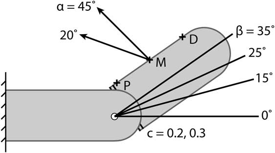
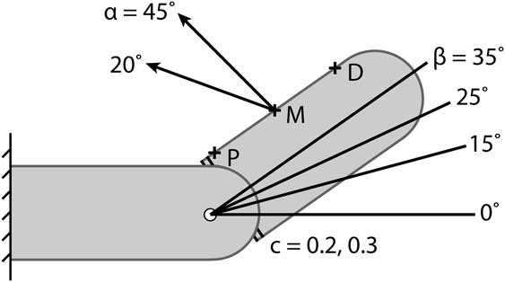
The hypothesis that joints between proximally concave centra are significantly more resistant to failure by rotation was tested by measuring how much weight applied to the proximal end of the free element was necessary for joint failure (Supplementary Fig. 2). The experimental design had several additional parameters (Fig. 5) that were varied to determine how different combinations affected the joint integrity. The tensile element supporting the free centrum, representing ligament, tendon, and muscle, was modeled as a piece of string, the length of which was varied to allow different insertion angles of the tensile element (20°, 45° to the horizontal; Fig. 5, α) and different angles of rotation of the free centrum (0°, 15°, 25°, 35°; Fig. 5, β). The insertion angles reflect uncertainty as to whether sauropods had a crocodylian-style supraspinal ligament that spanned each joint at a low angle (e.g., Schwarz et al. Reference Schwarz, Frey and Meyer2007) or an avian- or ungulate-style nuchal ligament spanning multiple segments at higher angles (e.g., Tsuihiji Reference Tsuihiji2004). The tensile element was attached with tape to the free centrum at one of three possible insertion sites: proximal to the joint (0.50 cm from the COR; Fig. 5, P), middle (3.25 cm from the COR; Fig. 5, M), or distal to the joint (6.00 cm from the COR; Fig. 5, D). These permitted assessment of the hypothesis that distal insertion sites result in medial rotation of the proximal end of the free element, as proposed by Fick (Reference Fick1890). Two different concavity depths were used (depth:height ratios of 0.2, 0.3; Fig. 5, c). The deepest concavity (depth:height ratio of 0.4) was not used because it only permitted rotation up to 15°; the intermediate-depth concavity, though designed to permit rotation up to 25°, could be rotated to 35° with only a barely visible opening of the joint space. For configurations using the less-stable shallow cotyle, an additional set of trials was conducted to determine whether the stabilizing effect of a distal load (e.g., an additional vertebra in series) would negate any stability difference between polarities. To do this, an additional 20 g (the approximate mass of an epoxy model) was suspended from the distal end of the free element at the position of the farthest insertion site from the joint.
Model parameters that were varied during the rotational stability experiments, depicted on a schematic proximally concave joint with the free centrum supported by a tensile element. The alternative, proximally convex polarity, is not pictured. The parameters are the insertion angle of the tensile element (α), the angle of rotation of the free element (β), the depth of the cotyle relative to its height (c), and the insertion site of the tensile element (black crosses). Another variable, the addition of weight to the distal end of the free element, is not shown. The white circle represents the center of rotation. D, distal to the joint; M, middle; P, proximal to the joint.
For conducting each trial, the elements were first placed in articulation, and it was observed whether the free element stayed in joint or whether it disarticulated under its own weight. If the configuration was stable, more weight was suspended from a loop of string affixed on the free element, for consistency of position, at the insertion site nearest the joint (Supplementary Fig. 2). Gram calibration weights were added in 5 g increments until disarticulation occurred or until 250 g had been applied, and the weight at which disarticulation occurred was recorded. When a 20 g weight was suspended from the distal end of the free element, the maximum weight applied to the proximal end was 230 g. When all measurements had been taken, comparisons were made between the results for each polarity, and the influence of each additional variable was compared between polarities. To determine whether a stability difference was significant (i.e., not attributable to model uncertainty), several trials were repeated at a later date; the weights recorded differed from the previous outcomes by up to 15 g. It was also noted that the convex model, bearing the additional weight of a condyle, weighed 11.8 g more than the lightest concave model. Therefore, a difference in stability between model configurations was considered significant only if the weight required for joint failure differed by more than 30 g.
Results
Stress Resistance
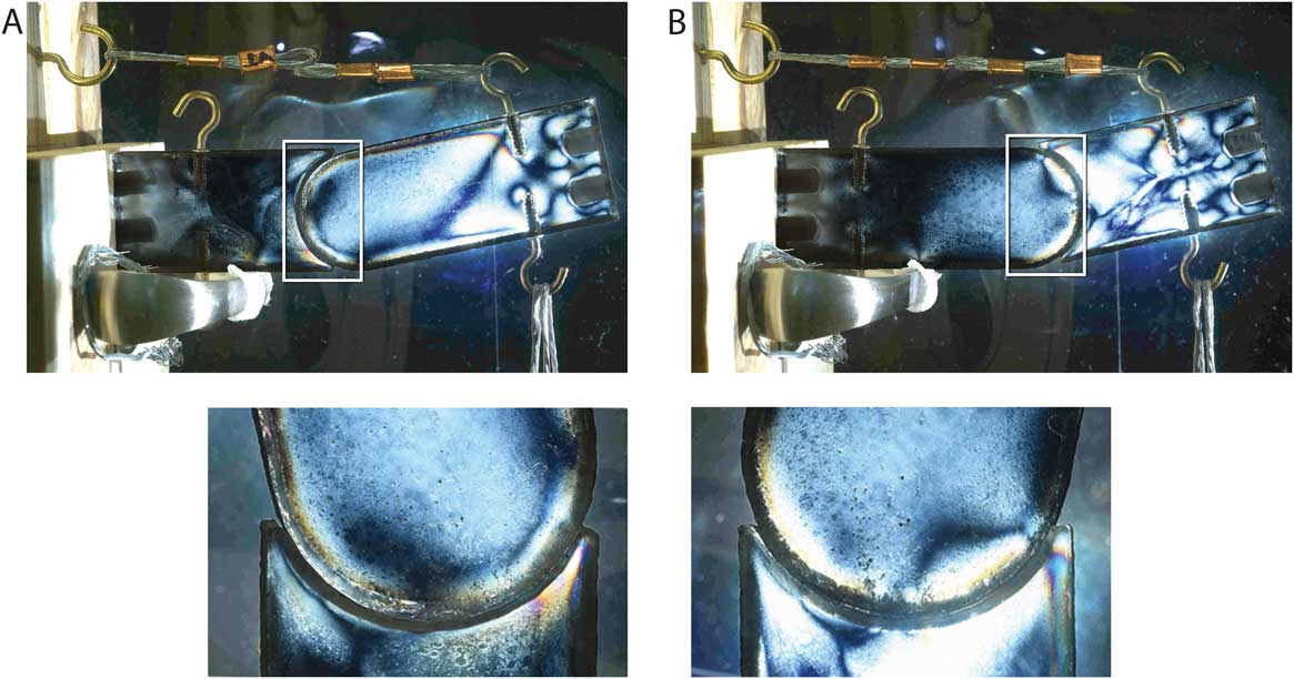
As revealed by the photoelastic analysis, strain on the concavo-convex joints was consistently concentrated in the cotylar rim, regardless of joint polarity (Fig. 6). The distribution and magnitude of strain was dependent on the insertion angle of the tensile element. With a horizontal tensile element, strain was concentrated in the ventral cotylar rim for joints between proximally convex centra (hereafter “proximally convex joints”; Fig. 6A), as predicted. For joints between proximally concave centra (hereafter “proximally concave joints”), strain was not evenly distributed across the articular surface; instead, it was concentrated in the dorsal cotylar rim (Fig. 6B). Within the sensitivity of the analysis, the magnitude of strain was consistent between the two polarities (Fig. 6, lower panels), with one exception. With the free element at a 25° angle of rotation, the strain magnitude was much greater in the proximally convex joint (low third-order interference colors) than the proximally concave joint (middle first-order colors). With a steeply angled tensile element (55° to the horizontal), no difference in strain magnitude was detected between the two polarities. Joints with a shallow cotyle exhibited a strain distribution as described above, but the magnitude of strain was much lower, barely exceeding the first-order gray colors of unstrained epoxy. The strain distribution differed with a deeper cotyle, as the proximally concave joint exhibited strain in both the dorsal and the ventral cotylar rim, and the strain magnitude was nearer to that of joints with a horizontal insertion.
Stress distribution on modeled concavo-convex joints, as visualized using photoelasticity. Proximally convex joints concentrate stress in the ventral cotylar rim (A). Proximally concave joints concentrate stress in the dorsal cotylar rim (B). When compared side by side (lower panels), the two polarities exhibit the same magnitude of strain, as indicated by the order of the interference colors observed. In the lower panels, the joints have been rotated to allow direct comparison of the stressed region of the cotylar rim.
Rotational Stability
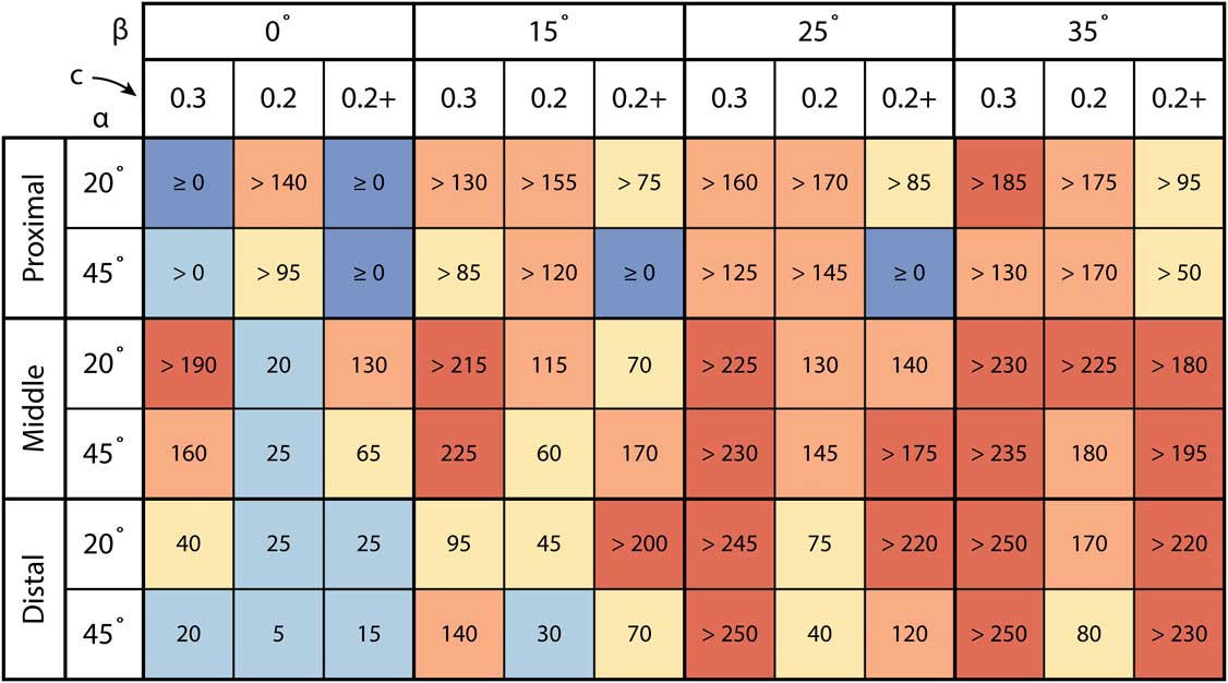
For any given combination of parameter states, proximally concave centra always required more weight for joint failure to occur than did proximally convex centra, except when both polarities were stable at the greatest weight applied (Fig. 7, Supplementary Table 1). The stability difference between the two polarities was significant (i.e., it exceeded the model uncertainty) for greater than 80% of the parameter state combinations (Fig. 7). Proximally concave joints were always stable without applied weight, and an applied weight significantly greater than zero was required for joint failure in all but three of the 72 variable combinations (Supplementary Table 1). Proximally convex joints were stable in an unloaded state for most variable combinations, but for exactly half of the combinations, the weight at which joint failure occurred was not significantly greater than zero (Supplementary Table 1). In a majority of the combinations with proximally concave joints, joint failure did not occur at the maximum applied weight, whereas this was true in only four combinations with proximally convex joints (Supplementary Table 1). Note that, as a result, the stability differences shown in Figure 7 are minima whenever the weight required for failure of the proximally concave joint remains unknown.
Stability differences between proximally concave and proximally convex joints across a range of biologically plausible loading conditions. Each cell indicates how much more weight (in grams) was required for joint failure to occur in the proximally concave joint than the proximally convex one. Warm colors indicate differences that exceeded the model uncertainty, and cool colors indicate differences that did not exceed the model uncertainty. The columns and rows indicate five additional variables: the insertion site of the tensile element (proximal, middle, distal), the insertion angle of the tensile element (α), the angle of rotation of the free element (β), the depth of the concavity relative to its height (c), and the addition of weight to the distal end of the free element (+). A > sign indicates that the measured value is a minimum because the proximally concave joint did not fail at the maximum applied weight (250 g with no distal loading or 230 g with distal loading). Values ≥0 are those for which neither polarity failed at the maximum load.

All five additional parameters examined (Fig. 5) were found to significantly influence joint stability (Supplementary Table 1). Deeper cotyles, tensile element insertion sites nearer to the joint, and the presence of additional weight at the end of the free element increased stability for both joint polarities. The difference in stability between the two polarities was generally higher in the more stable combinations (Fig. 7), most likely because joint failure imposed an absolute lower limit on the stability range, whereas no such upper limit exists. The other two variables had opposite effects depending on the joint polarity (Supplementary Table 1). Increasing the angle of rotation of the free element increased stability in proximally concave joints but decreased stability in proximally convex joints. As a result, the stability difference between joint polarities was least at the lowest angle of rotation (Fig. 7, β). Higher angles of tensile element insertion most often resulted in greater stability in proximally convex joints and lower stability in proximally concave joints, but counterexamples also occurred, and the difference was often less than the model uncertainty. The polarities were generally more similar in stability with a 45° angle of insertion, but the effect was smaller and less consistent than that of the angle of rotation (Fig. 7, α).
Discussion
Stress Resistance
The results of this analysis do not support the hypothesis of Troxell (Reference Troxell1925) that proximally concave (sauropod-type) centra distribute stress more evenly across the joint surface than do proximally convex centra. As seen in the epoxy models (Fig. 6), the cotylar rim is strained regardless of joint polarity, and no significant difference in the magnitude or distribution of strain is detected within the sensitivity of the photoelastic analysis. Troxell (Reference Troxell1925) assumed that forces acting along a series of procoelous caudal vertebrae would be parallel to the distal element in any given joint, and this assumption guided the experiment of Gosnold and Slaughter (Reference Gosnold and Slaughter.1977). The observed strain distribution in joints loaded as cantilevers indicates that forces act oblique to the free element, rather than parallel to it. This orientation is consistent with the resultant vector of the applied forces (i.e., gravity and tensile support). When the tensile element is horizontal, the resultant vector has a 45° angle to the ground. This is approximately orthogonal to the joint surface of the ventral cotylar rim for proximally convex centra and of the dorsal cotylar rim for proximally concave centra. When the tensile element is steeply angled, the resultant vector will have a shallower angle and a smaller magnitude, hence the lower observed strain in the models. Therefore, the strongly conserved pattern of cervical opisthocoely and caudal procoely in sauropods is not well explained by a differential vulnerability to cotylar rim fractures.
The only instance in which the strain magnitude differed between joint polarities occurred with a high angle of rotation, a horizontal insertion, and a shallow cotyle; in this case, the proximally convex joint experienced much greater strain. Observation of this joint revealed that the joint surfaces had lost contact with each other dorsally, creating a narrow gape. This gape appears to have resulted from the distal and ventral rotation and translation of the condyle, as discussed with regard to rotational instability. This outcome is unsurprising in a proximally convex joint having this angle of rotation, insertion angle, and cotyle depth, as this combination of parameter states is particularly unstable (see “Results”). As the condyle was pulled out of joint, the surface area of contact was reduced, increasing stress and, therefore, strain. It is plausible that proximally convex joints are more susceptible to cotylar rim fractures than the opposite polarity in the event of a partial joint failure.
The models are a simplification of real intervertebral joints; the only tensile support in the models represents the supraspinal or nuchal ligament. The consequences of the presence of other soft tissues must, therefore, be considered. In the living animal, it is possible that the action of the epaxial muscles would have redirected the resultant vector so the free element was loaded only in compression. As described by Crisco and Panjabi (Reference Crisco and Panjabi.1991), the human lumbar region requires active muscular stabilization to prevent buckling. In order to be effective for stabilization, the muscular force must be a “follower load”; that is, one that conforms to the lordotic curvature of the lumbar vertebrae rather than acting vertically on the column (Patwardhan et al. Reference Patwardhan, Havey, Meade, Lee and Dunlap.1999). The orientation of the follower load, however, is not parallel to either vertebra participating in a joint; instead, the angle is intermediate between the orientations of the two vertebrae, such that the compressive force acts oblique to each centrum. In addition, the human vertebral column is habitually held upright, such that the long axis of the vertebral column at any point is subparallel to the direction of gravity. In a cantilevered sauropod neck or tail, the vertebrae would have a higher angle to the direction of gravity, so the force necessary to load the vertebral column with a compressive follower load would have been much greater. Thus, although the stress on the cotylar rim in sauropod vertebrae could likely have been reduced by the action of the epaxial musculature, there is no basis to conclude that the stress would be preferentially eliminated from one joint polarity and not the other.
Rotational Stability
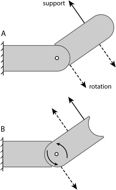
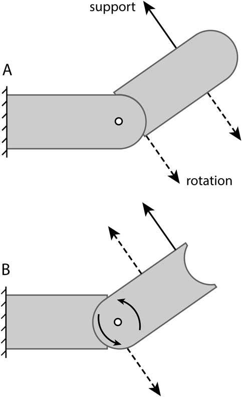
Proximally concave vertebral centra, such as the opisthocoelous cervical and procoelous caudal vertebrae of sauropods, are significantly more resistant to joint failure than proximally convex centra. The difference in stability between the joint polarities can be explained by the position of the center of rotation, which as noted by Troxell (Reference Troxell1925), is always within the condyle. In a proximally concave joint (Fig. 8A), the center of rotation is constrained to lie within the condyle of the more proximal, and therefore less mobile, element. As a result, the free element rotates about a point outside itself. The tensional force that supports the free element pulls it dorsally and proximally, holding the cotyle in joint with the condyle. A force directed toward the ground that is applied at the proximal end of the free element will tend to rotate the entire vertebra ventrally, a movement that is opposed by the tensile element. As demonstrated in the experiments, it is possible for a sufficiently large force acting close to the joint to pull the cotyle away from the condyle, causing joint failure. However, the forces required to do this are typically much greater than those causing joint failure in an equivalent proximally convex joint. In a proximally convex joint (Fig. 8B), the free element rotates about a point within itself. Consistent with the observations of Fick (Reference Fick1890), any dorsiflexion of the distal end of the free element will have the consequence of rotating the condyle ventrally and distally; that is, out and away from the cotyle. Conversely, if a force acts to rotate the condyle out of joint, the distal end of the centrum will rotate dorsally, and the tension in the supporting tensile element will be reduced.
Influence of the position of the center of rotation of a concavo-convex joint on rotational stability. In a proximally concave joint (A), the center of rotation (white circle) is within the fixed element, and the free element rotates about a point outside itself. For a force to pull the free element out of joint (dashed arrows), it must rotate both ends of the free element ventrally, which is directly opposed by the supporting tensile element (solid arrow). In a proximally convex joint (B), the center of rotation is within the free element. If a force pulls the proximal end down and out of joint, the distal end rotates dorsally, which is unopposed by the tensile element. As a result, sauropod-type proximally concave joints are less susceptible to joint failure by rotation.
It is possible to compensate for joint instability with particular morphologies and orientations of the vertebrae and tensile elements. Three of the parameters examined in our experiments bore the same relationship to stability for both joint polarities. Proximal tensile element insertion sites, distal loading of the free vertebra, and deeper cotyles conferred greater stability. A configuration of these parameter states that provides stability to proximally convex joints would increase stability as much or more for proximally concave joints (Fig. 7). The first two variables also involve trade-offs between joint stability and mechanical advantage, as proximal insertion sites decrease the leverage with which the tensile element supports the increasing distal load. Deeper cotyles could decrease joint mobility due to impingement, although a previous study of Alligator did not find a relationship between intervertebral joint morphology and range of motion (J. A. Fronimos and J. A. Wilson unpublished).
The angle of rotation of the free element and the angle of insertion of the tensile element affect the two joint polarities differently, although the insertion angle has an inconsistent influence. The two polarities are generally comparable in stability when the vertebrae are oriented horizontally, but the sauropod-type joints become more stable at higher angles of rotation, whereas the opposite type become less stable. The sauropod-type joints therefore permit a greater mobility to be maintained without sacrificing stability. Some uncertainty surrounds the insertion angle of the supraspinal or nuchal ligament in sauropod cervical vertebrae (e.g., Tsuihiji Reference Tsuihiji2004; Schwarz et al. Reference Schwarz, Frey and Meyer2007); high insertion angles are associated with an avian- or ungulate-type nuchal ligament that spans multiple segments (Tsuihiji Reference Tsuihiji2004; see also Dimery et al. Reference Dimery, Alexander and Deyst.1985). Insertion angles would be steepest in posterior cervical vertebrae, especially in taxa with anterior dorsal neural spines that are much taller than the cervical spines (e.g., Giraffatitan [Janensch Reference Janensch1950]). Osteological evidence led Tsuihiji (Reference Tsuihiji2004) to conclude that the sauropod nuchal ligament did not extend to the anterior cervical vertebrae; even if it did, the length of the neck would cause the insertion angles to be much lower than at the base of the neck. Proximally concave joints, which are generally more stable with lower insertion angles, would be most advantageous in longer necks.
The zygapophyses, cervical ribs, articular capsules, and epaxial musculature, which were not modeled in this study, also have the potential to stabilize joints that are otherwise susceptible to joint failure. These structures provide stabilizing influences regardless of the polarity of a joint, but in proximally convex joints the absence of support provided by the centrum articulations results in greater stress on other intervertebral structures. Zygapophyses limit vertebral rotation, which, depending on zygapophyseal orientation, can prevent the condyle from rotating out of articulation. Zygapophyses have a smaller surface area than do articulations between vertebral centra, so the same applied force results in greater stress. Soft tissues between vertebrae, including the articular capsule between centra and the intercostal ligaments joining cervical ribs, provide tensile forces resisting dislocation of centra. In each case, the sauropod-type polarity results in lower stress because the forces associated with joint stabilization are distributed across the greater surface area of the centrum articulation. A final mechanism for stabilizing proximally convex joints is active contraction of the epaxial muscles, but this is a less energetically efficient solution than the passive support provided by proximally concave joints. Thus, the strongly conservative pattern of cervical opisthocoely and caudal procoely in sauropods would have conferred greater intervertebral joint stability than the opposite polarity without sacrificing mobility, mechanical advantage, or energetic efficiency, and without increasing stress on components of the intervertebral joint system other than the centrum.
Exceptions to the Predominant Sauropod Pattern
Given the advantages of proximally concave intervertebral joints, the existence of exceptions to the dominant pattern requires explanation. These exceptions include the opisthocoelous caudal vertebrae of the titanosaur sauropod Opisthocoelicaudia (Borsuk-Bialynicka Reference Borsuk-Bialynicka1977) and the procoelous cervical vertebrae of crocodylians (e.g., Owen Reference Owen1859a) and pterosaurs (e.g., Owen Reference Owen1859b). How these joints were stabilized is more easily conjectured than what advantage they conferred. For Opisthocoelicaudia, it is possible that the tail was held in a consistently near-horizontal posture with a limited range of motion, as proposed by Borsuk-Bialynicka (Reference Borsuk-Bialynicka1977). Additionally, Borsuk-Bialynicka (Reference Borsuk-Bialynicka1977) noted that the cotyles of caudal centra in Opisthocoelicaudia are consistently deeper than the condyles are long. It should be noted that the sacricaudal joint in titanosaurs and crocodylians is also proximally convex. In Alligator, this joint is nearly immobile, but the cotyle depth is highly variable among crocodylians (J. A. Fronimos and J. A. Wilson unpublished). Posture and mobility cannot explain cervical procoely in crocodylians, as the crocodylian neck has an intrinsic curvature (Virchow Reference Virchow1914; J. A. Fronimos and J. A. Wilson unpublished) with joint mobility typical of the presacral spine (J. A. Fronimos and J. A. Wilson unpublished). In Alligator, cotyle depth is consistently less than condyle length (J. A. Fronimos and J. A. Wilson unpublished).
Procoelous cervical vertebrae in crocodylians might be stabilized by the combination of a short neck and a large, heavy skull. The short cervical centra result in more proximal insertion sites than in sauropods. The heavy terminal load of the skull might serve to hold the procoelous cervical vertebrae in articulation, a tendency that could also have facilitated cervical procoely in pterosaurs. These factors do not explain the condition in Opisthocoelicaudia. In Opisthocoelicaudia, the caudal neural arches resemble those of other titanosaurs, which have procoelous caudal vertebrae, in both the location of muscle insertion sites and the orientation of the zygapophyses. The caudofemoralis longus muscle, which would have imparted shear across the intervertebral joints, also had a similar extent in Opisthocoelicaudia compared to other titanosaurs, as evidenced by the serial extent of the transverse processes (see Gatesy Reference Gatesy1991). Thus, there does not appear to be a particular configuration of ligaments, muscles, and/or skeletal structures that provided additional stabilization. In addition, although the tail of Opisthocoelicaudia is short, there is no heavy terminal load at the distal end (Borsuk-Bialynicka Reference Borsuk-Bialynicka1977), and this morphology is shared by other titanosaurs for which we have complete tail sequences (e.g., Alamosaurus; Wilson Reference Wilson2002). Apart from the potential for active muscular stabilization, it appears that proximally convex cervical or caudal centra have been stabilized in different ways by different taxa.
Opisthocoelous caudal vertebrae and procoelous cervical vertebrae were also derived from different ancestral states in different taxa. Opisthocoelicaudia is nested phylogenetically among titanosaurs with procoelous caudal vertebrae (e.g., Salgado et al. Reference Salgado, Coria and Calvo1997; Wilson Reference Wilson2002), indicating that the joint polarity in the tail underwent an anatomical reversal. In contrast, the procoelous cervical vertebrae of crocodylians were derived from an amphicoelous ancestral condition (Salisbury and Frey Reference Salisbury and Frey2001); this seems also to be the case in pterosaurs, although the transition is less well understood (Sereno Reference Sereno1991). The repeated evolution of a seemingly suboptimal joint polarity in distinct taxa, from different functional precursors, suggests that such joints must confer some offsetting advantage under particular conditions. As noted by Borsuk-Bialynicka (Reference Borsuk-Bialynicka1977), a change in joint polarity can be explained if the fixed end and free end of a vertebral series are reversed. For example, if a sauropod adopted a tripodal posture during feeding or reproduction, the distal end of the tail would be braced against the ground and immobilized relative to the trunk. In this case, caudal opisthocoely would be the stable configuration and procoely comparatively unstable. Although this interpretation is mechanically plausible, there is no anatomical evidence to suggest that Opisthocoelicaudia was more likely to adopt a tripodal stance than other titanosaurs. Apart from caudal opisthocoely, the skeletal characteristics used to suggest a tripodal stance for Opisthocoelicaudia are shared with other titanosaurs, which have procoelous caudal vertebrae (Powell Reference Powell2003). Nopcsa (Reference Nopcsa1930) similarly applied the concept of a reversal of forces to explain presacral procoely in crocodylians and squamates. He reasoned that the sprawling posture and hind limb–driven locomotion of these animals would create forces acting from posterior to anterior. As a result, the head and shoulders would act as the fixed end of the vertebral column, and procoely would be favored. This hypothesis does not account for the diversity of locomotor patterns utilized by crocodylians (e.g., Renous et al. Reference Renous, Gasc, Bels and Wicker2002) and squamates (e.g., Russell and Bauer Reference Russell and Bauer1992). It might, however, offer an explanation for cervical procoely in crocodylians and pterosaurs. During crocodylian swimming and pterosaur flight, propulsion is generated posterior to the neck by the tail or the wings. Thus, the forces acting on the neck should be oriented as proposed by Nopcsa (Reference Nopcsa1930). These explanations remain conjectural in the absence of empirical data on how forces are distributed in the reptilian vertebral column during each of the different locomotor behaviors.
Rotational Stability in the Appendicular Skeleton
The earliest research into concavo-convex joint polarity (e.g., Fick Reference Fick1845; Henke and Reyher Reference Henke and Reyher.1874; Fick Reference Fick1890) was motivated to explain the patterns present in the human appendicular skeleton. If the polarity of concavo-convex intervertebral joints determines their rotational stability, it must be considered whether the same is true of appendicular joints. The tetrapod glenohumeral and femoro-acetabular joints exhibit a strong preferential joint polarity; in each, the stylopodium (viz., humerus, femur) bears a proximally facing condyle that fits into a socket on one or more girdle elements. That the cotyle in these joints faces away from the body seems, at first, to be at odds with the pattern found in the sauropod neck and tail. However, during standing and in the stance phase of locomotion, the limb is braced against the ground, and the body is free to rotate about it. Thus, the weight-bearing autopodium in contact with the ground is the fixed element and the body, which is the more mobile element, bears the concavity. This is mechanically equivalent to the condition in the sauropod neck and tail, in which the distal vertebra in each joint is more mobile and bears the cotyle.
Phylogenetic evidence supports the interpretation that the tetrapod glenohumeral and femoro-acetabular joint polarity provides stability when the limbs are braced against the ground, supporting the body. In the extant outgroups of Tetrapoda, the lungfishes and coelacanths, the glenoid and acetabulum are convex (Rosen et al. Reference Rosen, Forey, Gardiner and Patterson1981; Janvier Reference Janvier1996), indicating that the tetrapod condition evolved via a reversal of joint polarity. This reversal can be explained by the changing forces associated with the transition from fins to limbs. The nontetrapod polarity would be more favorable in a swimming animal because the distal end of the fin is mobile relative to the body; it is this mobile element that bears the cotyle. This also explains why the shoulder girdle bears a convexity in the basal actinopterygian Polypterus, another aquatic vertebrate with monobasal fins (Pollard Reference Pollard1892). As tetrapodomorphs began to use their fins in weight support and for terrestrial locomotion, a switch to the “tetrapod-type” joint polarity would have been necessary to maintain joint stability. This interpretation does not explain the retention of the concave glenoid in secondarily aquatic tetrapods. Nonetheless, further investigation of the functional significance of appendicular joint polarity in sarcopterygian evolution is merited.
The polarity of the human glenohumeral joint is sometimes altered surgically with the implantation of a reverse shoulder prosthesis. This structure is intended to compensate for rotator cuff injury by medializing the center of rotation, increasing the mechanical advantage of the deltoid (Grammont Reference Grammont1979). This is accomplished by replacing the glenoid fossa with a convex element and the humeral head with a concave element. Grammont (Reference Grammont1979) was inspired to invent this procedure by the evolutionary transition of human ancestors from quadrupedalism to bipedalism. As described by Baulot et al. (Reference Baulot, Sirveaux and Boileau.2011: p. 2426), “the freeing of the human upper limb resulted in a functional reversal of the roles of the humerus and the glenoid.” In humans, the hand is not involved in weight support and represents the free end of the forelimb under most conditions. The results of the present analysis suggest that a reverse shoulder joint should be capable of greater rotational stability than the normal anatomical orientation in humans. Conversely, it is expected that a comparable reversal applied to the human femoro-acetabular joint, if such a prosthesis existed, would have a destabilizing influence.
Conclusions
The functional significance of concavo-convex joint polarity has been a source of lingering uncertainty, including with regard to opisthocoelous and procoelous vertebrae. Physical modeling of concavo-convex joints reveals that proximally concave joints are significantly more resistant to joint failure. The strongly conserved pattern of cervical opisthocoely and caudal procoely in sauropod dinosaurs is, therefore, best explained as providing greater passive stabilization of the intervertebral joints than the opposite polarity. The sauropod configuration also would have permitted greater mobility, mechanical advantage of the supporting ligaments and muscles, and distal loading (i.e., a longer neck or tail) without sacrificing stability. The universal condition of cervical opisthocoely in sauropods may have been necessary for the evolution of large body size and neck hyperelongation. Among vertebrates, exceptions to the sauropod-type pattern may be dependent on limited mobility, short necks with large, heavy heads, or energetically costly muscular activity for stabilization. Cervical procoely and caudal opisthocoely could be advantageous if the loading direction of the intervertebral joints was reversed, as during the adoption of a tripodal stance or, perhaps, during particular forms of locomotion. Further research into the loading regimes of intervertebral joints during these behaviors is needed. The concept that concavo-convex joint polarity reflects particular loading regimes provides an explanation for some patterns of polarity in the appendicular skeleton, as in the case of the tetrapod glenohumeral and femoro-acetabular joints, and may offer new insights into the functional evolution of these joints.
Acknowledgments
We wish to thank S. O’Grady and S. O’Malley of the University of Michigan 3D Lab for their help with the design and printing of the models used in this study. W. Sanders provided advice and assistance with material selection and molding and casting. M. Cherney and T. Hudgins aided greatly in the design and construction of the experimental testing rigs. A. Rountrey provided access to equipment and space for conducting experiments. We are grateful to C. Abraczinskas for guidance on the execution of the figures. Access to the sauropod specimens pictured in Figure 1 was provided by D. Campos, R. Machado, and I. Yamamoto (Museu de Ciências da Terra). The color scheme in Figure 7 was developed using ColorBrewer, Version 2.0. We thank the University of Michigan Library’s Buhr Remote Shelving Facility staff for providing high-quality scans of Fick (Reference Fick1890); nineteenth-century literature was also provided by the HathiTrust Digital Library and the Biodiversity Heritage Library. Vielen Dank to J. Fahlke for input on German translations; the translation of Janensch (Reference Janensch1950) was provided through the Polyglot Paleontologist website (http://www.paleoglot.org) courtesy of G. Maier. We thank editor C. Jaramillo and two anonymous reviewers for their helpful feedback on this manuscript. This study represents part of the dissertation of J.A.F., and the authors thank committee members D. Fisher, P. Gingerich, and L. MacLatchy for their input throughout. This project benefited from discussions with University of Michigan Museum of Paleontology graduate students M. Cherney, J. El Adli, L. Joel, K. Loughney, E. Moacdieh, K. Purens, T. Smiley, V. Syverson, and M. Veitch, and many others in the Department of Earth and Environmental Sciences. Funding was provided by the University of Michigan Department of Earth and Environmental Sciences and by a University of Michigan Associate Professor Support Fund award to J.A.W.
Supplementary Material
Supplemental material deposited at Dryad: http://doi.org/10.5061/dryad.1p0q9