Case report
A 13-year-old boy, who had a history of angina since 10 years of age, was admitted for clinical and biological signs of rheumatic fever associated with chest pain. The clinical examination found heart sound with a discrete systolic murmur of mitral regurgitation. The electrocardiogram showed a microvoltage with diffuse repolarisation disorder. The chest radiography showed cardiomegaly with a cardiothoracic ratio of 0.57.
Biologically, he had inflammatory syndrome with a very high sedimentation rate, c protein-reactive level, and antistreptolysin O titre.
Transthoracic echocardiography showed circumferential pericardial effusion (15 mm) with anterosepto-apical hypokinesia of the left ventricle with systolic dysfunction, and a minimal mitral regurgitation.
The patient was treated by corticotherapy and antibiotherapy for 12 weeks with good clinical and biological outcomes.
After 1 year, the outcome was marked by orogenital aphthous ulceration and decreased visual acuity related to intermediate uveitis. The retinal angiography showed vasculitis and the pathergy test was positive. The late appearance of this symptom led to the right diagnosis of Behçet's syndrome.
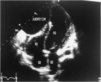
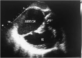
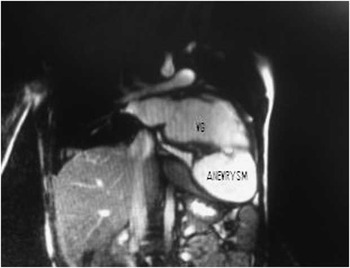
Transthoracic echocardiography showed a hypokinetic dilated cardiomyopathy left ventricular with septo apical and anterior akinesia and severe systolic dysfunction (EF = 38%), with a defect of the inferior septal with a collar measuring 11 mm communicating the left ventricle with a pseudo aneurysm measuring (75 × 80 mm) a seat of spontaneous contrast and small thrombi (Figs 1 and 2). Magnetic resonance imaging showed a giant pseudoaneurysm communicating with the left ventricle (Fig 3). The coronary computed tomography was normal.
Transthoracic echocardiography showing a giant left ventricular pseudoaneurysm in the apical four-chamber plane.

Transthoracic echocardiography showing a giant pseudoaneurysm communicating with the left ventricle.

Magnetic resonance imaging showing a giant pseudoaneurysm of the left ventricle.

The patient had undergone surgical treatment for the pseudoaneurysm with good outcomes.
In 1937, Hulusi Behçet, a Turkish dermatologist, first described a chronic autoimmune disease characterised by orogenital aphthous ulceration and uveitis.Reference Cakir, Eren, Ulka and Nazaroghi 1 The primary pathology is a vasculitis affecting the skin and joints, as well as pulmonary, gastrointestinal, urinary, and nervous systems.Reference Alkim, Gurkaynak, Sezgin, Oguz, Saritas and Sahin 2 Its vascular complications are most frequently manifested as thromboembolism in veins and pseudoaneurysm in arteries.Reference Silistreli, Karabay and Erdel 3
Although pseudoaneurysms are the most common form of arterial involvement in Behçet's disease, we could only find one case reported by Rolland et al.Reference Rolland, Bical and Laradi 4 with Behçet's disease and cardiac pseudoaneurysm. Occasional cases of cardiac pseudoaneurysms have been reported in association with rheumatoid arthritis,Reference Rittoo and Sutherland 5 like in our case; he was treated for rheumatoid arthritis and then he presented with symptoms of Behçet's disease complicated by a giant left ventricular pseudoaneurysm. There are no data on the long-term prognosis of patients with cardiac pseudoaneurysms in Behçet's disease; however, long-term survival could not be expected given the diffuse involvement of cardiac structure and vascular elements.Reference Gurgun, Ercan and Ceyhan 6 Considering its fatality and non-specific manifestations, one should consider cardiac pseudoaneurysms as a potential risk in any patient with Behçet's disease. Thanks to early diagnosis and surgery, our patient was treated successfully.