HISTORICAL NOTE
A cell sheath enveloping the body of the neurons in sensory and autonomic ganglia was mentioned for the first time by Valentin (Reference Valentin1836). In some illustrations of his paper, the nuclei of cells adjacent to the surface of the nerve cell body were clearly shown even though they were misinterpreted as pigment granules. Later Valentin (Reference Valentin1839) provided a more detailed description of this perineuronal sheath and gave a correct interpretation of the satellite glial cell (SGC) nuclei. There followed much debate as to whether or not the perineuronal sheath is a continuous envelope and whether it is built of discrete cells or has a syncytial structure (see Pannese, Reference Pannese1981 for more details); this dispute was brought to an end only when it became possible to examine peripheral ganglia under the electron microscope.
GENERAL ORGANIZATION OF THE SHEATH OF SGCs
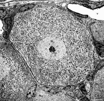
In the sensory and autonomic ganglia of adult vertebrates, SGCs envelope each nerve cell body, its dendrites (when present) and the initial segment of the axon within a sheath which is in turn completely surrounded by the interstitial connective tissue of the ganglion. Thus, a discrete unit consisting of a single nerve cell body together with its sheath is formed and each nerve cell body is completely isolated from every other (Fig. 1). However, clusters of two or three nerve cell bodies enclosed within a common connective envelope have also been found. The percentage of nerve cell bodies arranged in clusters is highest in neonates and decreases progressively with age. In the sensory ganglia of adult animals, the percentages of nerve cell bodies arranged in clusters range from 9.4 in the lizard, 5.6 in the rat to 4.3 in the rabbit (Pannese et al., Reference Pannese, Ledda, Arcidiacono and Rigamonti1991, Reference Pannese, Procacci, Ledda and Conte1993). Sometimes, the nerve cell bodies of a given cluster are separated from one another by a thin sheet of SGCs, in other cases they are in direct mutual contact.
Unit consisting of a nerve cell body (N1) and its sheath of SGCs (SC). This sheath is sharply separated from those enveloping the adjacent nerve cell bodies (N2–N5) by the interstitial connective tissue (ct) of the ganglion. Note that the chromatin is distributed differently in the nuclei of the SGCs and in those of the neurons. nu, nucleolus; v, blood vessel. Spinal ganglion of a young adult rabbit. Scale bar, 500 nm.

The sheath of SGCs may show a variously complicated pattern of organization. It may consist of one single layer of flattened cells or of several layers of cells that overlap in a complex fashion. Both the arrangement of the SGCs and the thickness of the sheath may vary from area to area within the same sheath. The sheath can in some places be as thin as a few dozen nanometers and, therefore, below the limit of resolution of the light microscope; this explains why the sheath may have appeared discontinuous to some optical microscopists. Although in electron microscope preparations the SGC sheath usually appears as a continuous structure, small gaps (from 0.06 to 0.75 µm in length in the rabbit) have occasionally been observed.
SGCs enveloping nerve cell bodies in sensory ganglia sometimes form a myelin sheath. Myelinated nerve cell bodies occur frequently in the acoustic and vestibular ganglia of all classes of vertebrates and occasionally in the other sensory ganglia of bony fishes (for a review see Scharf, Reference Scharf, Möllendorff and Bargmann1958). Electron microscope studies by Rosenbluth and Palay (Reference Rosenbluth and Palay1961), Rosenbluth (Reference Rosenbluth1962) and subsequently other authors showed that the perikaryal myelin may display either the compact structure exhibited by the myelin surrounding the axons or the characteristics of loose myelin. In the latter case, the perikaryal sheath is built by a varying number of lamellae (from 10 to 100 nm in thickness), each consisting of a layer of SGC cytoplasm bounded by its plasma membrane and separated from the nearby lamellae by a narrow space. Compact myelin has a highly regular pattern of organization, whereas in loose myelin the number and thickness of the lamellae and the width of the interval between the lamellae in a given sheath may vary from one region to another. The main characteristics of perikaryal loose myelin are similar to those of the sheaths enveloping certain axons of invertebrates and the developing myelinated nerve fibers of vertebrates. Perikaryal loose myelin differs from common multilayered perikaryal sheaths only in the thickness of the cytoplasmic layers and the location of the cell nuclei: in loose myelin, the cytoplasmic layers are thinner than in common multilayered perikaryal sheaths, and the nuclei of SGCs always lie in the outermost layer of the sheath.
Thus, a wide range of perikaryal sheaths can be found in sensory ganglia, namely, a simple layer of SGCs, multiple layers of SGCs, loose myelin, loose and compact myelin coexisting in the same sheath, and finally, compact myelin. Therefore, the perikaryal myelin sheaths are not basically different from the envelopes of SGCs usually found around the nerve cell bodies, but rather represent variants of them.
SHAPE AND STRUCTURE OF SGCs
As a rule, these cells are laminar and irregular in outline; the long, branching processes described in the past are likely the result of distortion of the cell shape due to technical procedures (Pannese, Reference Pannese1960). Even after long-term in vitro maintenance of sensory ganglia, SGCs display a shape quite similar to that observed in vivo (Shimizu, Reference Shimizu1965; Bunge et al., Reference Bunge, Bunge, Peterson and Murray1967; Waxman et al., Reference Waxman, Dichter, Hartwieg and Matheson1977); in cultures of dissociated ganglia, these cells often appear aggregated according to an epithelial-like pattern. In electron micrographs, SGCs often exhibit fine lamellar expansions and microvilli, which greatly increase their surface area.
In general, the SGC is mononucleate; binucleate SGCs have only occasionally been observed. The nucleus is often elliptic. Chromatin (Fig. 1) usually occurs in centrally located clumps, and in a peripheral band separated from the inner surface of the nuclear envelope by a fibrous lamina (Bunge et al., Reference Bunge, Bunge, Peterson and Murray1967; Pineda et al., Reference Pineda, Maxwell and Kruger1967). In the nuclei of neurons ensheathed by SGCs, by contrast, the chromatin is frequently widely dispersed (Fig. 1).
Free polysomes and cisternae of the granular endoplasmic reticulum are a common occurrence in the cytoplasm (Hess, Reference Hess1955; Wyburn, Reference Wyburn1958); these cisternae may be arranged in stacks (Pannese, Reference Pannese1960; Bunge et al., Reference Bunge, Bunge, Peterson and Murray1967). Tubules and vesicles of the agranular endoplasmic reticulum are less abundant. The Golgi apparatus and centrioles are usually localized in the juxtanuclear region; mitochondria are scattered in the cytoplasm. Autophagic vacuoles, lysosomes, lipofuscin granules and peroxisomes (Citkowitz and Holtzman, Reference Citkowitz and Holtzman1973) can also be observed.
Microtubules (20–30 nm in diameter) and intermediate filaments (about 10 nm in thickness) occur throughout the cytoplasm, generally lying more or less parallel to both the outer and inner surfaces of the satellite sheath. They appear more abundant around the axon hillock and the initial segment of the axon. Glial fibrillary acidic protein is present at low levels in perineuronal SGCs under normal conditions (Woodham et al., Reference Woodham, Anderson, Nadim and Turmaine1989; Ajima et al., Reference Ajima, Kawano, Takagi, Aita, Gomi and Byers2001) but increases markedly following axon injury (Woodham et al., Reference Woodham, Anderson, Nadim and Turmaine1989; Stephenson and Byers, Reference Stephenson and Byers1995; Vit et al., Reference Vit, Jasmin, Bhargava and Ohara2006) or compression of the ganglion (Zhang et al., Reference Zhang, Mei, Zhang, Ma, White and Donnelly2009). The glial fibrillary acidic protein of SGCs (and of Schwann cells) has the same molecular weight as that of astrocytes but its molecular identity is slightly different (Jessen et al., Reference Jessen, Thorpe and Mirsky1984).
Solitary cilia have been observed in SGCs of sensory ganglia (Pannese, Reference Pannese1964). The cilium, which usually originates in the juxtanuclear region from a basal body, projects into a narrow channel of the extracellular space formed by a deep invagination of the plasma membrane and may be directed towards the adjacent neuron or the connective tissue space. These cilia lack the axial pair of microtubules and only show the nine pairs of peripheral microtubules much as do those in neurons, in Schwann cells and in astrocytes. The finding of specific receptors on the solitary cilia of certain neurons (for a review see Pazour and Witman, Reference Pazour and Witman2003) supports the hypothesis that these cilia are sensory organelles. The cilia of SGCs may have a similar sensory function; however, there appear to be no data on the presence of specific receptors on the cilia of these cells.
Glycogen particles (25–30 nm in diameter) and lipid inclusions have been found in SGCs. In mammals and birds, lipid inclusions are usually scarce, and do not exceed 1 µm in diameter; in amphibians, by contrast, at certain periods of the year they can be very numerous and so large (several micrometers) as to distort the outline of the nucleus and even that of the whole cell. SGCs of sensory ganglia contain Ca2+-binding S100 proteins (Vega et al., Reference Vega, Del Valle-Soto, Calzada and Alvarez-Mendez1991; Sandelin et al., Reference Sandelin, Zabihi, Liu, Wicher and Kozlova2004), transporters for glutamate (Schon and Kelly, Reference Schon and Kelly1974a; Hösli and Hösli, Reference Hösli and Hösli1978; Berger and Hediger, Reference Berger and Hediger2000; Keast and Anderson, Reference Keast and Anderson2000), and for γ-aminobutyric acid (Schon and Kelly, Reference Schon and Kelly1974a, Reference Schon and Kellyb), as well as the enzyme glutamine synthetase (Miller et al., Reference Miller, Richards and Kriebel2002; Weick et al., Reference Weick, Cherkas, Härtig, Pannicke, Uckermann and Bringmann2003). This latter catalyzes the conversion of glutamate into glutamine. S100 proteins, the glutamate transporter GLAST and glutamine synthetase are used as molecular markers of SGCs.
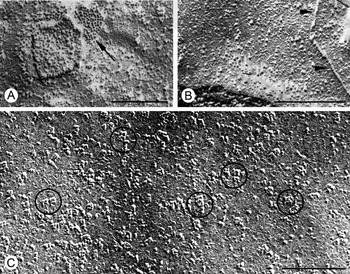
The plasma membrane of SGCs, like that of neuroglial cells in the central nervous system, appears thinner and less dense than that of neurons. Freeze-fractured plasma membranes of SGCs contain, in certain species, assemblies of orthogonally arranged particles (Fig. 2C); the center-to-center spacing of these particles is about 7 nm. Such assemblies were found, for example, in the SGC plasma membrane of the chick (Pannese et al., Reference Pannese, Luciano, Iurato and Reale1977) and rat (Gotow et al., Reference Gotow, Yoshikawa and Hashimoto1985) but not the frog (Matsumoto and Rosenbluth, Reference Matsumoto and Rosenbluth1986). Similar specializations also are found in astrocytes, ependymal cells and other cell types; however, the number per unit surface area of these assemblies is always lower in the plasma membrane of SGCs than in that of astrocytes. These assemblies in the plasma membranes of astrocytes and ependymal cells are composed of aquaporin-4 (Verbavatz et al., Reference Verbavatz, Ma, Gobin and Verkman1997; Rash et al., Reference Rash, Yasumura, Hudson, Agre and Nielsen1998), and are involved in cellular water transport. It is likely that the assemblies present in the SGC plasma membrane also consist of aquaporin-4. Another specialization can be seen in the freeze-fractured plasma membrane of SGCs. This specialization consists of strands, which appear as ridges on the P face (Fig. 2B), and as grooves on the E face. Since these strands show the same morphological characteristics as those composing tight junctions in other tissues, they could be interpreted as focal tight junctions (maculae occludentes) linking adjacent SGCs.
The K+ channels Kir4.1 and Sk3 (Hibino et al., Reference Hibino, Horio, Fujita, Inanobe, Doi and Gotow1999; Vit et al., Reference Vit, Jasmin, Bhargava and Ohara2006), and the acid-sensing ion channel 2 (Kawamata et al., Reference Kawamata, Ninomiya, Toriyabe, Yamamoto, Niiyama and Omote2006) have been identified in SGCs. There is also evidence suggesting that these cells express P2X (Zhang et al., Reference Zhang, Han, Faltynek, Jarvis and Shieh2005) and P2Y (Weick et al., Reference Weick, Cherkas, Härtig, Pannicke, Uckermann and Bringmann2003; Kobayashi et al., Reference Kobayashi, Fukuoka, Yamanaka, Dai, Obata and Tokunaga2006) receptors. SGCs also express receptors to some neurotransmitters (Bernardini et al., Reference Bernardini, Levey and Augusti-Tocco1999), to neurotrophins (Wetmore and Olson, Reference Wetmore and Olson1995; Pannese and Procacci, Reference Pannese and Procacci2002), to endothelins, to orexin-1, to bradykinin, to somatostatin and to laminin-2 (Masaki et al., Reference Masaki, Matsumura, Hirata, Yamada, Hase and Shimizu2001). Laminin-2 receptors are involved in the adhesion to the basal lamina. Finally, cell adhesion molecules have been demonstrated in SGCs (Mirsky et al., Reference Mirsky, Jessen, Schachner and Goridis1986). For a list of the molecules found in these cells, see Hanani (Reference Hanani2005).
Plasma membrane specializations of SGCs. Freeze-fracture replicas (preparation by E. Reale, L. Luciano, and E. Pannese). A gap junction (A, arrow), two ridges recalling strands of tight junctions (B, arrowheads), and assemblies of orthogonally arranged particles (C, some encircled) can be seen on the P face of the split plasma membrane of SGCs. The direction of the carbon-platinum shadowing is from bottom to top. Spinal ganglion of an adult fowl. Scale bars, 250 nm.

RELATIONSHIPS BETWEEN SGCs AND BETWEEN PERINEURONAL SHEATHS
In certain areas, adjacent SGCs contact simply, while in other areas within the same sheath their lamellar expansions overlap and interdigitate in a complex fashion. The gaps, from 15 to 20 nm in width, that intervene between the apposed plasma membranes of adjacent SGCs, open into the light space under the basal lamina at one end and into the narrow interval between the neuron and the sheath at the other. This potential pathway between the interstitial connective tissue and the neuronal surface usually follows a long and tortuous course, a condition that probably has some functional significance.
SGCs are linked to each other by adherent and gap (Fig. 2A) junctions (Pannese, Reference Pannese1974; Pannese et al., Reference Pannese, Luciano, Iurato and Reale1977, Reference Pannese, Luciano and Reale1978; Sakuma et al., Reference Sakuma, Wang, Asai, Tamaki, Amano and Mabuchi2001). The presence of gap junctions between adjacent SGCs has important functional consequences: dye coupling studies have shown that SGCs enveloping a given nerve cell body are functionally coupled (Hanani et al., Reference Hanani, Huang, Cherkas, Ledda and Pannese2002; Pannese et al., Reference Pannese, Ledda, Cherkas, Huang and Hanani2003; Huang et al., Reference Huang, Cherkas, Rosenthal and Hanani2005, Reference Huang, Hanani, Ledda, De Palo and Pannese2006). Information on the connexins that form these junctions in sensory ganglia is relatively scarce. Connexin43 has been identified in the perineuronal SGCs of rat trigeminal ganglion (Vit et al., Reference Vit, Jasmin, Bhargava and Ohara2006) and of mouse spinal ganglia (Procacci et al., Reference Procacci, Magnaghi and Pannese2008). However, it is not known whether the gap junctions linking perineuronal SGCs in sensory ganglia consist exclusively of connexin43 or contain other connexins. Reflexive adherent and reflexive gap junctions, i.e. junctions linking different portions of the same SGC, have also been observed.
As a rule, the sheath of SGCs enveloping a given neuron is completely separated from the neighboring perineuronal sheaths by connective tissue (Figs 1 and 3). Following axon injury, SGCs may extend processes that form bridges connecting previously separate perineuronal sheaths and the gap junctions between satellite cells increase markedly in number (Pannese et al., Reference Pannese, Ledda, Cherkas, Huang and Hanani2003). The marked increase in number of gap junctions correlates with the increase in the incidence of dye coupling among SGCs (Hanani et al., Reference Hanani, Huang, Cherkas, Ledda and Pannese2002). Therefore, SGCs, which in normal conditions are coupled only to other SGCs around a given neuron, following axon injury become extensively coupled to SGCs enveloping other neurons. The increased coupling following axon injury could provide the basis for medium- and long-distance communication between SGCs. Finally, coupling through gap junctions between SGCs significantly increases following compression injury of the ganglion (Zhang et al., Reference Zhang, Mei, Zhang, Ma, White and Donnelly2009).
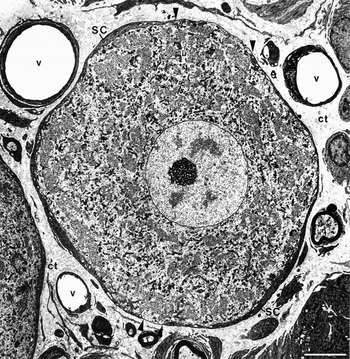
Unit consisting of a nerve cell body and its sheath of SGCs (SC) from an aged rabbit. Note two extensive gaps (between arrowheads) in the sheath of SGCs. ct, interstitial connective tissue of the ganglion; v, blood vessel. Spinal ganglion of an aged rabbit. Scale bar, 500 nm.

BOUNDARIES OF THE SGC SHEATH WITH THE NEURON AND WITH THE CONNECTIVE TISSUE
The boundary between neuron and SGCs is usually complicated by the presence of many projections from both the neuron and the SGCs (Hess, Reference Hess1955; Palay, Reference Palay and Windle1957; Wyburn, Reference Wyburn1958; and subsequently other authors). In single sections, the neuronal projections, usually finger-like, may sometimes appear continuous with the neuronal perikaryon; more frequently, they arise from the neuron at another level, thus appearing as discrete entities completely surrounded by SGCs or located along the interval between the neuron and its SGC sheath. Even in these cases, however, they can be identified because their cytoplasmic matrix is less dense and their plasma membrane thicker and denser than their counterparts in the adjacent SGCs.
SGC projections, which occur less frequently than neuronal ones, invaginate the neuronal profile to variable depths. These invaginations of the neuronal surface, which appear similar to those observed under the electron microscope in the large neurons of invertebrates (see, among others, Rosenbluth, Reference Rosenbluth1963; Coggeshall, Reference Coggeshall1967), possibly correspond to the trophospongium described by Holmgren (Reference Holmgren1901, Reference Holmgren1902).
The interval between the sheath of SGCs and the enclosed neuron shows a rather uniform width (about 20 nm), except where numerous projections arise from the neuron; here it appears uneven and enlarged. Button-like, adherent junctions have occasionally been observed at the boundary between neuron and SGCs (Pannese, Reference Pannese1969; Tennyson, Reference Tennyson and Himwich1970) but, as far as is known, tight or gap junctions have never been described at this interface.
In sensory ganglia, Ehrlich (Reference Ehrlich1886), Ramón y Cajal (Reference Ramón y Cajal1890) and Dogiel (Reference Dogiel1896, Reference Dogiel1897) described a plexus of unmyelinated fibers surrounding some nerve cell bodies. Although the presence of a perineuronal plexus of unmyelinated fibers in sensory ganglia was questioned by Van Gehuchten (Reference Van Gehuchten1892) and Retzius (Reference Retzius1894), the existence of such a plexus has been repeatedly confirmed over the last 20 years. The perineuronal unmyelinated fibers are currently regarded as autonomic fibers of extrinsic origin (for more details, see Matsuda et al., Reference Matsuda, Kobayashi, Terashita, Shimokawa, Shigemoto and Mominoki2005). In normal conditions, only a small proportion of ganglionic sensory neurons is surrounded by autonomic fibers. Sometimes these fibers are enclosed within the sheath of SGCs that envelope the sensory nerve cell body, and very rarely these fibers are directly apposed to the plasma membrane of the sensory nerve cell body, but never make synaptic contact with it. Since the number of autonomic fibers surrounding ganglionic sensory neurons increases notably after peripheral nerve injury, it has been suggested that this autonomic innervation of ganglionic sensory neurons may contribute to the generation and maintenance of some forms of chronic neuropathic pain.
It is important to emphasize that electron microscope studies have never convincingly demonstrated the presence of synaptic contacts on nerve cell bodies or their axons in sensory ganglia, either in situ or in organotypic cultures. Only in dissociated cell cultures have spinal ganglion cells been shown to develop rare synaptic contacts (Miller et al., Reference Miller, Varon, Kruger, Coates and Orkand1970; Lodin et al., Reference Lodin, Faltin, Booher, Hartman and Sensenbrenner1973).
The outer surface of the sheath of SGCs facing the connective tissue is covered by a basal lamina continuous from one SGC to the next; i.e. it bridges the 15–20 nm gaps between adjacent SGCs. Notwithstanding the presence of evaginations and invaginations, the outer surface of the sheath is generally smoother than that facing the neuron. The evaginations of SGCs appear as lamellar or finger-like cytoplasmic projections which extend into the connective tissue and are always surrounded by the basal lamina. In single sections, the profiles of these evaginations may sometimes be seen as discrete entities in the connective tissue since they arise from the SGC sheath at another level; in these cases, they may be distinguished from cytoplasmic processes of the connective tissue cells as the latter are devoid of a basal lamina.
Invaginations, which again always appear lined by a basal lamina, enter the SGC sheath to variable depths. Such invaginations are generally deeper, more numerous and more ramified around the initial segment of the axon than around the neuronal perikaryon; in the former location, they sometimes form a labyrinthine system which may nearly reach the axonal surface. In some amphibians and reptiles, the sheath of SGCs enveloping the initial segment of the axon is divided into two layers; the intervening cleft, which may contain collagen fibrils, communicates with the ganglionic connective tissue space.
QUANTITATIVE RELATIONSHIPS BETWEEN NERVE CELLS AND SGCs
Light microscope studies showed that in spinal ganglia the number of SGCs enveloping each nerve cell body is proportional to the volume of the latter (Pannese, Reference Pannese1960, Reference Pannese1964). Studies carried out with the electron microscope have revealed that in spinal ganglia the volume of the sheath of SGCs is directly proportional to both the volume and surface area of the nerve cell body (Pannese et al., Reference Pannese, Bianchi, Calligaris, Ventura and Weibel1972, Reference Pannese, Ventura and Bianchi1975). The surface of each nerve cell body is highly irregular, its area thus greatly exceeding that of a sphere of equal volume; this explains why the volume of the SGC sheath can be directly proportional to both the volume and the surface area of the nerve cell body.
As shown by the studies quoted above, a quantitative balance exists between nerve cell bodies and their associated SGC sheaths. The factors that maintain this balance are a matter of hypothesis. Neurons are known to differentiate earlier than SGCs during ganglionic development. Moreover, since in experimentally induced enlargement of the nerve cell body there follows an increase in volume of the SGC sheath (Pannese, Reference Pannese1963, Reference Pannese1964; Humbertson et al., Reference Humbertson, Zimmermann and Leedy1969), it is reasonable to suppose that the size and/or surface area of the nerve cell body influence and control the volume of the related sheath of SGCs. By comparing the quantitative results obtained from mammals and reptiles, it appears that, for a given size or surface area of the nerve cell body, the volume of the SGC sheath is greater in mammals than in reptiles (Pannese et al., Reference Pannese, Bianchi, Calligaris, Ventura and Weibel1972, Reference Pannese, Ventura and Bianchi1975). It seems, therefore, that in addition to the size and surface area of the nerve cell body, other factors, for example the metabolic rate of the ganglionic neuron, may influence the volume of the sheath of SGCs.
Mitotic SGCs and SGCs labeled with tritiated thymidine have been observed in sensory ganglia both during body growth (Pannese, Reference Pannese1969) and in normal adult animals (Pannese, Reference Pannese1960; Friede and Johnstone, Reference Friede and Johnstone1967). Under normal conditions, therefore, these cells retain their ability to divide also in adult life. The half life of the SGCs of spinal ganglia has been estimated at about 600 days in normal adult mice (Elson et al., Reference Elson, Ribeiro, Perelson, Simmons and Speck2004). The question arises as to whether SGCs arising by mitosis in the adult animal remain in situ and are added to the existing cell population, or whether they replace degenerated or otherwise lost SGCs. Since the size of the SGC population in a given sensory ganglion does not change appreciably between young adult and adult animals, it seems that new cells replace lost SGCs in these life periods, and the population of SGCs is renewed, although at a very slow rate. For the proliferation of SGCs in old animals, see Age-Related Changes section.
The rate of SGC proliferation in adult life increases under a variety of experimental conditions (e.g. after axonal injury, during neuronal hypertrophy, and following the administration of certain chemicals) as well as under some pathological conditions (see Pannese, Reference Pannese1981 for a more detailed account).
In various in vitro systems, SGCs of sensory ganglia were reported to proliferate (Pomerat et al., Reference Pomerat, Hendelman, Raiborn, Massey and Hydén1967; Lodin et al., Reference Lodin, Booher and Kasten1970; Varon et al., Reference Varon, Raiborn and Tyszka1973). The addition of nerve growth factor to the culture medium increases the mitotic activity in these cells (Ebendal, Reference Ebendal1975), an effect that is probably indirect being mediated by neurons.
Neurons and SGCs, which have been initially separated in dissociated cell cultures, show a clear tendency with time to reassociate and to reestablish their usual in vivo relationships (Waxman et al., Reference Waxman, Dichter, Hartwieg and Matheson1977). The mechanisms underlying this intercellular reaggregation may be surface recognition and/or attractive phenomena exerted by one cell type on the other. One can hypothesize that similar mechanisms are also at work during the in vivo development of sensory ganglia.
AGE-RELATED CHANGES
The structure of SGCs changes little with age. The Golgi apparatus of these cells in rabbit spinal ganglia undergoes neither structural changes, nor peripheral displacement with advancing age; both the total volume of the Golgi apparatus and the mean percentage of cytoplasmic volume occupied by this organelle decrease significantly in old animals compared to young adults (Ledda et al., Reference Ledda, Barni, Altieri and Pannese2003). The mitochondrial structure does not change either, whereas mitochondrial size increases progressively and significantly with advancing age. Both the mitochondrial mass and the mean percentage of cytoplasmic volume occupied by mitochondria decrease progressively and significantly with age (Martinelli et al., Reference Martinelli, Sartori, Ledda and Pannese2007). These findings suggest that the ability of SGCs to produce energy decreases with age and that the reduced ability of sensory neurons to respond to high-energy demands in old age (e.g. see Sylvia and Rosenthal, Reference Sylvia and Rosenthal1979) may be in part due to the diminished contribution of perineuronal SGCs.
In aged animals, the size of the population of SGCs in sensory ganglia is markedly lower than in earlier life periods (Pannese et al., Reference Pannese, Ledda, Martinelli and Sartori1997). In old age, therefore, the SGC proliferation does not seem sufficient to completely replace lost SGCs.
In rabbit spinal ganglia, the general organization of the sheath of SGCs in old animals (Fig. 3) is similar to that in young adults, whereas both the mean volume of the SGC sheaths and the mean value of the volume ratio between the SGC sheaths and the related nerve cell bodies are significantly lower in old animals (Pannese et al., Reference Pannese, Procacci, Ledda and Conte1996; Martinelli et al., Reference Martinelli, Sartori, De Palo, Ledda and Pannese2006). Since SGCs provide trophic and protective support for neurons, it is likely that the significant decrease in their number and the marked quantitative reduction in the volume ratio between the SGC sheaths and the related neurons has negative consequences for neuronal activity.
Furthermore, the gaps in the SGC sheaths are significantly more frequent and extensive (from 0.06 to 7.7 µm in length) in aged rabbits (Fig. 3) than young adults (Pannese et al., Reference Pannese, Procacci, Ledda and Conte1996). Since the nerve cells in sensory ganglia lack the protection of a vascular barrier such as is present in the central nervous system, only the sheath of SGCs controls the traffic of substances from the blood to the ganglionic neurons. Therefore, the greater presence and extent of these gaps in old animals may render the sensory neurons more susceptible to damage by harmful substances from the circulation.
The gap junctions between perineuronal SGCs increase in number with age in rabbit spinal ganglia, whereas the mean size of individual gap junctions remains constant with age (Martinelli et al., Reference Martinelli, Sartori, De Palo, Ledda and Pannese2005). Both the number of gap junctions and the dye coupling between SGCs increase (Huang et al., Reference Huang, Hanani, Ledda, De Palo and Pannese2006), whereas connexin43 expression decreases (Procacci et al., Reference Procacci, Magnaghi and Pannese2008) in mouse spinal ganglia in senescence. A possible explanation for this discrepancy is that in these ganglia additional connexin types other than connexin43 contribute to the gap junctions between SGCs. Further investigations are necessary to determine whether the gap junctions linking perineuronal SGCs in spinal ganglia contain other connexins in addition to connexin43. Permeability characteristics of gap junctions depend on what connexins are present (for reviews see Bruzzone et al., Reference Bruzzone, White and Paul1996; Kumar and Gilula, Reference Kumar and Gilula1996; Nicholson et al., Reference Nicholson, Weber, Cao, Chang, Lampe and Goldberg2000). Hence, the identification of other connexins at the gap junctions between SGCs may provide information on the permeability of these junctions and lead to a better understanding of the functional roles of these cells in spinal ganglia.
ACKNOWLEDGEMENTS
The author thanks Professor L. Luciano (Medizinische Hochschule Hannover, Germany) for helpful discussion concerning orthogonally arranged particles, and for invaluable help in preparing the figures.
Statement of interest
There is no conflict of interest.