Introduction
Sinonasal papillomas are benign mucosal neoplasms. They fall into three categories based on their specific location and histological characteristics: inverted papillomas, fungiform papillomas and oncocytic papillomas. These papillomas are derived from the Schneiderian membrane (the ectodermally derived mucosa lining the nasal cavities and paranasal sinuses),Reference Norris1 and account for 0.4–4.7 per cent of all sinonasal tumours.Reference Lampertico, Russell and MacComb2
Fungiform papillomas account for 45–50 per cent of all sinonasal papillomas.Reference Cheng, Ueng, Chen, Chang and Chen3 They usually present as a unilateral exophytic mass involving the anterior portion of the nasal septum. Patients commonly have symptoms of nasal obstruction and epistaxis. In contrast to inverted and oncocytic papillomas, fungiform papillomas are not considered premalignant, and complete surgical removal remains the mainstay of treatment.
We present a rare case of a bilateral fungiform papilloma with a synchronous verrucous carcinoma of the nasal septum, along with a review of the literature regarding malignant changes in fungiform papillomas.
Case report
A 60-year-old man presented to our out-patient department. His chief complaints were of a bilateral nasal mass along with nasal obstruction and occasional blood-stained nasal discharge, for the previous eight months. There were no associated visual complaints, headache or anosmia.
The patient's anterior rhinoscopic examination revealed a bilateral exophytic, warty, pinkish mass (approximately 1.5 × 1 cm) involving the anterior part of both nasal cavities. On probing the mass, it was found to be attached by a broad base to the cartilaginous part of both sides of the nasal septum. The rest of the endonasal structures and osseo-cartilaginous framework were within normal limits. Systemic examination did not reveal any abnormalities.
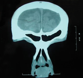
The diagnostic nasal endoscopy findings were consistent with the anterior rhinoscopy findings. Apart from routine investigations, the patient underwent a computed tomography (CT) scan of the paranasal sinuses and a punch biopsy. The CT scan revealed a soft tissue density along the anterior part of both sides of the nasal septum, without septal erosion (Figure 1). The punch biopsy findings were suggestive of a fungiform papilloma. On the basis of these findings, a diagnosis of a bilateral fungiform papilloma was established.
Coronal computed tomography scan of paranasal sinuses showing a soft tissue density along the anterior part of both sides of the nasal septum, without septal erosion.

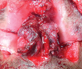
In view of the bilateral involvement of the nasal septum, an external rhinoplasty approach was planned. Intra-operatively, the mass was found to involve mucoperichondrium on both sides, with adhesions to the underlying septal cartilage in places; however, the cartilage was grossly intact (Figure 2).
Intra-operative photograph depicting bilateral fungiform papilloma with normal underlying cartilage (white arrow).

The mass was excised en bloc, including a part of the underlying septal cartilage, taking wide surgical margins. In view of the reported high recurrence rates for this tumour, reconstructive procedures were planned for a later date. The patient's post-operative period was uneventful.
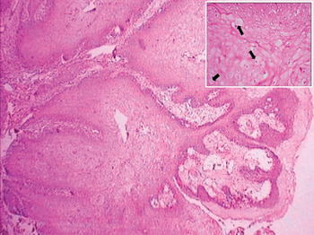
Histological analysis of the surgical specimen revealed fungiform papilloma with coexistent verrucous carcinoma (Figure 3). Since the margins of the specimen were tumour-free, the patient did not undergo any further treatment; however, he was counselled accordingly. At the time of writing, seven months after surgery, the patient was disease-free and was attending regular follow up.
Photomicrograph showing fungiform papilloma. Some rete pegs are large and bulbous, with broad, pushing margins, indicating verrucous change within the papilloma (H&E; ×40). Inset shows koilocytosis in superficial epithelial cells (black arrows), indicating a human papilloma virus cytopathic effect (×400).

Discussion
Fungiform papillomas are also known as septal papillomas, everted papillomas, papillomatosis and Ringertz tumours.Reference Barnes, Tse, Hunt, Brandwein, Curtin, Boeffetta, Barnes, Eveson, Riechart and Sidransky4 They commonly arise from the nasal septum and are generally solitary and unilateral. Bilateral involvement is exceptional.Reference Barnes, Tse, Hunt, Brandwein, Curtin, Boeffetta, Barnes, Eveson, Riechart and Sidransky4
Typical presenting symptoms of fungiform papillomas are unilateral nasal obstruction, epistaxis, an asymptomatic nasal mass and, occasionally, headache. Unusually, our patient had a bilateral nasal mass. He presented with bilateral nasal obstruction and occasional blood-stained nasal discharge.
On gross appearance, fungiform papillomas are exophytic, papillary or warty; grey or pink or tan, non-translucent growths that are attached to the nasal septum by a relatively broad base. Histologically, they have branching fibrovascular stalks covered by hyperplastic non-keratinising squamous to transitional-type epithelium. Excessive and atypical mitoses are not features of fungiform papillomas; when present, these should raise suspicion of squamous cell carcinoma.Reference Zarbo, Torres, Gomez and Pilch5
Histological examination of our patient's lesion revealed a mass lined by stratified squamous epithelium displaying acanthosis and papillomatosis, with extensive koilocytic atypia of the lining. Towards the base, the rete pegs had broad, pushing margins. However, no stromal invasion was seen. These findings correlate with a diagnosis of fungiform papilloma with verrucous carcinoma.
Fungiform papillomas have a possible aetiological link with human papilloma virus, especially types 6 and 11.Reference Sarkar, Visscher, Kintanar, Zarbo and Crissman6 Other postulated aetiologies are allergy, chronic infectious rhinosinusitis and exposure to toxic substances.
• Fungiform papillomas have been described as benign mucosal neoplasms that usually present as a unilateral exophytic nasal mass
• A rare case is presented of a bilateral fungiform papilloma with synchronous verrucous carcinoma
The diagnosis of fungiform papilloma mandates a detailed clinical history investigating environmental exposure, noxious habits, allergies and associated diseases, supplemented with a complete otorhinolaryngological examination. Endoscopic and radiological (i.e. CT and magnetic resonance imaging) examinations are fundamental for tumour study. They assist the evaluation of disease extent, including any erosion of underlying bone, and also aid surgical planning. On CT scanning, fungiform papilloma appears as a soft tissue density along the nasal septum, with little, if any, evidence of septal erosion. Biopsy and histopathological examination confirm the diagnosis.
Malignant transformation of inverted papilloma and cylindrical papilloma is well known; incidences range from 2 to 27 per cent and 4 to 17 per cent, respectively.Reference Sarkar, Visscher, Kintanar, Zarbo and Crissman6 In contrast, the general consensus in most of the literature is that malignant change in fungiform papillomas is rare (Appendix 1). This low association of fungiform papillomas with malignancy distinctly contrasts with that of inverted papilloma, and may be related to location rather than the growth pattern of fungiform papillomas per se.Reference Zarbo, Torres, Gomez and Pilch5 However, there have been studies reporting rare cases of malignancy in fungiform papillomas; the majority have been associated with invasive squamous cell carcinoma.Reference Norris1, Reference Zarbo, Torres, Gomez and Pilch5, Reference Buchwald, Franzman and Toss11
There have been two previously reported cases of verrucous squamous carcinoma of the nasal septum.Reference Pothula and Jones12 In both of these, previous surgical treatment for assumed squamous papilloma had resulted in multiple recurrences, necessitating formal surgical resection. Our patient probably represents the third reported case of a verrucous carcinoma of the nasal septum, but the first presenting with a bilateral mass and a biopsy showing a fungiform papilloma with coexistent verrucous carcinoma.
Seven months after surgery, there had been no recurrence of disease in our patient. This is likely to be the result of complete lesion removal with wide surgical margins.
Conclusion
Although fungiform papillomas are generally not premalignant, occasionally malignant transformation may occur. The recurrence rate of fungiform papillomas is high (22–42 per cent),Reference Zarbo, Torres, Gomez and Pilch5 mandating extensive removal of the lesion. The best opportunity for successful control of papillomas lies with early and complete surgical removal.
Appendix 1. Published comments on malignant transformation of fungiform papilloma
‘Fungiform papilloma are quite unlikely to undergo malignant transformation’Reference Norris1
‘Malignant change in exophytic papilloma is exceptional’Reference Barnes, Tse, Hunt, Brandwein, Curtin, Boeffetta, Barnes, Eveson, Riechart and Sidransky4
‘Whether fungiform papilloma serves as [a] precursor to papillary squamous carcinoma is unproved’Reference Zarbo, Torres, Gomez and Pilch5
‘Malignancy is not associated with fungiform papilloma’Reference Hyams7
‘Everted papilloma shows a benign behavior’Reference Michaels, Hellquist and Michaels8
‘An associated carcinoma [occurring] with septal papillomas is so rare that it must be regarded as a medical curiosity’Reference Batsakis, Blitzer, Lawson and Friedman9
‘Fungiform papilloma has essentially no risk of invasive carcinoma’Reference Lewis, Humphrey, Dehner and Pfeifer10
‘Exophytic papilloma does not have any malignant potential’Reference Buchwald, Franzman and Toss11