INTRODUCTION
Haemogregarines are apicomplexan protozoans and were first reported from the blood of fish by Laveran & Mesnil (Reference Laveran and Mesnil1901) in northern France. Siddall (Reference Siddall1995) classified fish haemogregarines in three genera, including Cyrilia Lainson, 1981 from freshwater fish, and Desseria Siddall, Reference Siddall1995 and Haemogregarina (sensu lato) Danilewsky, 1885 from freshwater and marine fish. This classification relied on haemogregarine development in fish and vector (leech) hosts although such complete development was known for only a few species (see Davies & Johnston, Reference Davies and Johnston2000). Members of the genera Cyrilia and Haemogregarina are partially characterized by intraerythrocytic division, whereas those of the genus Desseria lack this property (see Siddall, Reference Siddall1995).
Haemogregarina (sensu lato) bigemina Laveran & Mesnil, Reference Laveran and Mesnil1901 is a fish haemogregarine that appears in peripheral blood films as intraerythrocytic trophozoites, meronts undergoing binary fission, and paired gamonts, characteristic of the species (Davies et al., Reference Davies, Smit, Hayes, Seddon and Wertheim2004). Remarkably, it is an apparently cosmopolitan haemogregarine, recorded from the blood of at least 96 species of teleost fish, across 70 genera and 40 families (Davies et al., Reference Davies, Smit, Hayes, Seddon and Wertheim2004). On the south and west coasts of South Africa, H. bigemina is found in intertidal fish of the families, Blenniidae, Clinidae and Gobiesocidae (see Davies et al., Reference Davies, Smit, Hayes, Seddon and Wertheim2004; Hayes et al., Reference Hayes, Smit, Seddon, Wertheim and Davies2006). In addition, developmental stages of H. bigemina occur in haematophagous juveniles (pranizae) of the gnathiid isopod, Gnathia africana Barnard, 1914 from South Africa, indicating that the haemogregarine is likely transmitted by arthropods rather than leeches (Davies & Smit, Reference Davies and Smit2001).
A further four fish haemogregarines are currently known from South Africa: Haemogregarina (sensu lato) koppiensis Smit & Davies, Reference Smit and Davies2001 from the evil eye pufferfish, Amblyrhynchotes honckenii Bloch, 1795 (see Smit & Davies, Reference Smit and Davies2001, Reference Smit and Davies2005); Desseria zei Smit & Davies, Reference Smit and Davies2006 from Zeus capensis Valenciennes, 1835 (see Smit & Davies, Reference Smit and Davies2006); a Desseria sp. from flathead mullet, Mugil cephalus Linnaeus, 1758 (see Smit et al., Reference Smit, Eiras, Ranzani-Paiva and Davies2002); and Haemogregarina curvata Hayes, Smit, Seddon, Wertheim & Davies, Reference Hayes, Smit, Seddon, Wertheim and Davies2006 from a variety of intertidal fish and the marine leech Zeylanicobdella arugamensis De Silva, 1963 (see Hayes et al., Reference Hayes, Smit, Seddon, Wertheim and Davies2006).
These latter four haemogregarines and H. bigemina are known only from the cold temperate west coast or the warm temperate south coast of South Africa. In these locations and, likely influencing the life cycles of marine animals, the cold Benguela Current (southern Atlantic Ocean) flowing up the west coast, mixes with the warm Agulhas Current (Indian Ocean) moving down the east coast, providing the relatively warm temperate conditions seen on the south coast (Branch et al., Reference Branch, Griffiths, Branch and Becckley2002). However, the current paper reports for the first time haemogregarines in intertidal teleost fish from the subtropical east coast of South Africa, where the Agulhas Current provides warm water from the tropics and likely accompanying changes in the marine life (Branch et al., Reference Branch, Griffiths, Branch and Becckley2002). Haemogregarines from this locality are compared with H. bigemina and the European fish haemogregarine originally named Haemogregarina quadrigemina Brumpt & Lebailly, Reference Brumpt and Lebailly1904. Unusual developmental stages are observed in the fish, as well as remarkable changes to some host erythrocytes, and a new species of haemogregarine is named. Possible haemogregarine development is observed in pranizae of the gnathiid, Gnathia pilosus Hadfield, Smit & Avenant-Oldewage, Reference Hadfield, Smit and Avenant-Oldewage2008.
MATERIALS AND METHODS
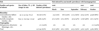
Intertidal fish of three species in two teleost families were collected from rock pools in March, June and September 2006, February 2007 and October 2008 at Tinley Manor (29o27′S 31o17′E) and Sheffield Beach (29o29′S 31o15′E) on the east coast of South Africa, north of Durban (see Table 1). Fish were captured using hand-held nets or a baited hook-and-line, identified using Smith & Heemstra (Reference Smith and Heemstra1995), measured (total length (TL)), examined for haematophagous ectoparasites (see below), and kept in small 20 l aerated aquaria. Prior to further examination, fish were anaesthetized with clove oil (Griffiths, Reference Griffiths2000; Chanseau et al., Reference Chanseau, Bosc, Galiay and Oules2002), and slides with thin blood films (one per fish) were prepared from caudal vein blood, taken using a 25 gauge needle. Films were air dried, fixed with methanol, stained with phosphate-buffered Giemsa solution, and screened with a 100× oil immersion objective on a Zeiss Axioplan 2 photomicroscope (Smit & Davies, Reference Smit and Davies1999; Davies & Smit, Reference Davies and Smit2001). Microscopic images of blood protozoans were captured and measured with Zeiss Axiovision 4.7; measurements were calculated as means ± standard deviation (range). Fish caught on the east coast were also hosts to the ectoparasitic blood-sucking juvenile (praniza) stages of Gnathia pilosus (see Hadfield et al., Reference Hadfield, Smit and Avenant-Oldewage2008). Gnathiids were collected from fish with brushes or broad mouthed pipettes, drained of residual seawater on absorbent paper and each was crushed, smeared, fixed and stained for screening either immediately or at 1–30 days post-feeding (d.p.f.) on fish blood. Methods for preparing and screening gnathiids d.p.f. followed Davies & Smit (Reference Davies and Smit2001) and Hayes et al. (Reference Hayes, Smit, Seddon, Wertheim and Davies2006). Following examination, fish were released at the site of capture.
Fish identity, mean length ± SD and range (in mm), number infected with haemogregarina (prevalence %) and date of capture.

TL, total length; SD, standard deviation.
RESULTS
Infections with haemogregarines
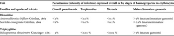
Haemogregarines were found in two members of Blenniidae, the horned rockskipper Antennablennius bifilum Günther, 1861 and the maned blenny Scartella emarginata Günther, 1861, and a single member of the Triptrygiidae, the hotlips triplefin Helcogramma obtusirostre Klunzinger, 1871. Parasites were restricted to erythrocytes, with trophozoites, meronts, and gamonts present, but no intraleucocytic stages (see Laird, Reference Laird1953). Prevalence of haemogregarines in host fish was 67% overall; A. bifilum showed 77% prevalence, S. emarginata, 53%, and the single H. obtusirostre was also parasitized (Table 1). Parasitaemias involved <1% of erythrocytes in A. bifilum and S. emarginata, and ~2% of erythrocytes in H. obtusirostre (Table 2). Overall, trophozoites and meronts occurred in <0.1% of erythrocytes, while gamonts occupied ~1% of erythrocytes (Table 2). Gnathiids (Gnathia pilosus) were the only haematophagous ectoparasites found on the fish, and their abundance varied among the three fish hosts (Table 3). Prevalence of gnathiids on fish was 31% overall; A bifilum showed 44% prevalence, S. emarginata 12% and the single H. obtusirostre was also parasitized (Table 3). Of the three stages of pranizae found on fish (P1, P2 and P3), P1 juveniles were the most prevalent (80%), with P2 and P3 stages contributing 18% and 2% respectively (Table 3).
Fish species, parasitaemias with haemogregarines and percentages of haemogregarine life stages found in erythrocytes.

Classification of Gnathia pilosus pranizae from fish by stage collected from March 2006 to October 2008. P1, Praniza 1; P2, Praniza 2; P3, Praniza 3.

SYSTEMATICS
MATERIAL EXAMINED
Voucher material: in the collection of the South African Museum, Cape Town (Antennablennius bifilum blood film SAM A25102 with trophozoites, meronts, mature gamonts from Tinley Manor (29o27′S 31o17′E), collected by M.L. Ferreira, 22 March 2006. Scartella emarginata blood film SAM A25103 with trophozoites, meronts, mature gamonts from Tinley Manor, collected by M.L. Ferreira, 24 March 2006. Antennablennius bifilum blood film SAM A A25104 with trophozoites, meronts, mature gamonts from Sheffield Beach (29o29′S 31o15′E), collected by M.L. Ferreira, 14 February 2007. Scartella emarginata blood film SAM A25105 with trophozoites, meronts, mature gamonts from Sheffield Beach, collected by M.L. Ferreira, 15 February 2007). Other material: in the collection of N.J. Smit (80 A. bifilum blood films; 42 S. emarginata blood films).
DESCRIPTION
Trophozoites
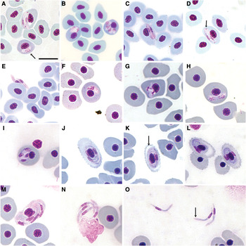
In both fish species (Antennablennius bifilum and Scartella emarginata) intraerythrocytic trophozoites occurring singly in erythrocytes; elongate, with broad anterior and narrower, roundly pointed, posterior pole (Figure 1A). From A. bifilum 5.5 ± 0.6 (4.5–6.3) by 2.1 ± 0.4 (1.3–2.8) µm (N = 20), from S. emarginata 5.0 ± 0.9 (3.2–6.8) by 1.9 ± 0.5 (1.1–3.0) µm (N = 20). Cytoplasm stained light blue. Nucleus situated predominantly centrally within the trophozoite body, stained purple, with chromatin loosely arranged. Nuclei 2.7 ± 0.6 (1.7–4.1) by 1.6 ± 0.4 (1.1–2.6) µm (N = 20) in A. bifilum, 2.4 ± 0.4 (1.8–3.3) by 1.4 ± 0.4 (0.7–2.4) µm (N = 20) in S. emarginata.
Micrographs of Giemsa stained blood films from Antennablennius bifilum Günther, 1861 and Scartella emarginata Günther, 1861 showing the Haemogregarina bigemina-like organism (A–E), and of Helcogramma obtusirostre Klunzinger, 1871 illustrating the Haemogregarina kunegemina sp. nov. (F–O). (A.) Trophozoite (arrowed) and paired, mature gamonts in host erythrocyte above within parasitophorous vacuole (visible just below host nucleus); (B) meronts producing two or four individuals (the latter seen only in S. emarginata); (C, D) paired matured intracellular gamonts (arrow indicating granule at posterior extremity); (E) Extracellular gamonts; (F) trophozoite; (G–I) meronts apparently producing two or four merozoites; (J, K, L) four mature gamonts within host erythrocytes demonstrating spiked periphery and de-haemoglobulinization (arrow in K); (M) eight gamonts in erythrocyte; (N, O) extracellular gamonts (arrow indicating pinkish cap). Scale bar: A–O = 10 µm.

Meronts
Meronts mostly singly in erythrocytes in both fish species, occasionally in pairs, elongate, one pole slightly broader than the other (Figure 1B). From A. bifilum 6.1 ± 0.8 (5.2–8.0) by 2.5 ± 0.4 (1.8–3.6) µm (N = 20), from S. emarginata 5.7 ± 0.9 (3.3–7.1) by 2.6 ± 0.6 (1.3–4.0) µm (N = 20). Cytoplasm stained pale blue, without granules. Nuclei stained purple, often with stranded chromatin, 2.9 ± 0.4 (1.9–3.6) by 1.7 ± 0.4 (1.0–2.5) µm (N = 20) from A. bifilum, 2.5 ± 0.6 (1.5–3.8) by 1.8 ± 0.6 (0.7–2.7) µm (N = 20) from S. emarginata. Meronts apparently producing two merozoites in A. bifilum, but two to four in S. emarginata (Figure 1B). Individual merozoites 3.6 ± 0.8 (2.1–4.6) by 2.3 ± 0.5 (1.9–3.0) µm (N = 10), with nuclei, each 2.4 ± 0.6 (2.0–3.8) by 1.0 ± 0.4 (0.8–2.1) µm (N = 10).
Gamonts
Gamonts in pairs in erythrocytes (Figure 1C, D) in both fish species, with free gamonts resembling those in erythrocytes (Figure 1E); slightly broader anterior than posterior poles, anterior generally rounded, occasionally pointed; posterior straight or re-curved and always narrow; in A. bifilum 8.0 ± 0.5 (6.7–8.9) by 1.3 ± 0.2 (0.8–1.8) µm (N = 50) in S. emarginata 9.0 ± 1.0 (5.4–10.2) by 1.4 ± 0.3 (0.7–2.3) µm (N = 50). Cytoplasm stained pale blue or pinkish, granule at posterior extremity for some gamonts from S. emarginata. Nuclei with loosely arranged chromatin, situated approximately halfway or more posterior in parasite body, 2.7 ± 0.4 (1.9-3.6) by 1.0 ± 0.2 (0.6–1.3) µm (N = 50) from A. bifilum, and 2.6 ± 0.6 (1.6–5.0) by 1.0 ± 0.2 (0.6–1.4) µm (N = 50) from S. emarginata. Tight-fitting parasitophorous vacuole visible in some instances (Figure 1A).
REMARKS
In Antennablenius bifilum and Scartella emarginata blood films, mature gamonts appeared paired within erythrocytes, resembling those of H. bigemina (see Laveran & Mesnil, Reference Laveran and Mesnil1901; Davies et al., Reference Davies, Smit, Hayes, Seddon and Wertheim2004). Trophozoites from A. bifilum and S. emarginata were close to each other in size, while meronts from A. bifilum were slightly larger than in S. emarginata. Importantly, meronts from S. marginata produced four as well as two merozoites, whereas those from A. bifilum apparently gave rise to only two. This former characteristic is not typical of H. bigemina from its type hosts (see Davies et al., Reference Davies, Smit, Hayes, Seddon and Wertheim2004) and is not reported from other South African infections of this haemogregarine (Smit & Davies, Reference Smit and Davies1999; Davies & Smit, Reference Davies and Smit2001; Hayes et al., Reference Hayes, Smit, Seddon, Wertheim and Davies2006). Furthermore, there was no evidence of mixed H. bigemina-like and H. kunegemina sp. nov. infections in the current study to account for the extra merozoites seen in S. emarginata (see below). However, division of an H. bigemina-like organism in Australian fish also produces occasionally up to 4 individuals in a single erythrocyte (Smit et al., Reference Smit, Grutter, Adlard and Davies2006), highlighting apparent variations in the development of this haemogregarine across continents.
Gamonts in A. bifilum (6.7–8.9 by 0.8–1.8 µm) were overall smaller than in S. emarginata, (5.4–10.2 by 0.7–2.3 µm). The latter may have represented more mature stages, because of their increased length and width, a granule at the posterior pole, and a visible parasitophorous vacuole in some instances; thus their appearance was similar to that of mature H. bigemina gamonts in a variety of New Zealand intertidal fish (Laird, Reference Laird1953), although gamonts from New Zealand material were somewhat longer (9.1–14.9 by 1.0–2.2 µm) than those from S. emarginata. The type material for H. bigemina described by Laveran & Mesnil (Reference Laveran and Mesnil1901) in intertidal blennies Lipophrys pholis (Linnaeus, 1758) and Coryphoblennius galerita (Linnaeus, 1758) from St Martin's cove, near Cape de la Hague, France also demonstrated longer gamonts (12 by 1.5–2 µm) than those in S. emarginata and division produced only two individuals.
Smit & Davies (Reference Smit and Davies1999) reported H. bigemina from Clinus superciliosus (Linnaeus, 1758) and Clinus cottoides Valenciennes, 1836 from Jeffreys Bay on the south coast of South Africa, and gamonts from A. bifilum and S. emarginata from the east coast are close in size to those from both C. superciliosus (9.0–13.2 by 1.2–2.2 µm) and C. cottoides (9.8–12.3 by 1.6–2.5 µm). Similar H. bigemina infections were later documented from De Hoop Nature Reserve, also on the south coast, infecting Blennioclinus brachycephalus (Valenciennes, 1836), Chorisochismus dentex (Pallas, 1769), C. superciliosus, C. cottoides, and Parablennius cornutus Linnaeus 1758 (see Davies & Smit, Reference Davies and Smit2001; Hayes et al., Reference Hayes, Smit, Seddon, Wertheim and Davies2006) and from Clinus agilis Smith, 1931 at Mouille Point on the west coast of South Africa (Hayes et al., Reference Hayes, Smit, Seddon, Wertheim and Davies2006). In the present study, it is concluded that the haemogregarine found in A. bifilum and S. emarginata, despite its unusual development in the latter fish species, is also likely H. bigemina, making this the first record of this haemogregarine in these fish hosts and the first report of it from the east coast of South Africa.
TYPE MATERIAL
Hapantotype: in the collection of the South African Museum, Cape Town (Helcogramma obtusirostre blood film SAM25106 with trophozoites, meronts, mature gamonts from Sheffield Beach (29°29′S 31°15″E), collected by M.L. Ferreira, 27 October 2008).
DIAGNOSIS
Gamonts intraerythrocytic, encircling host nucleus. These stages predominantly in fours, with vacuoles near pinkish cap and occasionally, granules posteriorly. Haemogregarine-infected erythrocytes elongated when gamonts present compared with uninfected cells. Host erythrocytes usually with a spiny perimeter, and others de-haemoglobulinized.
DESCRIPTION
Trophozoites
Intraerythrocytic trophozoite stage observed only once (Figure 1F); elongate, with broad anterior, and narrower pointed posterior, 6.9 × 1.8 µm (N = 1). Cytoplasm stained light blue. Nucleus situated towards the posterior pole, stained purple, 2.3 × 1.6 µm (N = 1).
Meronts
Meronts of irregular shape (Figure 1G–I) occurring singly within erythrocytes, 8.5 ± 1.5 (8.4–10.1) by 8.4 ± 1.9 (7.0–17.2) µm (N = 3). Cytoplasm stained pale blue and chromatin of developing merozoite nuclei, deep purple. Meronts apparently producing two and four merozoite nuclei (Figure 1H, I). Individual merozoites 3.5 ± 0.8 (2.6–4.6) by 2.0 ± 0.4 (1.3–2.4) µm (N = 10), with nuclei, each 2.6 ± 0.6 (2.0–3.3) by 1.6 ± 0.3 (1.3–1.9) µm (N = 10).
Intracellular gamonts
Four gamonts normally in erythrocytes (Figure 1J–L), exceptionally eight (Figure 1M). These stages difficult to measure, owing to overlapping and encircling of host nucleus, but ~12.4 × 0.7 µm. Cytoplasm stained blue. Gamont nucleus elongate, narrow, purple stained, 3.7 ± 0.3 (3.3–4.1) by 1.0 ± 0.2 (0.8–1.3) µm (N = 7) (Figure 1M). Little or no parasitophorus vacuole evident.
Extracellular gamonts
Slender forms, slightly broader anteriorly, curved or bent at ~120o, occurring singly, in pairs, or fours; individuals (Figure 1N, O) 12.9 ± 0.7 (11.5–14.1) by 1.2 ± 0.2 (0.9–1.5) µm (N = 20). Cytoplasm stained bluish, sometimes with vacuoles near pinkish cap (Figure 1O, arrowed), occasionally a few granules posteriorly. Nucleus in probable posterior half of parasite body; nucleus elongate, narrow, stained purple, 3.2 ± 0.3 (2.5–3.9) µm by 1.1 ± 0.1 (0.9–1.4) µm (N = 20) (Figure 1N, O).
Haemogregarine infected erythrocytes were elongated when gamonts were present compared with uninfected cells. Many of these erythrocytes had spiny perimeters (Figure 1J–L), while others were de-haemoglobulinized (Figure 1K (arrowed), L). Infected cells measured 15.4 ± 2.5 (13.2–26.0) µm in length and 8.4 ± 1.9 (7.0–17.2) µm (N = 26) in width, whereas uninfected cells were 11.7 ± 0.9 (10.2–13.3) µm by 8.2 ± 1.2 (6.0–11.1) µm (N = 25).
ETYMOLOGY
This parasite broadly resembles Haemogregarina quadrigemina Brumpt & Lebailly, Reference Brumpt and Lebailly1904 (see below), thus the new species epithet is derived from the Zulu word kune which means ‘four’, referring to the four gamonts present in the erythrocytes, replacing the quadri of quadrigemina.
REMARKS
Haemogregarina kunegemina sp. nov. observed in Helcogramma obtusirostre broadly resembles Haemogregarina quadrigemina, as it was described originally from its type host Callionymus lyra Linnaeus, 1758 at Luc-sur-Mer, France (Brumpt & Lebailly, Reference Brumpt and Lebailly1904). The organism from C. lyra, like H. kunegemina, normally produces four comma-shaped, individuals in each parasitized erythrocyte. In H. quadrigemina, however, these stages are larger (17 by 1.8 µm, compared with ~12.9 by 1.2 µm) and arranged in a barrel-like formation. On the other hand, and unlike H. quadrigemina, H. kunegemina gamonts have vacuoles near an anterior cap, and parasitized erythrocytes commonly have spiny perimeters, and some show de-haemoglobinization. Siddall (Reference Siddall1995) regarded H. quadrigemina as a junior synonym of Haemogregarina callionymi Brumpt & Lebailly, Reference Brumpt and Lebailly1904, a shorter, stouter haemogregarine (12 by 2.5 µm), believing the former to be merogonic stages of the latter, since both were reported from C. lyra. However, not only would it be unusual for haemogregarine merozoites (H. quadrigemina) to exceed gamonts (H. callionymi) in length, but also, if the life cycle of H. quadrigemina (currently unknown) proves similar to that of H. bigemina, then the slender forms resulting from division in erythrocytes would be gamonts, not merozoites (Davies & Smit, Reference Davies and Smit2001). The relationship between the two haemogregarines from C. lyra (if any) is obviously complex and requires further study but, for the purposes of this paper, the name H. quadrigemina is retained.
Another haemogregarine, Haemogregarina clavata Neuman, 1909, found originally in Buglossidium luteum (Risso, 1810), at Naples, Italy (Neumann, Reference Neumann1909) is also characterized by having four mature gamonts in host erythrocytes. However, these measure 32 µm long by 2.5 µm (Neumann, Reference Neumann1909), greatly exceeding the size of H. kunegemina gamonts. Furthermore, this species was synonymized by Siddall (Reference Siddall1995) with Haemogregarina simondi Laveran & Mesnil, Reference Laveran and Mesnil1901, which produces eight gamonts in each parasitized erythrocyte (Laveran & Mesnil, Reference Laveran and Mesnil1901).
Haemogregarina kunegemina was found in only one H. obtusirostre on the east coast and the significance of the eight gamonts observed in one erythrocyte is not understood currently, although it may represent atypical development. Clearly, H. kunegemina most closely resembles H. quadrigemina in appearance, but we consider the differences in size and remarkable effects on the host cells, to be sufficient to regard the former as a species new to science.
MATERIAL EXAMINED
Voucher material: in the collection of the South African Museum, Cape Town (squash preparation of Gnathia pilosus praniza SAM A25107, with free gamonts, oocysts, merozoite-like stages from Tinley Manor (29o27′S 31o17′E), collected by M.L. Ferreira, 10 October 2008). Other material: in the collection of N.J. Smit (12 squash preparations of Gnathia pilosus pranizae).
DESCRIPTION
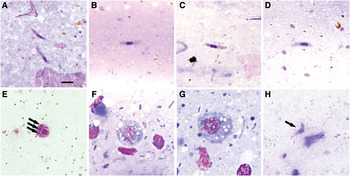
Free haemogregarine gamonts (Figure 2A–D) were observed up to 20 days post-feeding (d.p.f.) in squashes of P1 and P2 stages that had fed on A. bifilum and H. obtusirostre, but none was found in pranizae from S. emarginata. These likely immature gamonts measured 6.2 ± 1.5 (0.7– 8.5) by 1.0 ± 0.2 (0.7–1.6) µm (N = 22). Suspected pairing of gamonts (syzygy) (Figure 2E) was recorded up to 15 d.p.f. At this stage, the microgamont wrapped itself around the macrogamont, with the microgamont nucleus replaced by individual microgamete nuclei (see Davies & Smit, Reference Davies and Smit2001). Oocysts (Figure 2F, G) were observed in pranizae from S. emarginata from 3 d.p.f., and were rounded, pale blue stained, with a vacuolated cytoplasm, and had a centrally placed nucleus staining deep magenta. These stages measured 9.7 ± 0.6 (9.1–10.7) by 9.6 ± 0.9 (8.3–11.3) µm in diameter (N = 7). No sporozoites or meronts were observed. However, merozoite-like stages were found in pranizae that fed on all three hosts (A. bifilum, S. emarginata and H. obtusirostre), 26 d.p.f. Two merozoites (Figure 2H) were 4.6 by 1.5 µm, and 5.0 by 1.7 µm; the remainder were too poorly preserved to measure. Many smears contained a bacterial flora, presumed to comprise the symbionts needed to help digest fish blood in these pranizae (Davies, Reference Davies1995).
Micrographs of Giemsa stained smears from Gnathia pilosus that fed on Antennablennius bifilum, Scartella emarginata and Helcogramma obtusirostre. (A) Gamonts freed from erythrocytes; (B–D) free gamonts; (E) syzygy, showing macrogamont (right) encircled by microgamont with microgamont nuclei (arrows); (F, G) oocysts; (H) merozoite-like stage (arrow). Scale bar = 5 µm.

REMARKS
The association of haemogregarines, intertidal fish and gnathiid isopods was examined by Davies & Smit (Reference Davies and Smit2001) and Gnathia africana was considered the likely vector of H. bigemina in South Africa. Antennablennius bifilum, S. marginata and H. obtusirostre caught during the current survey were hosts to praniza stages of G. pilosus. It was also evident that haemogregarine stages, particularly gamonts, were present in some of these pranizae and immature oocysts found in in G. pilosus were similar to those in G. africana, which measured 8.3–10.8 µm across (Davies & Smit, Reference Davies and Smit2001). Suspected merozoites were however smaller than first, second and third generation merozoites found in G. africana (see Davies & Smit, Reference Davies and Smit2001), but these results suggest, yet again, that gnathiids may act as vectors for fish haemogregarines. However, the current data were insufficient to firstly, identify the species of haemogregarine(s) accurately in the gnathiid, and secondly, to propose a life cycle for the apicomplexans.
ACKNOWLEDGEMENTS
The financial assistance of the National Research Foundation (grant number FA2005022500012) towards this research is hereby acknowledged. The Centre for Aquatic Research, University of Johannesburg funded a research visit by one of us (M.L.F.) to Kingston University, UK.