Significant outcomes
-
Patients with hypochondriasis showed abnormal ReHo values in DMN and insula.
-
The resting-state alterations of insula are closely related to cognitive dysfunction in HS.
-
The combination of the ReHo in FG and insula ideally distinguish HS from SZ.
Limitations
-
The study was cross-sectional, thereby limiting causal inference.
-
The study had a relatively small sample size.
-
Hypochondriasis is a chronic and easily misdiagnosed disease, and the results of hypochondriasis patients are interfered by treatment.
Introduction
Hypochondriasis (HS), generally regarded the same as pathological health anxiety, is defined as a distorted belief in fears and suspicions of having serious diseases, which is the result of an excessive focus on one’s body or a misconstruction of body’s signals (Abramowitz et al., Reference Abramowitz, Olatunji and Deacon2007). It is a remarkably common and persistent disease that results in long-standing suffering even disability and higher medical care utilisation and costs, posing a heavy burden on public health institutions and society (Barsky et al., Reference Barsky, Orav and Bates2005; Eilenberg et al., Reference Eilenberg, Frostholm, Schroder, Jensen and Fink2015; Tyrer, Reference Tyrer2018). Although the diagnosis of HS has been replaced by new concepts of somatic symptom disorder and illness anxiety disorder in the DSM-5 and obsessive-compulsive and related disorders in the ICD-11 (Stein et al., Reference Stein, Kogan, Atmaca, Fineberg, Fontenelle, Grant, Matsunaga, Reddy, Simpson, Thomsen, Van Den Heuvel, Veale, Woods and Reed2016), there is still some controversy to the nosologically status and conceptualisation of HS (Bailer et al., Reference Bailer, Kerstner, Witthoft, Diener, Mier and Rist2016; Starcevic, Reference Starcevic2015; van den Heuvel et al., Reference Van Den Heuvel, Veale and Stein2014). Notably, investigators constantly paid little attention to the neuropathology of HS, and there are limited studies about the changes in the volume of grey matter and brain function (Atmaca et al., Reference Atmaca, Yildirim, Sec and Kayali2010; Garcia-Campayo et al., Reference Garcia-Campayo, Fayed, Serrano-Blanco and Roca2009).
A growing body of research recently indicated some potential links between schizophrenia (SZ) and HS. Patients with SZ showed a higher rate of co-occurring with HS, compared to the known prevalence rate in the general population, and overall functions of SZ were negatively correlated with the severity of HS symptoms (Grassi et al., Reference Grassi, Poli, Cantisani, Righi, Ferrari and Pallanti2014). A 13-year follow-up study in South Korea found that hypochondriac status in patients with SZ may be a reliable indicator for poor outcomes (Kobayashi & Kato, Reference Kobayashi and Kato2004). Several studies suggested that both patients with HS or SZ have a biased cognitive schema, always producing an erroneous judgement (Beck & Rector, Reference Beck and Rector2005; Gorgen et al., Reference Gorgen, Hiller and Witthoft2014; Nowak et al., Reference Nowak, Sabariego, Switaj and Anczewska2016; Weck et al., Reference Weck, Gropalis, Hiller and Bleichhardt2015). In addition, current researches have suggested that pituitary volumes change in HS and SZ patients, which suggests that neuroimaging could be the keystone to better understand SZ and HS (Atmaca, Reference Atmaca2014; Atmaca et al., Reference Atmaca, Yildirim, Sec and Kayali2010; Upadhyaya et al., Reference Upadhyaya, El-Sheikh, Macmaster, Diwadkar and Keshavan2007).
As functional magnetic resonance imaging (fMRI) is popularly used to study psychiatric diseases, resting-state fMRI (rs-fMRI) has attracted more and more attention as a new method for evaluating brain function, especially reflecting spontaneous neuronal activities. In this regard, regional homogeneity (ReHo), a data-driven whole-brain analysis algorithm, is able to examine regional hemodynamic responses, which reflect regional functional connectivity and homogeneity (Jiang & Zuo, Reference Jiang and Zuo2016; Zang et al., Reference Zang, Jiang, Lu, He and Tian2004). It is considered as an appropriate and convenient method of data analysis in exploring the regional activity of the human brain during resting state. Resting-state brain activities in SZ are known to be abnormal. Patients with SZ typically show decreased ReHo in the right fusiform gyrus (FG), left middle temporal gyrus (MTG), and right superior parietal lobule compared with healthy controls (HC) (Gao et al., Reference Gao, Lu, Shi, Ming, Xiao, Sun, Yao and Xu2018). ReHo value in right inferior parietal lobule (IPL) and left paracentral lobule significantly decreases in patients with SZ when compared with HC (Wang et al., Reference Wang, Zhang, Lv, Wu, Fan, Zhao and Guo2018). However, it remains unclear whether patients with HS have altered local synchronisation at rest, impeding a comprehensive understanding of the relationship between HS and SZ. Therefore, this present rs-fMRI study was conducted to compare local resting-state brain activities using ReHo analysis in patients with SZ or HS and well-matched HC, and to explain the correlation between ReHo values of a certain brain region and cognitive function.
First, we are exploring local synchronisation of patients with HS, SZ, and HC at rest, leading to a more in-depth understanding of the pathogenesis of HS and the relationship between HS and SZ. Second, the differences were measured in brain function between them, and whether abnormal brain activity is relevant to cognitive function. Third, we predicted that the abnormal ReHo would optimally differentiate HS from SZ with high sensitivity and specificity. Support vector machine (SVM), a supervised learning algorithm, has been widely applied in recognising subtle patterns in complex data sets for its four advantages, namely strong theoretical foundation, accuracy, flexibility, and large application range (Pavlidis et al., Reference Pavlidis, Wapinski and Noble2004). So, it was used to explore the potential utility for ReHo in the differentiation of HS and SZ.
Methods
Subjects
A total of 44 drug-naïve patients (aged 18–60), recruited from the First Affiliated Hospital of Zhejiang University, were included in this study: 21 patients with HS and 23 drug-naive patients with SZ. The HS in our study believed that they were infected with a serious disease, subsequently turned to the Internet for help, and gradually formed a network group where they interacted with each other. As a result, their clinical symptoms were increasingly similar, and they repeatedly sought medical treatment in the infectious department of various hospitals in China. They were then transferred to the psychiatric department. All patients were diagnosed as HS or SZ according to the Structured Interview for DSM-IV Axis I disorders by two professional psychiatrists. Twenty-five HC who underwent their yearly physical examination in the same hospital voluntarily participated in this study during the same period. The exclusion criteria were also pre-established: significant organic diseases, history of neurological disorders or any structural brain abnormalities, current alcohol and drug abuse, a full-scale IQ score < 80, any contraindications for undergoing an MRI scan. Due to excessive head movement, an HC was excluded from the final data set. Finally, 68 participants were included: 21 HS, 23 first-episode, treatment-naïve SZ, and 24 HCs.
Participants and their legal guardians voluntarily joined this study with written informed consent after being well informed about the procedure and purposes of this research. Ethical approval was obtained from the Local Ethics Committee at the First Affiliated Hospital, College of Medicine, Zhejiang University, China.
MRI data acquisition
The MRI data were collected from a Philips Achieva 3.0T TX MRI system. Head was placed in a soft head coil to limit movement, while all participants were supposed to keep still and relax during the whole scanning. The scanning sequences were as follows: (1) rs-fMRI scan with a fast-field echo-echo planar imaging sequence: TR/TE = 2000/35 ms, flip angle (FA) = 80°, slice thickness/gap = 5.0/1.0 mm, voxel size = 2.4 × 2.4 × 5.0 mm3, matrix = 100 × 100, FOV = 240 × 240 mm2, 24 slices. The rs-fMRI scan lasted for 6 min and 48 s, and we collected a total of 200 image volumes; and 2 three-dimensional high-resolution T1-weighted images with a fast-field echo sequence: 150 slices, TR/TE = 7.5/3.7 ms, matrix = 240 × 240, slice thickness = 1 mm, FOV = 240 × 240 mm2, voxel size = 1 × 1 × 1 mm3, FA = 80.
Cognitive test
The MATRICS consensus cognitive battery (MCCB), a standardised measurement tool for evaluating the cognitive function, was conducted for every participant. It includes speed of processing (SP), attention/vigilance (AV), working memory (WM), verbal learning (VeL), visual learning (ViL), social cognition (SC), reasoning and problem-solving (RPS). This version of MCCB contained some cultural modifications and excluded the letter–number span test (Shi et al., Reference Shi, Kang, Yao, Ma, Li, Liang, Cheng, Xu, Shi, Xu, Zhang, Franklin, Heaton, Jin and Yu2015). The overall composite score (OC) was extracted from the seven domains described above to show cognitive function.
Image data pre-processing and ReHo analysis
All MRI image preprocessing and analyses including the calculation of the Kendall’s coefficient concordance were performed using the Advanced DPARSF (http://www.restfmri.net) and SPM8 (http://www.fil.ion.ucl.ac.UK/spm) toolkits. To avoid errors caused by adapting to the MRI environment, the first 10 volumes were deleted. The remaining volumes were corrected for slice time delay and head motion, and no one was excluded based on the criteria that head movement could not be more than 2.0 mm of translation and 2.0° of rotation. Then, the images were processed by spatial normalisation (EPI template), filtering (0.01 – 0.1 Hz), and detrend in turn. Meanwhile, the nuisance variables were regressed from each voxel’s time course, including white matter and CSF signals, and 24 head motion covariates (translations, rotations, their derivatives, and quadratic terms). In addition, the ReHo analysis was performed using the rs-fMRI Data Analysis Toolkit V 1.7 (http://www.restfmri.net). The technical details of ReHo calculation were reported in our previous publication (Zhao et al., Reference Zhao, Huang, Tang, Ni, Pan, Yan, Fan, Xu and Luo2017). Finally, ReHo maps for each participant were smoothed using an isotropic Gaussian kernel at a full width and at a half maximum of 6 mm to reduce spatial noise.
SVM classification analysis
In this study, SVM was performed using Feature Explorer (FAE, V 0.2.2, https://github.com/salan668/FAE) on Python (3.5.4, https://www.python.org) to build the predictive model. In the classification model, the training data set had 44 cases with 21 HS patients and 23 SZ patients were classified as HS and SZ based on the results of the ReHo value. All the feature variables were normalised and tested for their similarities (removed if pairwise correlations >0.86) and between-group differences. The final feature variables entered into the classification models were the ReHo values showing the differences between the groups. The SVM is the classifier for building the predictive model, and its kernel function makes use of hyperplane for separating the cases with HS or SZ. In view of the relatively small sample size, k-fold cross-validation method (Abdel-Nasser et al., Reference Abdel-Nasser, Rashwan, Puig and Moreno2015; Varoquaux et al., Reference Varoquaux, Raamana, Engemann, Hoyos-Idrobo, Schwartz and Thirion2017), with k = 5, was employed for testing the predictive performance of the SVM model. This method has been adopted in studies that did not involve recruiting an independent sample for model testing. Under this method, all the patients were randomly classified into five subgroups. The four subgroups (training set) were used to fit the parameters of each model and the left-out subgroup (test set) was used to estimate the HS and SZ predictive performance. Receiver operating characteristic (ROC) curve using Feature Explorer (FAE, V 0.2.2, https://github.com/salan668/FAE) on Python (3.5.4, https://www.python.org) presented the performances of the SVM model. The accuracy, sensitivity, specificity, positive predictive value (PPV), and negative predictive value (NPV) were computed for the cut-off value that maximises the Yorden index.
Statistical analysis
The data were performed with the software of SPSS (V 22.0, IBM), and p < 0.05 was considered to be significant in all tests. Chi-square test and one-way ANOVA were conducted to investigate the differences of demographic and clinical characteristics of subjects. One-way ANOVA was also applied to compare the differences in ReHo amongst the three groups with GRF correction (voxel p < 0.01, cluster p < 0.05). And then, post hoc analysis was performed between any pair of the three groups [false discovery rate (FDR) correction, p < 0.05]. The correlations between ReHo value and cognitive functions were analysed using Spearman’s correlation analysis.
Results
Characteristics of subjects
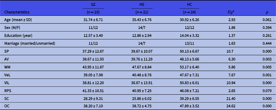
Demographic and clinical characteristics of participants are presented in Table 1. There was no significant difference amongst the three groups in age, gender, marriage, or years of education. Both HS and SZ patients showed lower scores in SP, AV, WM, VeL, ViL, SC, OC of MCCB than HC group (all p < 0.01), but no significant difference was observed between the SZ and HS groups. Furthermore, there was no significant difference for RPS amongst HS, SZ, and HC groups (p = 0.079).
Demographic and clinical information of all participants

HC, healthy controls; HS, patients with hypochondriasis; SZ, schizophrenic patients. SP, speed of processing; AV, attention/vigilance; WM, working memory; VeL, verbal learning; ViL, visual learning; SC, social cognition; RPS, reasoning and problem-solving; OC, Overall composite score in the seven categories of MCCB.
Group differences in ReHo
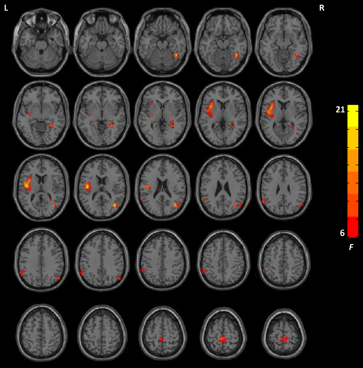
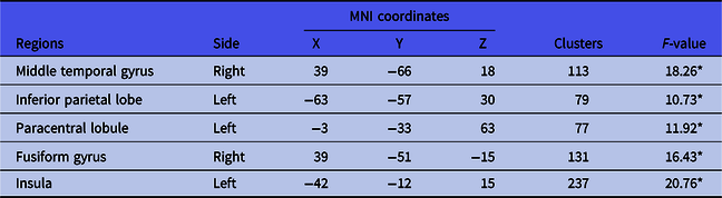
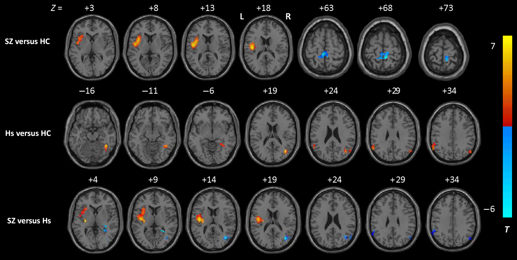
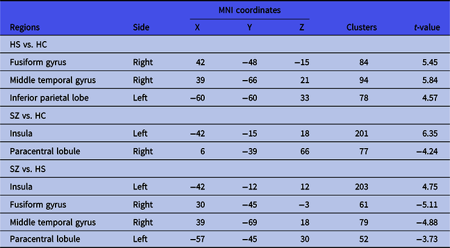
There was no significant difference of ReHo in right MTG, left IPL and left paracentral lobule, right FG, left insula amongst HS, SZ, and HC groups (Fig. 1 and Table 2). We observed an increased ReHo values in right FG, right MTG, and left IPL of HS when compared to HC group (Fig. 2 and Table 3), and SZ exhibited significantly increased ReHo in left insula, but decreased ReHo values in right paracentral lobule than HC. Moreover, the patients with HS had significantly higher ReHo values in FG, MTG, and paracentral lobule, but lower in insula than SZ (Fig. 2 and Table 3).
Regional homogeneity (ReHo) differences amongst hypochondriac patients (HS), schizophrenic patients (SZ), and healthy controls (HC). The colour bar represents F values of the ANOVA analysis.

The results of one-way ANOVA analysis amongst the three groups (GRF corrected, voxel p < 0.01, cluster p < 0.05, two-tailed)

* p < 0.05.
The result of post hoc analysis. The colour regions represent the level of ReHo.

The results of post hoc tests (FDR corrected, p < 0.05, cluster > 30 voxels)

HC, healthy controls; HS, patients with hypochondriasis; SCZ, schizophrenic patients.
The correlations between MCCB scores and ReHo
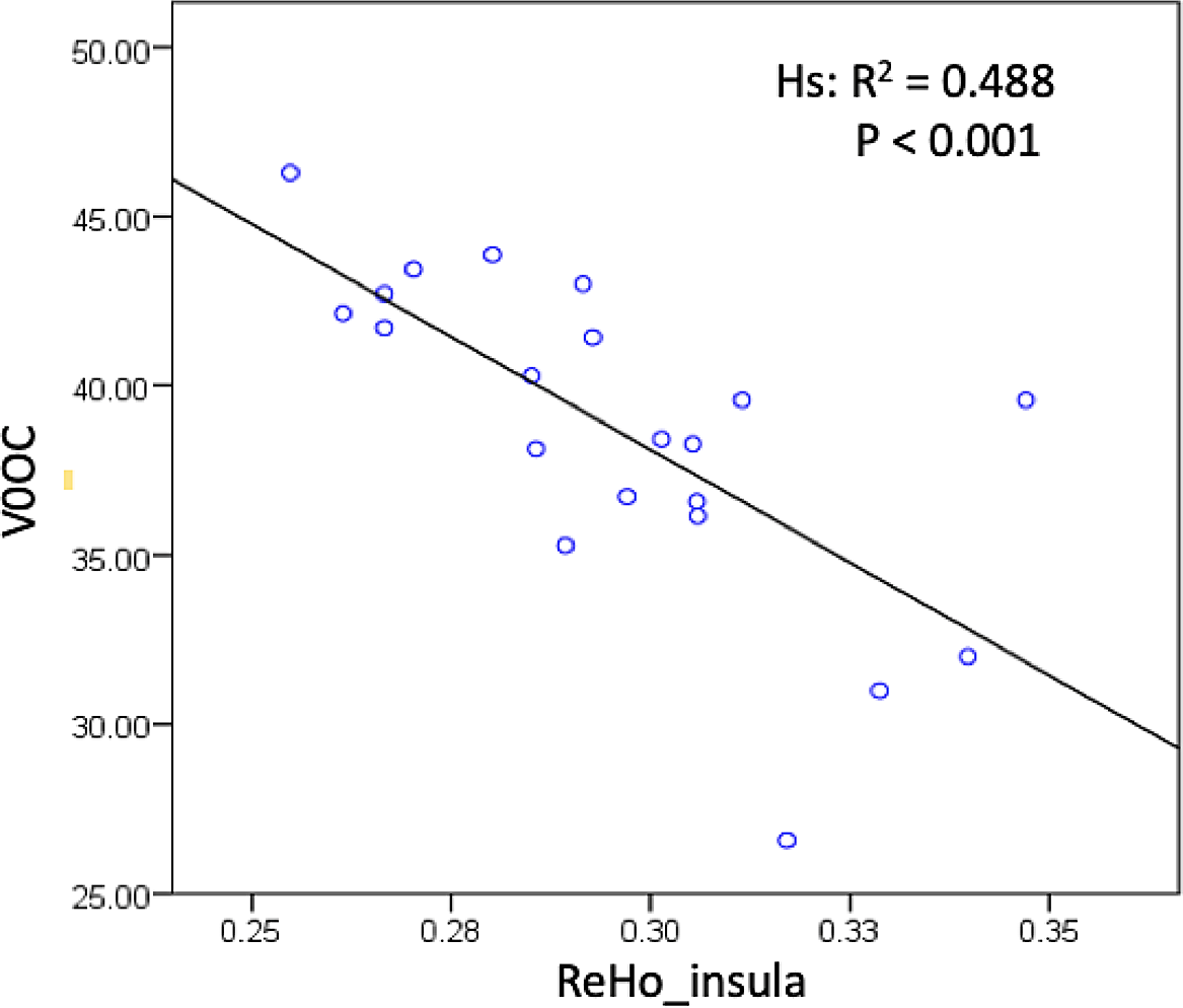
The ReHo values of brain regions with significant alternation were extracted to investigate the relationship between MCCB scores and ReHo value. The results revealed a significant negative correlation between MCCB scores and ReHo in left insula of HS (p < 0.01) (Fig. 3). However, no significant correlation was found in any brain region of patients with SZ (all p > 0.05).
The correlations between the MATRICS consensus cognitive battery (MCCB) scores and ReHo of HS. HS, patients with hypochondriasis; OC, overall composite score in the seven categories of MCCB.

SVM analyses: identifying potential imaging biomarkers
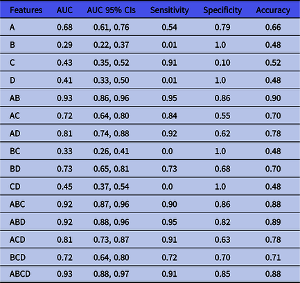
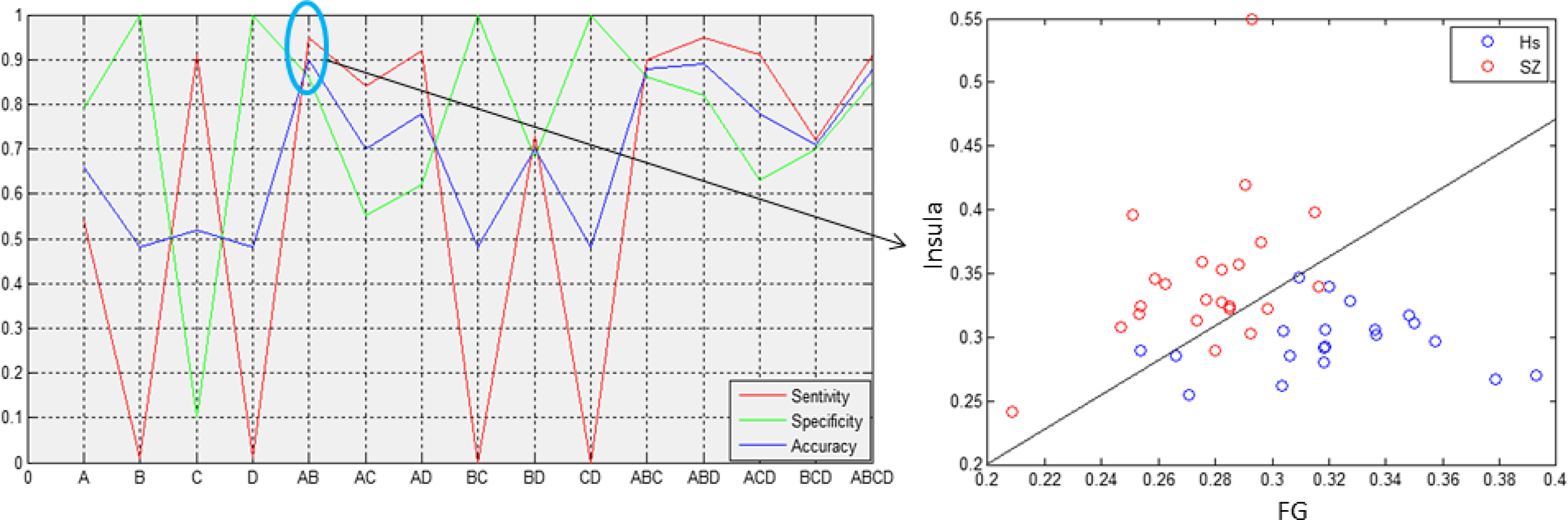
SVM analyses were performed to investigate whether ReHo values could distinguish SZ patients from HS patients. No brain region individually provided a satisfactory result with high sensitivity, specificity, and accuracy. But after combining ReHo values in various regions, we observed some optimistic results, especially in the combination of insula and FG, which gave a specificity of 0.86, sensitivity of 0.95, and accuracy of 0.90 (Table 4, Fig. 4).
The results of SVM analysis based on the selected features

MTG: medial temporal gyrus; AUC: area under curve; CIs: confidence intervals.
A, B, C, and D represent the mean ReHo of FG, insula, IPL, and MTG, respectively.
Performance for ReHo-based classification. Accuracy, sensitivity, and specificity curves for the combination of ReHo values in the five brain regions were labelled with different colours. A, B, C, and D represent the mean ReHo of FG, Insula, IPL, and MTG, respectively. FG, fusiform gyrus; MTG, medial temporal gyrus; IPL, inferior parietal lobe.

Discussion
In the present study, both HS and SZ groups of patients had shown cognitive impairment in SP, AV, WM, VeL, ViL, SC, OC compared with HC, but there was no significant difference between SZ and HS group in any domain of MCCB, which was partly consistent with previous studies (Liu et al., Reference Liu, Wang, Jin, Lyu, Liu, Guo, Shi, Meyers, Wang, Zhao, Wu, Smith and Davis2019; Schwenzer & Mathiak, Reference Schwenzer and Mathiak2012; Rachman, Reference Rachman2012). We found significantly higher ReHo in the right FG, right MTG, left IPL of HS than HC, and increased ReHo in left insula, but decreased ReHo values in right paracentral lobule of patients with SZ compared with HC. Besides, the patients with HS had significantly higher ReHo value in FG, MTG, and paracentral lobule, but lower in insula than SZ. Meanwhile, there was a negative correlation between the ReHo value in left insula and cognitive function. SVM analysis showed the combination of the ReHo in FG and insula could clearly distinguish HS from SZ.
We observed that the patients with HS had higher ReHo values in MTG than HC group. MTG connecting to the frontal lobe and other parts for language, primarily involved in understanding insight, language, and auditory processing (Bonilha et al., Reference Bonilha, Hillis, Hickok, Den Ouden, Rorden and Fridriksson2017; Fan et al., Reference Fan, Zhong, Gan, Liu, Niu, Liao, Zhang, Tan, Yi and Zhu2017), and is a key component of language centre (Xu et al., Reference Xu, Wang, Fan, Li, Zhang, Hu and Jiang2015). Previous studies suggested that MTG, a phylogenetically late-developing and hominoid-specific brain region (van Swam et al., Reference Van Swam, Federspiel, Hubl, Wiest, Boesch, Vermathen, Kreis, Strik and Dierks2012), contributes to the development and existence of auditory verbal hallucinations (Chen et al., Reference Chen, Liang, Pu, Song, Mwansisya, Yang, Liu, Liu, Shan and Xue2015). This suggestion was latterly proved by a strong negative correlation between the thickness of MTG and hallucinatory severity (Cui et al., Reference Cui, Liu, Song, Lipnicki, Li, Xie, Chen, Li, Lu, Lv, Wang, Yan, Yan, Zhang, Zhang and Jiang2018). Besides, HS patients also showed higher ReHo values in IPL. The latter is considered to be one of the key network hubs with wide participation in bottom-up perception, SC, spatial attention, and episodic memory (Caspers et al., Reference Caspers, Eickhoff, Geyer, Scheperjans, Mohlberg, Zilles and Amunts2008; Igelstrom & Graziano, Reference Igelstrom and Graziano2017; Rugg & Vilberg, Reference Rugg and Vilberg2013; Sestieri et al., Reference Sestieri, Corbetta, Romani and Shulman2011). These two regions are both located in the default mode network (DMN) (Lagioia et al., Reference Lagioia, Van De Ville, Debbane, Lazeyras and Eliez2010; Whitfield-Gabrieli et al., Reference Whitfield-Gabrieli, Thermenos, Milanovic, Tsuang, Faraone, Mccarley, Shenton, Green, Nieto-Castanon, Laviolette, Wojcik, Gabrieli and Seidman2009), one of the most active networks during resting state, which is involved in internally directed cognition (Sestieri et al., Reference Sestieri, Corbetta, Romani and Shulman2011), attention to internal emotional states (Raichle & Snyder, Reference Raichle and Snyder2007), monitoring the internal thoughts (Mason et al., Reference Mason, Norton, Van Horn, Wegner, Grafton and Macrae2007). Abnormal ReHo in brain regions within the DMN has been reported in patients with somatisation disorder and related disorders in the previous study. Increased ReHo in the DMN including the IPL, MPFC, and supramarginal gyrus might be associated with the aetiology of patients with persistent somatoform pain disorder (Stankewitz et al., Reference Stankewitz, Sorg, Von Kalckreuth, Schulz, Valet, Neufang, Zimmer, Henningsen, Gundel, Wohlschlager and Tolle2018). Patients with somatisation disorder also show increased Cohe-ReHo in the DMN (Ou et al., Reference Ou, Liu, Chen, Pan, Wu, Su, Zhang, Zhao and Guo2018), which positively correlated to the personality (Song et al., Reference Song, Su, Jiang, Liu, Yao, Dai, Long, Yu, Liu, Zhang, Zhang, Xiao and Guo2015). The aforementioned studies were consistent with our study. HS may be attributable to the abnormalities of DMN (Ou et al., Reference Ou, Liu, Chen, Pan, Wu, Su, Zhang, Zhao and Guo2018), and its over-activation may be one of the primary causes why patients pay too much attention to their body signals and insist they are ill. Perhaps that also partly explains why HS is prone to discomfort at break. Whereas, no significant abnormity was found in these two brain regions in the SZ of our study, which contradicts previous reports that ReHo of patients with SZ decreased ReHo of the DMN, including the MTG, right IPL, and right middle frontal gyrus (Liu et al., Reference Liu, Zhang, Lv, Wu, Zhao and Guo2018; Zhao et al., Reference Zhao, Yao, Lv, Zhang, Han, Chen, Ren, Jin, Li and Sui2018). A possible reason is the difference in illness duration.
Decreased ReHo value in the paracentral lobule of SZ was observed in our study, which was consistent with previous evidence (Wang et al., Reference Wang, Zhang, Lv, Wu, Fan, Zhao and Guo2018; Liu et al., Reference Liu, Zhang, Lv, Wu, Zhao and Guo2018). As a junction of the frontal lobes and the parietal lobes, the paracentral lobule not only played an important role in sensorimotor innervation of the lower extremities (Spasojević et al., Reference Spasojević, Malobabic, Pilipović-Spasojević, Djukić-Macut and Maliković2013), but also involved micturition and defecation (Wang et al., Reference Wang, Li, Zhang, Wang, Feng, Liang, Wei, Zhang, Li and Chen2019). The paracentric lobules in SZ patients were related to the function of implicit motor sequence learning (Exner et al., Reference Exner, Weniger, Schmidt-Samoa and Irle2006; Hirjak et al., Reference Hirjak, Wolf, Stieltjes, Hauser, Seidl, Schroder and Thomann2014). But no significant abnormalities were observed in this brain region in HS and HC. Therefore, altered ReHo value in the paracentral lobule might be a relatively specific character for subjects with SZ.
Increased ReHo of FG was found in HS, indicating people with HS at rest had increased neural activity in FG. To be noted, FG is a hominoid-specific pivotal structure for selectively high-level visual processes such as word recognition, face perception, memory (Grill-Spector et al., Reference Grill-Spector, Kourtzi and Kanwisher2001; Henson et al., Reference Henson, Shallice and Dolan2000; Kanwisher et al., Reference Kanwisher, Mcdermott and Chun1997). Previous studies suggested reduction of FG volume may underlie delusional misidentification syndrome and poor performance in facial recognition, implying that the FG could further play a critically vital role in severe cognitive dysfunctions and social deficits (Hudson & Grace, Reference Hudson and Grace2000; Takahashi et al., Reference Takahashi, Zhou, Nakamura, Tanino, Furuichi, Kido, Kawasaki, Noguchi, Seto, Kurachi and Suzuki2011). Interestingly, a previous report showed abnormalities of the FG might contribute to reduced conscientiousness and extraversion (Onitsuka et al., Reference Onitsuka, Nestor, Gurrera, Shenton, Kasai, Frumin, Niznikiewicz and Mccarley2005). Therefore, it may be related to the personality traits of patients and the clinical characteristics that the HS patients constantly seek social support. Several studies have demonstrated that progressive changes in FG volume were associated with negative symptoms in SZ (Onitsuka et al., Reference Onitsuka, Shenton, Kasai, Nestor, Toner, Kikinis, Jolesz and Mccarley2003; Takahashi et al., Reference Takahashi, Suzuki, Zhou, Tanino, Hagino, Niu, Kawasaki, Seto and Kurachi2006). Gao et al. proved that chronic SZ also had hypoactivation and hypometabolism in the bilateral FG (Gao et al., Reference Gao, Lu, Shi, Ming, Xiao, Sun, Yao and Xu2018). And a meta-analysis verified decreased ReHo in the left FG in chronic SZ when compared to HC (Xiao et al., Reference Xiao, Wang, Liu, Meng, He and Luo2017). But our study did not show ReHo changes in FG for SZ, suggesting that the abnormalities of neural activity in this region may be in part related to the development of SZ.
Higher ReHo values in SZ insula either compared to HS or HC group, our finding was consistent with a previous study that patients with psychosis when experiencing auditory hallucinations which showed increased blood flow and metabolism in the insula (Sommer et al., Reference Sommer, Diederen, Blom, Willems, Kushan, Slotema, Boks, Daalman, Hoek, Neggers and Kahn2008). This evidence supported a hypothesis that insula dysfunction probably results in endogenous sensory information being mistaken for an external factor, which leads to hallucinations in patients with SZ (Yu et al., Reference Yu, Hsieh, Wang, Liu, Liu, Hwang, Chien, Hwu and Tseng2013). To be noted, insula is responsible for processing external and internal information, further generating subjective feelings (Gasquoine, Reference Gasquoine2014), which are largely based on a special anatomical position in connection to the anterior cingulate cortex, orbitofrontal cortex, the frontal lobe, the parietal and temporal cortices (Gogolla, Reference Gogolla2017; Wylie & Tregellas, Reference Wylie and Tregellas2010). Moreover, it is considered to be critical for the regulation of the autonomic nervous system according to interoceptive information (Critchley et al., Reference Critchley, Rotshtein, Nagai, O’doherty, Mathias and Dolan2005; Griffiths et al., Reference Griffiths, Tadic, Schaefer and Resnick2007; Walter et al., Reference Walter, Kolbaske, Patejdl, Steinhagen, Abu-Mugheisib, Grossmann, Zingler and Benecke2013). In addition, we found that HS ReHo value in insula shows a negative correlation with OC of MCCB, which suggested the role of dynamic switching between the central executive network and the DMN has a strong correlation with the cognition (Menon & Uddin, Reference Menon and Uddin2010). Therefore, altered local synchronisation may be employed as an imaging marker for the severity of cognitive deficits in patients with HS.
SVM analysis suggested that the combination of ReHo in insula and FG for correctly classifying patients by diseases had the specificity of 0.86, sensitivity of 0.95, and accuracy of 0.90. Thus, the combination of the ReHo values could serve as a potential imaging marker to differentiate patients with HS from SZ.
Limitations
There are still several limitations in this study. First, because it is difficult in China to recruit patients with HS who always visit other departments of hospitals, the sample size of our study was relatively small, which may cause some false-positive results. Multicentre research with a larger sample size might be a trend to increase the reliability in the future. Second, suboptimal imaging parameters with larger slice thickness (5mm) lead to relatively rough spatial resolution, which may further exacerbate the risk of some details being overlooked. Third, altered brain microstructure underlying ReHo value abnormalities remains unclear. Up till now, few studies provide a powerful hypothesis for the functional significance of ReHo, and it is still unclear how ReHo alternation leads to regional functional connectivity. Finally, the effects of treatment on ReHo of the patients are inevitable, longitudinal follow-up studies could be adopted to observe their dynamic changes as the disease progresses, and get a better understanding of the function of each brain region.
Conclusions
In sum, our findings suggested that the increased spontaneous neural activity in MTG, FG, and IPL might be specific for HS, and the abnormalities of DMN might play a key role of the pathophysiology of HS. The alterations in resting state of insula are closely related to cognitive dysfunction in HS. Furthermore, the combination of the ReHo in FG and insula was a relatively ideal indicator to distinguish HS from SZ.
Acknowledgements
We gratefully acknowledge the participation of the study participants.
Author contributions
MH and DX were involved in the design of this study and contributed to the writing of the manuscript. KJ performed the study and wrote the manuscript. ZS, GF, FP, and SH were screening the proper patient subjects and in charge of clinical information, JL and HL are responsible for statistical analysis. DS, JC, and ZZ performed neuroimaging data analysis.
Financial support
This study was supported by National Key Research and Development Program of China (2016YFC1307005, 2016YFC1306900, and 2017YFC1308502), the China National Nature Science Foundation (No. s81471734), Basic Public Welfare Research projects in Zhejiang province (LGF18H090003) and major subject of Zhejiang Province (2015C03054).
Conflicts of interest
The authors report no conflicts of interest.
Ethical standards
The authors assert that all procedures contributing to this work comply with the ethical standards of the relevant national and institutional committees on human experimentation and with the Helsinki Declaration of 1975, as revised in 2008.
Supplementary material
To view supplementary material for this article, please visit https://doi.org/10.1017/neu.2021.9.