I. INTRODUCTION
The unique three-dimensional nanoarchitecture of apatite crystals contributes to the remarkable mechanical properties of human teeth and is important for protection against dental decay. Enamel is relatively stable in the healthy oral environment, although there is a dynamic equilibrium between demineralization and remineralization at the tooth–pellicle and plaque–saliva interfaces.Reference Aoba1 The net mineral gain induced by saliva is small, and the process is slow because of the low ionic concentration gradient between saliva and tooth lesions; thus, early caries lesions occur when bacterial plaque, cariogenic microflora, or dietary carbohydrates are presentReference van Houte2,Reference Dorozhkin and Epple3 or where the dissolution rate of calcium phosphates is greater than the recombination rate. The most common and conventional treatment for clinically detectable caries is to replace the decayed tissue with artificial restorative substitutes, such as amalgam, metal alloys, ceramics, or polymer composite materials. However, differences in thermal expansion between natural enamel and artificial substitute materials, and the stresses caused by diet and mastication can cause the interfacial gap between the tooth and the substitute material to gradually expand, allowing bacteria to accumulate, thereby causing secondary caries.
A preferred approach is to promote the remineralization of early small dental erosion before they progress into full lesions; this involves the use of casein phosphopeptide-amorphous calcium phosphate (CPP-ACP) nanocomplexes,Reference Reynolds4–Reference Hannig and Hannig8 hydroxyapatite nanoparticles,Reference Cai and Tang9 or amorphous calcium phosphates.Reference Tung and Eichmiller10 Peptides have also been used in biomimetic processes for hard tissue remineralization.Reference Kirkham, Firth, Vernals, Boden, Robinson, Shore, Brookes and Aggeli11,Reference Segman-Magidovich, Grisaru, Gitli, Levi-Kalisman and Rapaport12
Dentin phosphoprotein (DPP) is the most abundant noncollagenous extracellular matrix component of dentin. The highly anionic DPP is specific to calcium ions, and the numerous repetitive nucleotide sequences of aspartic–serine–serine in DPP promote the formation of hydroxyapatite.Reference George, Bannon, Sabsay, Dillon, Malone, Veis, Jenkins, Debra and Neal13 A synthetic aspartic–serine–serine peptide is reported to promote the deposition of the calcium phosphate mineral and improve the nanomechanical properties of acid-eroded human enamel in simulated body fluid and artificial saliva.Reference Hsu, Chung, Yang, Shi and Wu14,Reference Chung, Li and Hsu15 Another study demonstrates that nonphosphorylated leucine-rich amelogenin peptides guide amorphous calcium phosphate transformation into crystalline apatite structure, whereas phosphorylated leucine-rich amelogenin peptides stabilize amorphous calcium phosphate.Reference Norcy, Kwak, Wiedemann-Bidlack, Beniash, Yamakoshi, Simmer and Margolis16
We designed a nonphosphorylated triplet-repeating asparagine–serine–serine (3NSS) peptide, not abundantly found in nature dental tissue, which is expected to attract aqueous ions present in saliva and stabilize crystalline apatite crystals. To simulate an oral cavity remineralization environment, we chose artificial saliva rather than a supersaturated simulated body fluid. This study investigated surface roughness, the deposited mineral phases, the subsurface microstructure, and the surface nanomechanical behaviors of newly deposited apatites to assess the effectiveness of the 3NSS peptide for the remineralization of tooth enamel in vitro.
II. EXPERIMENT
Twenty adult human third molars were collected after informed consent as approved by the Commission for Medical Ethics of China Medical University. The specimens were cleaned from debris and stored in distilled water with 0.5% thymol solution until use. Each whole tooth was embedded in long-cure epoxy resin (Leco, St. Joseph, MI) and then longitudinally sectioned using a diamond blade (Accutom 50; Struers, Cleveland, OH). The size of enamel slab was 3 × 3 × 1 mm. The enamel surfaces were polished sequentially using silicon carbide followed by 1 and 0.3 μm Al2O3 (Buehler, Lake Bluff, IL). All polished samples were individually sonicated in distilled water for 5 min to remove the residual abrasives.
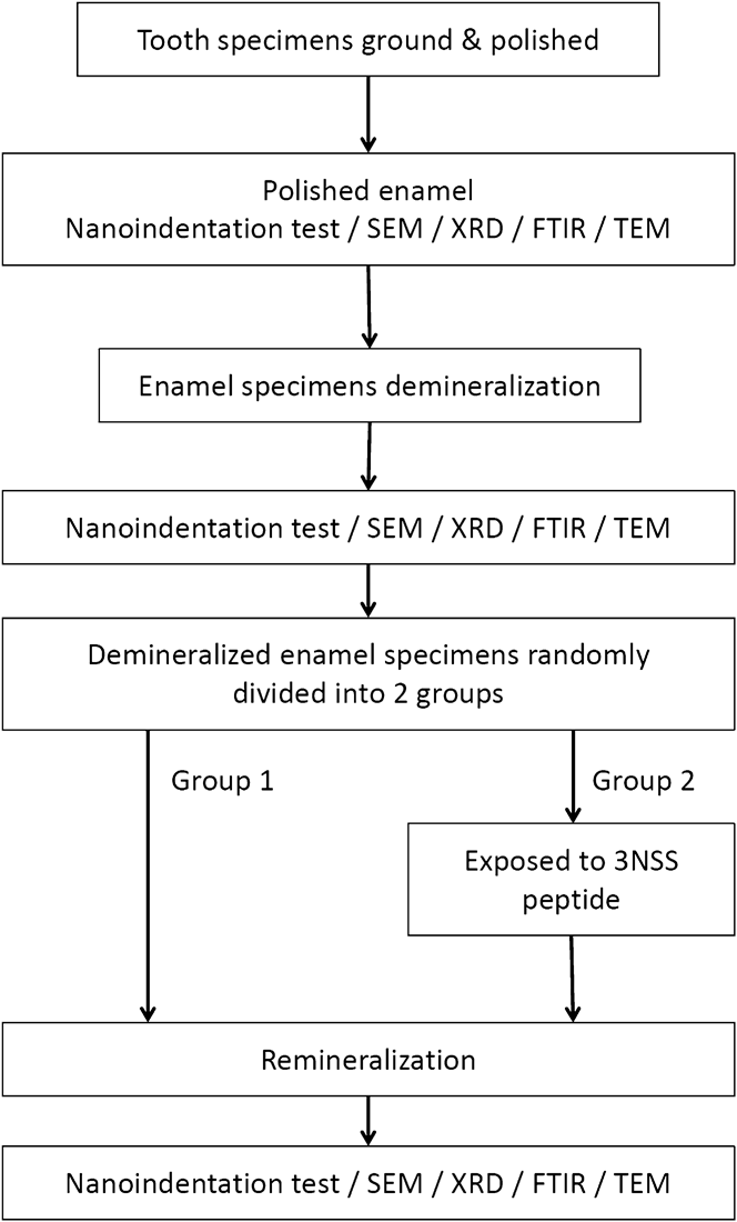
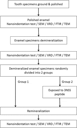
To simulate erosive enamel, the polished enamel surfaces were demineralized with 34% phosphoric acid gel (pH < 1; Dentsply International Inc., Milford, DE) for 30 s to create aggressive lesions, and then rinsed thoroughly with distilled water to terminate the demineralization reaction. The demineralized specimens were randomly assigned to 2 groups. The peptide was dissolved in a HEPES buffer, and the concentration of peptide was adjusted to 33.3 μM. Control group samples were directly incubated in artificial saliva at 37 °C for 7 days; the experimental specimens were exposed to the 3NSS peptide for 60 min followed by immersion in artificial saliva for 7 days. The artificial saliva contains 6.84 mM of NaCl, 5.37 mM of KCl, 5.44 mM of CaCl2·2H2O, 4.42 mM of NaH2PO4·2H2O, 0.21 mM of Na2S·9H2O, and 16.65 mM of urea, and the pH value of solution was adjusted to 7.0 with 1 M NaOH.Reference Holland17 The artificial saliva is supersaturated with regard to the hydroxyapatite (degree of saturation, DS > 1). A flow chart showing the study procedures is shown in Fig. 1.
Flow chart of the study procedures.

Atomic force microscopy (AFM; Veeco Instruments, Santa Barbara, CA) was operated in tapping mode using a nonconductive silicon nitride tip at a scanning rate of 1 Hz to map enamel topography. Areas of 10 × 10 μm on the enamel surface were scanned 3 times to determine average surface roughness (R a), root mean squared surface roughness (R q), and maximum surface roughness (R max) for each sectioned sample. X-ray diffraction (XRD; PANalytical X'Pert Pro, Almelo, Netherlands) with Cu Kα radiation (λ = 1.54 Å) over a 2θ range of 25–55° was used to identify the phases and crystallinity of newly deposited minerals observed on the enamel surface. Additionally, the XRD ω-value was set to 3.0° so that x-ray penetration depth was roughly the thickness of the newly deposited mineral layer. Attenuated total reflectance-Fourier transform infrared (ATR-FTIR; Bruker Optics, Billerica, MA) spectra were acquired; sectioned specimens were scanned 32 times at a resolution of 1 cm−1, from 4000 to 600 cm−1. The ultrathin enamel specimens were prepared by a focused ion beam system (Nova 200; FEI Co., Hillsboro, OR) with a protective layer of Pt coating on the top surface. The microstructure and selected area electron diffraction patterns were analyzed by high-resolution transmission electron microscopy (JEM-2100F, JEOL Co., Japan) with an accelerating voltage of 200 kV.
Nanoindentation testing was conducted using an MTS XP nanoindenter (Agilent Technologies Inc., Vista, CA) with a Berkovich diamond tip. Before each test, the nanoindenter was calibrated using a fused silica block. Nanohardness and elastic modulus were determined from the oscillation of the resulting depth signals by a continuous stiffness measurement (CSM) technique and calculated using equations developed by Oliver and Pharr,Reference Oliver and Pharr18
 $$H = {P_{\max }}/A\quad ,$$
 $$1/{E_{\rm{r}}} = [(1 - {{\rm{\nu }}^2})/E + (1 - {{\rm{\nu }}_{\rm{i}}}^2)/{E_{\rm{i}}}]\quad ,$$
 $${E_{\rm{r}}} = (S/2{\rm{\beta }}){({\rm{\pi }}/A)^{0.5}}\quad ,$$
where P max is the maximum load, A is the contact area; H is the nanohardness, E is the elastic modulus, and ν is the Poisson's ratio of the specimen; E i and ν i are the elastic modulus and Poisson's ratio for the diamond indenter. E r is the reduced modulus that depends on the slope of the upper portion of the load–displacement unloading curve (S), and β is a shape constant for the indenter. From the oscillation of the resulting depth signals, the contact stiffness is continuously measured so that the values of hardness and elastic modulus can be obtained along the varying indent depth. Data were collected from penetration depths of 250–750 nm to minimize the influence of surface roughness and the substrate effect arising from the underlying native enamel. After the various experimental treatments, AFM prescan was conducted to ensure that there was enough smooth surface for the diamond tip to locate initially, and 20 indents were made for each sectioned enamel specimen (n = 10, each group). The organic matrix phase in enamel is significantly sensitive to the strain rate,Reference Zhou and Hsiung19 so that a constant strain rate of 0.05 s−1 was set in the loading process for all tests.
The data were expressed as mean ± SD (standard deviation). Comparative studies of means were performed using one-way analysis of variance, and the statistical significance from the demineralized enamel was accepted with P < 0.05.
III. RESULTS AND DISCUSSION
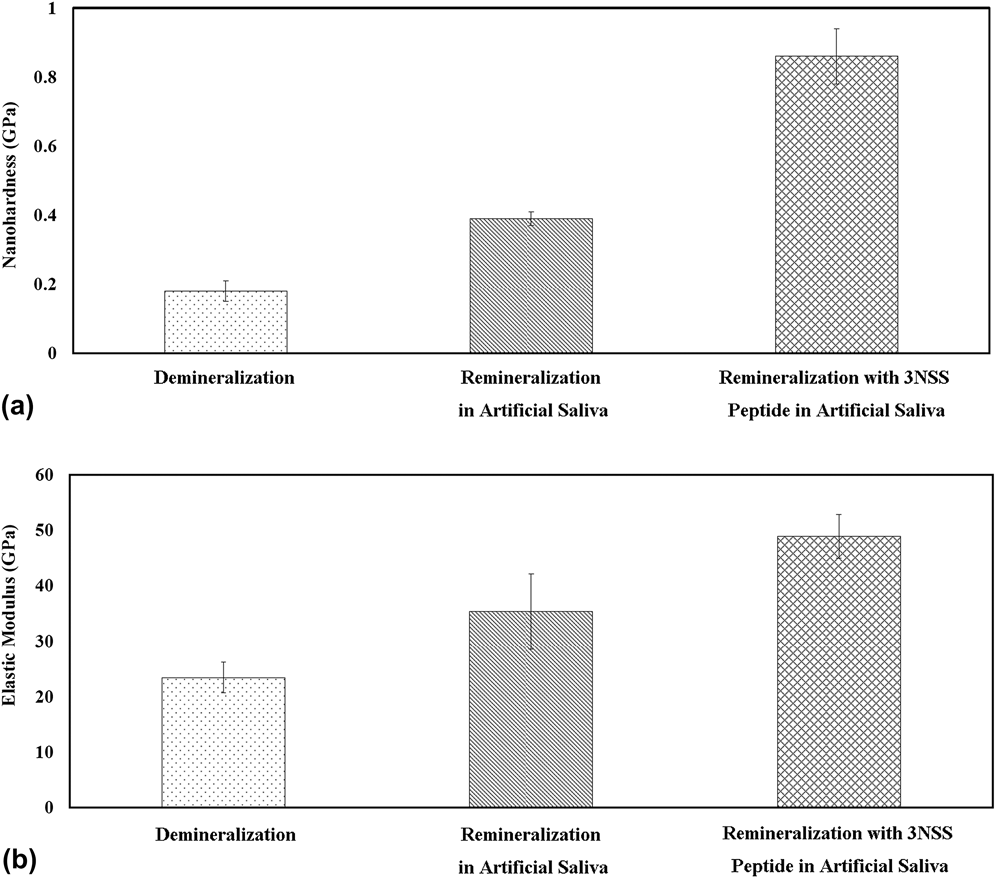
Figure 2 shows the nanohardness and elastic modulus measured at various treatment stages. The average surface nanohardness of enamel decreased from 4.36 to 0.18 GPa after demineralization (P < 0.0001). The average nanohardness of demineralized enamel remineralized in artificial saliva increased to 0.39 GPa (P < 0.001). In contrast, the average nanohardness increased to 0.86 GPa by exposing the demineralized enamel to 33.3-μM 3NSS peptide and artificial saliva (P < 0.001).
The average surface (a) nanohardness and (b) elastic modulus of human enamel at various treatment stages.
The average elastic modulus substantially changed from 80.65 to 23.45 GPa (P < 0.0001) after demineralization [Fig. 2(b)]. The average elastic modulus of demineralized enamel remineralized in artificial saliva solely increased from 23.45 to 35.39 GPa (P < 0.05), whereas preexposure to 33.3-μM 3NSS peptide increased the elastic modulus to 48.91 GPa (P < 0.05). These results indicated that the mechanical and elastic properties of acid-demineralized enamel were effectively improved with the aid of the 3NSS peptide during the biomimetic process.
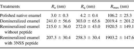
The average surface roughness (R a), root mean squared surface roughness (R q), and maximum surface roughness (R max) of the enamel at various treatment stages are listed in Table I. The average surface roughness considerably increased from 3.0 ± 0.3 to 241.0 ± 56.6 nm when the enamel was demineralized by phosphoric acid gel. Immersion in artificial saliva resulted in reductions in average surface roughness (R a = 215.0 ± 36.0 and 207.3 ± 30.4 nm for the enamels remineralized without and with the 3NSS peptide, respectively), and the magnitude of reduction increased slightly in the presence of the 3NSS peptide.
The average surface roughness (R a), root mean squared surface roughness (R q), and maximum surface roughness (R max) of the enamel at various treatment stages.
XRD results for various treatment stages shown in Fig. 3 indicate that the main inorganic minerals of the newly deposited enamel were hydroxyapatite phases; however, the crystallinity apparently decreased after remineralization, which was also observed in most of the aqueous biomimetic systems. The ATR-FTIR spectra (Fig. 4) show bands at approximately 1073, 1019, and 958 cm−1 characteristic of  ${\rm{PO}}_{\rm{4}}^{{\rm{3}} - }$ groups, which together with carbonate absorption bands appearing at roughly 1542, 1492, 1457, and 874 cm−1 were supportive of carbonated hydroxyapatite composition.Reference Moriguchi, Yano, Nakagawa and Kaji20,Reference Reyes-Gasga, García-García, Arellano-Jiménez, Sanchez-Pastenes, Tiznado-Orozco, Gil-Chavarria and Gómez-Gasga21
XRD patterns of polished native enamel, demineralized enamel, remineralized enamel immersed in artificial saliva solely, and remineralized enamel treated with the 3NSS peptide prior to the immersion of artificial saliva.
ATR-FTIR spectra of polished native enamel, demineralized enamel, remineralized enamel immersed in artificial saliva solely, and remineralized enamel treated with the 3NSS peptide prior to the immersion of artificial saliva.
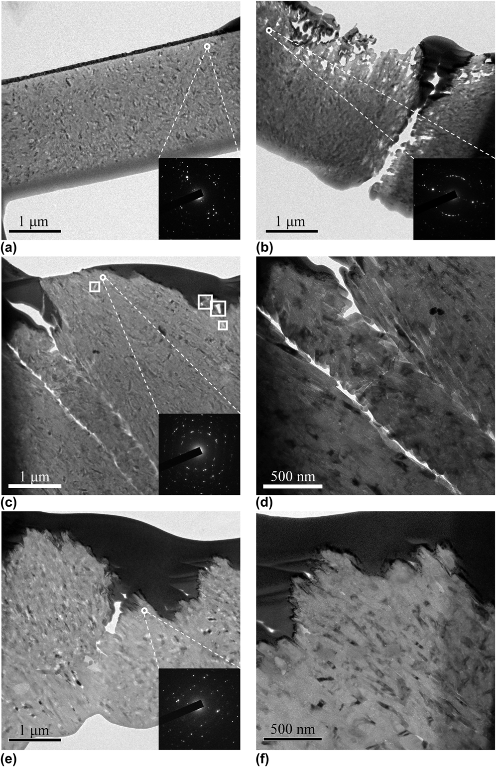
A smooth top surface with close-packed hydroxyapatite crystals was observed in the polished native enamel in Fig. 5(a). Following demineralization, the top surface of the enamel became significantly rougher and presented large pores with submicro depths of roughly 1 μm. Moreover, most of the minerals in the enamel interrod were etched away, particularly near the top surface. There was a distinct vacant band with a width of 200–300 nm, and the crystal arrangement was less packed at the boundary of the cavity [Fig. 5(b)]. In the presence of the 3NSS peptide, the large submicro pores were filled with nanocrystalline hydroxyapatites [diffraction patterns; inset in Fig. 5(e)]. No obvious pores were present [Fig. 5(f)], and the vacant enamel interrod was completely reconstructed, without any deep cavities in the enamel [Fig. 5(e)]. The elongated hydroxyapatite crystals exhibited an ordered arrangement. By contrast, enamel remineralized in artificial saliva without the 3NSS peptide presented a few remaining nanopores [marked by rectangles in Fig. 5(c)], and the vacant band still present, although it had narrowed to a width of approximately 50 nm [Fig. 5(d)].
The transmission electron micrographs of (a) polished native enamel, (b) demineralized enamel, (c, d) remineralized enamel immersed in artificial saliva solely, and (e, f) remineralized enamel treated with 3NSS peptide prior to the immersion of artificial saliva.
The dental cavity self-remineralization effect could be improved by modifying the functional groups on the enamel surface.Reference Toworfe, Composto, Shapiro and Ducheyne22 The charged C- and N-side groups in the 3NSS peptide have a significant affinity for anions and cations in saliva. The peptide interacted with these ions to deposit new minerals, particularly near the vacant enamel interrod, where a greater concentration of peptide had aggregated. The greater interrod hydroxyapatite crystallization rate compared with that of the enamel rod resulted in a greater decrease in overall surface roughness (Table I). Additionally, the peptides completely reconstructed acid-erosive pores close to the top surface [Fig. 5(f)] and in the deep interrod regions [Fig. 5(e)], with a dense arrangement of hydroxyapatite crystals. By contrast, several nanopores remained and the vacant gap was still present [Fig. 5(c)], albeit partially repaired, in the enamel treated without the 3NSS peptide.
The artificial saliva used in this study contains no carbonate salts; thus, the characteristic carbonate bands observed in the ATR-FTIR spectra of remineralized enamel probably originated from the underlying native enamel. We identified the individual phosphate and carbonate bands in ATR-FTIR spectra of the remineralized samples (Supplementary); the relative phosphate- and carbonate-integrated area ratios were 3.35 and 4.07 for the enamel remineralized without the peptide and in the presence of the 3NSS peptide, respectively. The greater phosphate/carbonate ratio ( ${\rm{PO}}_{\rm{4}}^{{\rm{3}} - }/{\rm{CO}}_{\rm{3}}^{{\rm{2}} - }$) implies that more hydroxyapatite crystals deposited on the erosive enamel surface, resulting in a weakening intensity of the underlying carbonate group's absorption.
The quality of hard tissue is dependent on the sum of its characteristics, including the microstructure, mineral density, surface roughness, and morphology.Reference Hsu, Chung, Yang, Shi and Wu14,Reference Chung, Li and Hsu15,Reference Fong, Sarikaya, White and Snead23–Reference Hsu, Chung, Yang, Shi and Wu27 The mechanical behaviors of the tissue integrate all these factors; therefore, nanoindentation was used to assess the degree of remineralization using the equation:
 $${\rm{[(}}{H_{{\rm{remineralized \;enamel}}}} - {H_{{\rm{demineralized \;enamel}}}}{\rm{)/}}({H_{{\rm{polished \;native \;enamel}}}} - {H_{{\rm{demineralized \;enamel}}}}{\rm{)]}} \times {\rm{100\% }}\quad {\rm{,}}$$
where H represents the nanohardness value.Reference Cury, Simões, Del Bel Cury, Gonçalves and Tabchoury28,Reference Bertassoni, Habelitz, Marshall and Marshall29 Incubation in artificial saliva solution for 7 days, the nanohardness of both the control and experimental groups showed increases. The degree of recovery was 5.02% for the control (without peptide) and was 16.27% for enamels treated with 33.3-μM 3NSS peptide. The degree of nanohardness recovery was 3 times greater, and the recovery of elastic modulus was approximately 2 times greater for enamel remineralized in the presence of the 3NSS peptide than for the enamel without peptide treatment. This result demonstrated that the nanomechanical and elastic properties of demineralized enamel can be improved by adding the 3NSS peptide to promote remineralization.
Measuring the full width at half maximum height and applying Scherrer's equation to calculate the crystalline size of the newly deposited hydroxyapatites for the control samples, the average crystalline sizes calculated from the (211) and (300) diffraction peaks were found to be 14.73 and 16.91 nm, respectively. The average crystal sizes of enamel remineralized in the presence of the 3NSS peptide were 11.37 and 11.95 nm for the (211) and (300) diffraction peaks, respectively. Smaller grain size can resist plastic deformations at grain boundaries more effectively, thus providing greater nanohardness.
Casein phosphopeptide stabilizes amorphous calcium phosphate; however, the 3NSS peptide used in this study sequesters calcium and phosphate ions from saliva to provide its biomimetic effect. Use of CPP-ACP provides a 46.25% recovery of enamel hardness,Reference Srinivasan, Kavitha and Loganathan30 whereas treatment with the 3NSS peptide yields a recovery of 16.27%. This apparent difference in performance may have occurred because of the aggressive erosion created using a strong phosphoric acid (pH < 1), in contrast to the smaller erosion commonly created by low-concentration acidic beverages (pH = 2.3). However, our results demonstrated that peptide-induced remineralization was not limited to the enamel surface, but extended into the enamel subsurface, which were the same results investigated in CPP-ACP remineralization systems.Reference Reynolds4
IV. CONCLUSIONS
Nonphosphorylated 3NSS peptides promoted the deposition of small-grain hydroxyapatite crystals for the complete reconstruction of erosive interrod and pores in the enamel subsurface. The resulting small deposited crystal sizes, increased quantities of hydroxyapatite formation, and reductions in surface roughness provided improvements in the mechanical properties of acidic demineralized enamel over those of the controls not exposed to the 3NSS peptide. The degree of nanohardness recovery was 3 times greater, and the recovery of elastic modulus was approximately 2 times greater for enamel remineralized in the presence of the 3NSS peptide than for the enamel without peptide treatment.
ACKNOWLEDGMENTS
The authors are grateful to National Science Council (Grant Nos. NSC 99-2221-E-035-039 and NSC 100-2221-E-035-008) for the financial support and Industrial Technology Research Institute for the collaboration. We also thank Dr. Chia-Chan Hsu and Dr. Ren-Jye Wu for the support in analysis.