Accurate diagnosis and appropriate monitoring are critical in the management of infants with pulmonary arterial hypertension.Reference Hansmann1 Pulmonary arterial catheters are occasionally used to monitor the pulmonary arterial pressure of patients undergoing cardiac surgery. The placement of pulmonary arterial catheters in infants may be performed by cardiac surgeons; however, this is more challenging in infants who have not recently undergone cardiac surgery. Swan-Ganz catheters are often used in critically ill adults, but not in infants due to size limitations. Hence, echocardiography is commonly used to estimate pulmonary arterial pressure in such patients. The pulmonary arterial pressure is estimated based on tricuspid regurgitation velocity or a crescendo deformation of the left ventricle. However, estimated pulmonary arterial pressure is only obtained intermittently and is often inaccurate in the absence of tricuspid regurgitation. Herein, we present the case of a 3-month-old infant with severe pulmonary arterial hypertension who was successfully treated with the help of continuous real-time monitoring of pulmonary arterial pressure using a tip deflecting steerable microcatheter.
Case report
A male baby (birth weight, 2.6 kg) was born at a gestational age of 39 weeks. Foetal ultrasound screenings showed no abnormalities. The baby was admitted to the neonatal intensive care unit due to cyanosis with systemic oxygen saturation of 80%. On admission, echocardiography revealed supracardiac total anomalous pulmonary venous connection with narrowing of the vertical vein, a flattened interventricular septum, and no tricuspid regurgitation. He developed respiratory distress and cyanosis, but he did not require intubation or sedation. A chest X-ray showed the cardiothoracic ratio of 0.52, with diffuse pulmonary congestion. Subsequently, he underwent corrective surgery at 7 days of age.
After a median sternotomy, the common pulmonary vein was directly anastomosed to the left atrium while he was on cardiopulmonary bypass. Postoperative echocardiography showed a modest increase in pulmonary arterial pressure with a tricuspid regurgitation pressure gradient of 30 mmHg, while his systemic blood pressure was 74/47 mmHg. He was discharged at 29 days of age.
At the age of 2 months, echocardiography showed narrowing of the right pulmonary vein draining into the left atrium and suprasystemic pulmonary arterial pressure, with a tricuspid regurgitation pressure gradient of 82 mmHg and a systemic blood pressure of 72/42 mmHg. Arterial blood gas analysis showed that oxygen and carbon dioxide partial pressures were 64 and 37 mmHg, respectively. Cardiac catheterization showed a pulmonary artery pressure of 75/35 mmHg (mean pressure of 53 mmHg); the ratio of pulmonary to systemic arterial pressure was 1.01; a mean pulmonary artery wedge pressure was 18 mmHg, and a pulmonary arterial resistance was 9.04 Wood units·m2. As these findings were consistent with recurrent pulmonary venous obstruction, he underwent reconstructive surgery of the right pulmonary veins using the sutureless technique at the age of 3 months. A lung biopsy obtained at the second operation revealed medial hypertrophy and intimal hyperplasia of the pulmonary arteries; however, there was no evidence of pulmonary veno-occlusive disease. Postoperatively, echocardiography showed trace tricuspid regurgitation that was not enough to estimate pulmonary arterial pressure; however, it showed a flattened interventricular septum, which was suggestive of pulmonary hypertension. He was treated with inhalational nitric oxide and a combination of tadalafil and bosentan. Despite these treatments, echocardiography showed the deterioration of tricuspid regurgitation and a high pressure gradient of 61 mmHg (systemic blood pressure of 87/45 mmHg), without findings of recurrent pulmonary venous obstruction. He continued to be admitted.
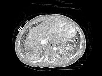
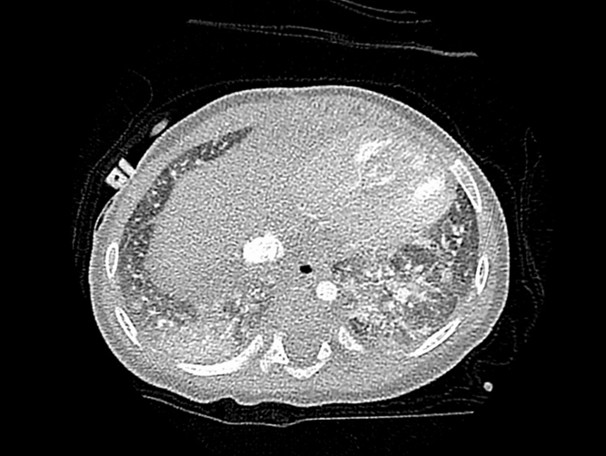
We performed cardiac catheterization again at the age of 4 months, which showed elevation in a pulmonary arterial pressure to 63/32 mmHg (mean pressure of 45 mmHg). The ratio of a pulmonary to systemic mean arterial pressure was 1.50, and a pulmonary arterial resistance was 5.99 Wood units·m2. The right and left pulmonary artery wedge pressures were 11 and 8 mmHg, respectively. He was diagnosed with pulmonary arterial hypertension and treated with additional doses of selexipag. However, he developed pulmonary congestion, and computed tomography showed mosaic pattern, which suggested the possibility of pulmonary veno-occlusive disease (Fig 1). We then discontinued the administration of pulmonary vasodilators and inhaled nitric oxide.
High-resolution computed tomography shows that patchy ground-glass opacities and mosaic pattern of lung attenuation.
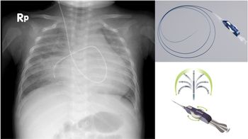
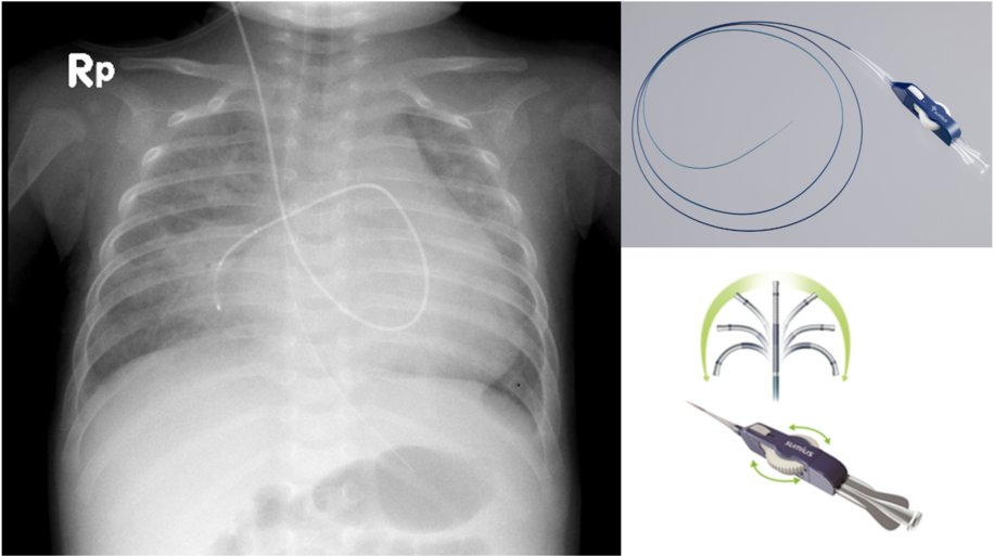
We performed continuous monitoring of the pulmonary arterial pressure using a tip deflecting microcatheter (Leonis Mova®, SUMITOMO BAKELITE CO, LTD. Tokyo, Japan) that was characterized by a 0.025-inch internal lumen and a tip that could be bent in any direction by the proximal handle. This microcatheter was introduced via a 4-French sheath through the right internal jugular vein and placed in the right pulmonary artery using fluoroscopic imaging in the catheter laboratory (Fig 2 and Supplemental Movie S1). We simultaneously obtained systemic arterial pressure at the radial artery and pulmonary arterial pressure, via the microcatheter at the intensive care unit, which showed a pulmonary arterial pressure as high as 109/59 mmHg (mean pressure of 74 mmHg) and a ratio of pulmonary to systemic arterial pressure of 1.43. Then, he was intubated and supported by synchronized intermittent mandatory ventilation with an inspiratory oxygen fraction of 0.6, respiratory rate of 35 per minute, and a positive end-expiratory pressure of 5 mmHg, while inhaled nitric oxide was administrated again. Blood gas analysis showed that the partial pressure of carbon dioxide was 39 mmHg. Due to the respiratory care, pulmonary arterial pressure was dramatically decreased to 50/24 mmHg (mean pressure of 33 mmHg) (Supplemental Fig S2) Unfortunately, the catheter had to be removed four days later due to methicillin resistant staphylococcus aureus bacteraemia associated with catheter infection. He was extubated 2 weeks later, because echocardiography showed the flattened interventricular septum and no deterioration of pulmonary hypertension. He has been continued receiving inhalational nitric oxide on admission, while tadalafil and bosentan were administrated orally.
A chest X-ray after the placement of a tip deflecting microcatheter in the right pulmonary artery is shown in the right panel. A tip-deflecting microcatheter, Leonis Mova®, is characterized by a soft and thin shaft and a tip that can be remotely bent in any direction by the proximal handle (shown in the left panel). The copyright of the picture of a microcatheter, Leonis Mova®, belongs to, and is approved by SUMITOMO BAKELITE CO, LTD.
Discussions
We present a notable case of an infant with severe pulmonary arterial hypertension in whom pulmonary arterial pressure was continuously monitored by a tip deflecting microcatheter in the intensive care unit.
Pulmonary arterial pressure is commonly monitored using a balloon-tipped end-hole catheter or a Swan-Ganz catheter in the management of adults and children with pulmonary arterial hypertension, heart failure, and cardiac surgery.Reference von der Brelie, Bein and Schirmer2–Reference Damen and Wever4 In a previous report on pulmonary arterial monitoring using balloon-tipped pulmonary arterial catheters in children after cardiac surgery, catheter-related complications involved infection in 8%, carotid artery puncture in 10%, and severe hypotension in 4%.Reference Damen and Wever4 The present case showed that a microcatheter could be used to continuously monitor pulmonary arterial pressure even in infants. Microcatheters are commonly inserted selectively into peripheral vessels to administer contrast agents, drugs, and embolic substances in variety of clinical settings.Reference Inaba, Arai, Sone, Aramaki, Osuga, Tanaka and Kanemasa5–Reference Keulers, Nikoubashman, Mpotsaris, Wilson and Wiesmann7 Among patients with congenital heart disease, they are usually used to embolize aortopulmonary collaterals or introduce into anatomically complex small vessels.Reference Inaba, Arai, Sone, Aramaki, Osuga, Tanaka and Kanemasa5 Although there are several of kinds of microcatheters, a tip deflecting microcatheter is characterized by a soft and thin shaft and a tip that can be remotely bent in any direction by the proximal handle, which allows this microcatheter to be placed into small and complex peripheral vessels with or without a guidewire.Reference Keulers, Nikoubashman, Mpotsaris, Wilson and Wiesmann7 Although we used a 4-French sheath and a 0.025-inch guidewire in the present case, it is possible to place a tip deflecting microcatheter in the pulmonary artery via an 18-guage needle without any guidewire.
There are several advantages of using a tip deflecting microcatheter for continuous monitoring of pulmonary arterial pressure in infants. First, this microcatheter is soft and thin and, thus, does not easily traumatize intracardiac structures, such as the tricuspid and pulmonary valves. Second, other catheters could easily get entrapped and produce erroneous pressure measurements, which could be identified by unusual pressure contours, whereas a tip deflecting catheter does not easily get trapped by deflecting a tip even in a small pulmonary artery and, thus, could provide reliable pulmonary arterial pressure in infants. The microcatheter was not designed to be placed in the body for several days and could easily get infected as was seen in this case; however, this complication could be avoided by placing the microcatheter appropriately at the neck and maintaining strict aseptic procedures.
The pathophysiology of pulmonary hypertension in neonates and infants is multifactorial, and therefore, multidisciplinary approach is necessary in its management. Currently, as there are various pulmonary vasodilators available, the choice of pulmonary vasodilator should depend on the underlying pathophysiology.Reference Kim, McSweeney, Lee and Ivy8 In this case, we suspected pulmonary veno-occlusive disease because of the clinical manifestations and features of pulmonary congestion after the administration of selexipag. However, real-time continuous monitoring of pulmonary arterial pressure in the intensive care unit suggested that the respiratory support was the most effective treatment in the present case.
In conclusion, a tip deflecting microcatheter is an alternative to a balloon-tipped catheter and feasible for continuous real-time monitoring of pulmonary arterial pressure in the intensive management of neonates and infants with pulmonary arterial hypertension. Continuous monitoring of pulmonary arterial pressure using a tip deflecting microcatheter yields precise pulmonary haemodynamics and allows for individualized targeted therapy for neonates and infants with pulmonary arterial hypertension.
Acknowledgements
The copyright of the picture of a microcatheter, Leonis Mova®, belongs to, and is approved by SUMITOMO BAKELITE CO, LTD.
Financial support
This research received no specific grant from any funding agency, commercial or not-for-profit sectors.
Conflicts of interest
None.
Supplementary material
To view supplementary material for this article, please visit https://doi.org/10.1017/S1047951120001286