Implications
In pig production, pre-slaughter fasting is used to reduce abattoir hygiene problems. Sometimes the fasting time is extended due to long journey times or a prolonged stay at the slaughterhouse before the slaughter, despite the fact that current legislation (EC, 2009) protects animals against lack of feeding during their stay. The results of this study suggest that during fasting there is an increase of lipolysis and the longer the pre-slaughter fasting, the more lipolysis is stimulated. Lipolysis can lead to an increase of the oxidative processes primarily responsible for jeopardizing pork organoleptic characteristics. Long fasting times not only compromise the well-being of the animals but can also be a cause of a qualitative worsening of the pork produced.
Introduction
Adipose tissue, the main energy reserve in animals, is composed of adipocytes embedded in a matrix of connective tissue with a highly developed vascular system. The main metabolic function of adipose tissue is to accumulate lipids by synthesis of triacylglycerols and lipid mobilization through hydrolysis of triacylglycerols.
The adipose tissue of growing and finishing pigs, whose nutritional requirements are completely satisfied, is in a state of lipogenesis, but during fasting or in conditions of stress, lipolysis prevails, in order to exploit fatty acids as a source of energy for peripheral tissues (Guesnet and Demarne, Reference Guesnet and Demarne1987). The stimulation of lipolysis is under neural and hormonal control. During short-term fasting and low-calorie diets an increase of catecholamine induces lipolysis through the stimulation of the adrenergic receptors in the adipose cells (Sengenes et al., Reference Sengenes, Stich, Berlan, Hejnova, Lafontan and Pariskova2002).
Lipolysis is a mechanism that is always active in adipose cells (Guesnet and Demarne, Reference Guesnet and Demarne1987), so a quantity of free fatty acids (FFAs) are always present near the triacylglycerols in adipose tissue. This quantity is normally very small, but increases significantly during fasting (Fernandez et al., Reference Fernandez, Mourot, Mounier and Ecolan1995). In a situation of lipogenesis, fatty acids resulting from lipolysis are mainly re-esterified into triacylglycerols; in cases of increased energy demand, they are released into the bloodstream (Mersmann and MacNeil, Reference Mersmann and MacNeil1985).
Triacylglycerols are hydrolysed to FFAs and glycerol through the activity of many enzymes, in particular hormone-sensitive lipase (HSL) (Raclot, Reference Raclot2003) and adipose triglyceride lipase (ATGL) (Langin, Reference Langin2006); ATGL and HSL remain active over the 12 months of maturation of the ham (Motilva et al., Reference Motilva, Toldrá, Aristoy and Flores1993; Coutron-Gambotti and Gandemer, Reference Coutron-Gambotti and Gandemer1999; Vestergaard et al., Reference Vestergaard, Schivazappa and Virgili2000; Xiao et al., Reference Xiao, Zhang, Yang, Ma, Ahn, Li, Lei and Du2010).
In the cytoplasm of adipocytes, triacylglycerols accumulate in the form of fat droplets; the most polar are triacylglycerols with high content of polyunsaturated fatty acids (PUFA) which are more abundant on the surface than in the core of the droplets (Raclot, Reference Raclot2003). During stimulation of lipolysis, lipases are transferred from the cytosol to the droplet surface and, consequently, they preferentially hydrolyze the most polar triacylglycerols on the lipid–water interface.
According to Raclot (Reference Raclot2003) and Lafontan and Langin (Reference Lafontan and Langin2009) the fatty acids that leave adipose cells more rapidly are short-chain or unsaturated fatty acids. This means that the difference in mobilization is associated not only with polarity but also with the solubility of fatty acids. Monin et al. (Reference Monin, Hortos, Diaz, Rock and Garcia-Regueiro2003) showed that lipolysis occurs in pig muscle, even postmortem, with an increase in the total percentages of FFAs, particularly PUFA. The increase of FFAs and their degree of unsaturation could lead to an increase of the processes of oxidation in fat (Girard, Reference Girard1982; Allen, Reference Allen1987; Coutron-Gambotti and Gandemer, Reference Coutron-Gambotti and Gandemer1999; Xiao et al., Reference Xiao, Zhang, Yang, Ma, Ahn, Li, Lei and Du2010). During the curing period, the cover fat of raw ham is subjected to oxidation, which, if too accentuated, can jeopardize the organoleptic characteristics of the cured ham. The literature contains only few reports on the selectivity of fatty acids mobilization from triacylglycerols in adipose tissue of different species of animals (Raclot and Groscolas, Reference Raclot and Groscolas1993 and Reference Raclot and Groscolas1995; Nieminen et al., Reference Nieminen, Käkelä, Pyykönen and Mustonen2006a and Reference Nieminen, Rouvinen-Watt, Collins, Grant and Mustonen2006b; Price et al., Reference Price, Krokfors and Guglielmo2008). Mersmann and MacNeil (Reference Mersmann and MacNeil1985) showed an increase of plasma FFAs in fasted pigs. Some researchers (Coutron-Gambotti and Gandemer, Reference Coutron-Gambotti and Gandemer1999; Xiao et al., Reference Xiao, Zhang, Yang, Ma, Ahn, Li, Lei and Du2010; Volpelli et al., Reference Volpelli, Cino, Comellini, Ielo, Minelli and Lo Fiego2011) found that during curing there is an increase in the percentage of unsaturated fatty acids in the FFAs. Several studies have shown the effects of the fasting time on the productive performances and on meat quality. Some studies have shown that increasing the time of food deprivation before slaughter reduces the incidence of PSE pork (Murray and Jones, Reference Murray and Jones1994) and increases the risk of DFD with fasting time longer than 22 h (Guàrdia et al., Reference Guàrdia, Estany, Balash, Oliver, Gispert and Diestre2005). It also improves meat colour (D’Souza et al., Reference D’Souza, Dunshea, Warner and Leury1998) and pH (Eikelenboom et al., Reference Eikelenboom, Bolink and Sybesma1991; Turgeon and Bergeron, Reference Turgeon and Bergeron2002; Sterten et al., Reference Sterten, Frøystein, Oksbjerg, Rehnberg, Ekker and Kjos2009). Kephart and Mills (Reference Kephart and Mills2005) and Beattie et al. (Reference Beattie, Burrows, Moss and Weatherup2002) reported a slight decrease (−1%) of carcass weight in pigs fasted for 20 and 24 h, respectively. However, there are no available data on the variation of the acidic fraction during fasting before slaughtering in pigs.
The subcutaneous fat of pigs is constituted by two (Camara et al., Reference Camara, Mourot and Février1996) or more layers (Apple et al., Reference Apple, Maxwell, Galloway, Hamilton and Yancey2009) with different chemical compositions and different metabolic activity. In fact it should not be considered as a single entity, but rather as two separate tissues (Camara et al., Reference Camara, Mourot and Février1996). The outer layer is richer in unsaturated fatty acids (Malmfors et al., Reference Malmfors, Lundstrom and Hansson1978) and in collagen tissues with greater organization of the connective structure surrounding the adipocytes (Lo Fiego et al., Reference Lo Fiego, Tedeschi, Santoro and Nanni Costa1987) that gives it a greater consistency compared with the inner layer. The lipogenic activities are greater in the inner layer than in the outer layer (Camara et al., Reference Camara, Mourot and Février1996; Warnants et al., Reference Warnants, Van Oeckel and Boucque1999).
The aims of this study were to determine the FFA and triacylglycerols composition of pig fat subcutaneous tissue to analyse the effects of pre-slaughtering fasting time on lipolysis and to discover whether lipolysis continues after slaughtering.
Material and methods
Animals and sampling
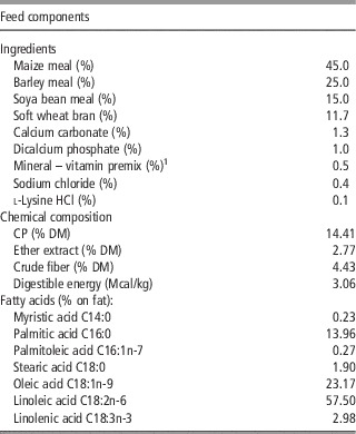
The study involved 16 barrows pigs (Large White×Landrace) of 48 kg±4.12 initial live weight subdivided into four boxes of four pigs each; they were wet-fed (water : feed ratio 3 : 1) and the feed was restricted (1.8 kg/head day at start; increasing by 100 g/head per day per week for 12 weeks until 3.0 kg/head per day as the maximum daily feed intake until slaughtering). A water nipple was available for each box. Lipid, crude fiber and protein (n×6.25) content of the feed were determined according to AOAC (1990) and the fatty acids profile was determined according to Folch et al. (Reference Folch, Lees and Sloane-Stanley1957). The formulation, used for the duration of the experimental period (from 48 kg to slaughter), is reported in Table 1. The average daily gain during the entire trial was 715±74.5 g/day (ADG) and feed : gain ratio was 3.6±0.05 kg of feed/kg of weight gain.
Feed ingredients and dietary fatty acid composition

1Content per kg of feed: vitamin A 15 000 IU; vitamin D3 2000 IU; vitamin E 50 mg; vitamin K 2.5 mg; vitamin B1 2 mg; vitamin B2 5 mg; pantothenic acid 15 mg; vitamin B6 4 mg; vitamin B12 0.03 mg; niacin 25 mg; folic acid 1 mg; biotin 0.15 mg; choline 346 mg, Cu 15 mg; Fe 15 mg; Mn 25 mg; Co 0.4 mg; I 1.5 mg; Zn 100 mg; Se 0.1 mg.
The pigs were slaughtered at 167±12.5 kg live weight in a slaughterhouse according to pig slaughtering rules. Before slaughter, the pigs were subdivided into two homogeneous groups, one subjected to 12 h and the other to 60 h of fasting (fasting time). The two fasting times were selected as follows: fasting time 12 h is the minimum fasting time for pigs delivered to slaughterhouse and slaughtered the same day; 60 h is the maximum fasting time: 24 h of fasting time in the period before the pigs are loaded for transport, at most 24 h during transport (EC, 2005), and at most 12 h in the waiting pen of the slaughterhouse (EC, 2009). For each pig, backfat thickness was measured on half carcass in correspondence to the cranial point of the Gluteus Medius muscle (thickness mm 36.4±4.77 for 12 h fasting and 36.6±4.50 for 60 h fasting); thereafter the backfat was sampled in the same position; at ham trimming 24 h later the ham covering fat was sampled under the Femur head; both outer and inner layers were sampled for each pig and each sampling time. Samples were stored at –80°C until analysis.
Lipid extraction
The fat of subcutaneous tissue was chopped into small pieces (~0.5 cm3), placed in individual microwave-resistant glass containers, and heated in a MDS-2000 laboratory microwave oven (CEM Corporation, Matthews, NC, USA), 650 W power, 2450 MHz microwave frequency, for 10 min at 33% power. The resulting liquid fat was filtered through anhydrous sodium sulphate (De Pedro et al., Reference De Pedro, Casillas and Miranda1997).
Separation of free and esterified fatty acids
Totally, 100 μl of internal standard solution (pentadecanoic acid, C15:0 Catalogue no. P6125; Sigma-Aldrich Corp., St. Louis, MO, USA) were added to the fat (500 mg accurately weighed), the sample was dissolved in 1.5 ml solvent mixture (hexane-diethyl ether 1 : 1) and added to a column prepared by filling a glass column with 1.5 g alumina previously activated with 10% water. Neutral lipids were eluted with 8 ml solvent mixture, and FFA were extracted with 6 ml diethyl ether containing 3% formic acid in a 10-ml tube with a teflon-lined screw cap (Lambertini and Della Casa, Reference Lambertini and Della Casa1994).
Gas chromatographic analyses and identification of fatty acid methyl esters (FAME)
Extracts were dried under a nitrogen stream and then methylated with 3 N HCl/methanol in a boiling water bath. FAME were analysed in a Mega 5160 gas chromatograph (Carlo Erba, Milan, Italy) equipped with flame ionization detector and automatic injection system (A200S; Carlo Erba). The column was a Restek Stabilwax-DA fused silica capillary column (20 m×0.32 mm i.d., phase thickness 0.25 μm). Carrier gas was hydrogen, flow rate 5 ml/min. Samples were injected in to the split mode at a split ratio of 1 : 15. Both injector and detector temperatures were set at 250°C. Initial oven temperature was 180°C, immediately raised to 5°C/min to 240°C. Nelson-Perkin Elmer software was used for data analysis. Peaks were identified by comparison with known standards (Supelco Inc., Bellefonte, PA, USA).
Hydrolysis index (HI)
According to (Raclot and Groscolas, Reference Raclot and Groscolas1993) to compare the mobilization of all fatty acids, the ratio between their percentage in FFA to that in triacylglycerols was calculated; called ‘relative mobilization’. A ratio greater, equal, or lower than one means that the fatty acid is, respectively, more, equally, or less intensively mobilized than the total fatty acids.
For total saturated fatty acids (SFA), monounsaturated fatty acids (MUFA) and PUFA, the HI was calculated as follows:
 $${{{\rm Percentage \ of\ total \ free \ fatty \ acids}} \over {{\rm Percentage \ of \ total \ esterified \ fatty \ acids}}}$$
Statistical analysis
The data for the inner and outer backfat layer, were analysed using the procedure Mixed Linear Model according to the following model:
 $$Y_{{ijkm}} =\mu {\plus}A_{i} {\plus}B_{j} {\plus}\left( {AB} \right)_{{ij}} {\plus}C_{k} {\plus}E_{{ijkm}} $$
where Y is the dependent variable considered, μ the overall mean, A i the fixed effect of fasting (j=1, …, 2); B j the fixed effect of sampling time (k=1, …, 2); (AB) ij the effect of the interaction (fasting, sampling time); C k the random effect of pig (k=1, …, 16) E ijkm the random residual.
The Bonferroni t-test (α=0.01; 0.05) was used for pair-wise comparisons of variables differing by P⩽0.05. The Bonferroni test, used same error terms as the corresponding F tests.
The means were estimated using the procedure LSMEANS.
The variance component estimation method was restricted maximum likelihood, and the type of covariance matrix was compound symmetry.
All statistical analysis were made using the SAS System, 9.3 release for Windows (SAS, 1997).
Results
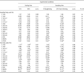
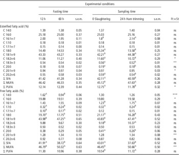
Tables 2 and 3 show the percentage of fatty acids subdivided by layers; the percentage of each fatty acid is compared with total fatty acids in the two fractions examined (esterified and free), and for each experimental factor (fasting and sampling time).
Effect of fasting and sampling time on esterified and free fatty acid of backfat outer layer (% on total fatty acids)

SFA=saturated fatty acids; MUFA=monounsaturated fatty acids; PUFA=polyunsaturated fatty acids.
Significance levels: ns=not significant; *P<0.05; **P<0.01; ***P<0.001.
Within a row, values with different superscript alphabets differ significantly: P⩽0.01 (capital letters) or P⩽0.05 (small letters).
Ft×St: interaction fasting time×sampling time.
Effect of fasting and sampling time on esterified and free fatty acid of backfat inner layer (% on total fatty acids)

SFA=saturated fatty acids; MUFA=monounsaturated fatty acids; PUFA=polyunsaturated fatty acids.
Significance levels: ns=not significant; *P<0.05; **P<0.01; ***P<0.001.
Within a row, values with different superscript alphabets differ significantly: P⩽0.01 (capital letters) or P⩽0.05 (small letters).
Ft×St: interaction fasting time×sampling time.
The percentage variation of the fatty acid in the two layers shows the same trend. Fasting time did not alter the esterified fatty acids composition either in the outer or in the inner layer. Among FFAs the fasting time caused, in both layers, a decrease of SFA, in particular a significant decrease of C14:0 and C17:0, and an increase of MUFA (C18:1n-9), whereas the PUFA did not show any difference (only the C18:3n-3 in outer layer showed a significant increase).
According to the sampling time the percentage of esterified fatty acids showed, in both layers, the same differences: a decrease of SFA (C18:0) an increase of MUFA (C16:1n-7 and C18:1n-9) and a decrease of PUFA (C18:2n-6 and C18:3n-3). The percentage of the FFA showed in both layers the same variation: a decrease of SFA (C17:0, C18:0 and C20:0, C14:0 and C16:0 only in outer layer) an increase of MUFA (C16:1n-7 and C18:1n-9) and an increase of PUFA (C18:2n-6 and C18:3n-3 only in outer layer).
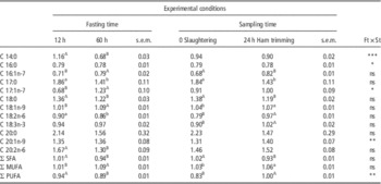
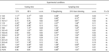
Tables 4 and 5 show the effects of pre-slaughter fasting (12 or 60 h) and sampling time after slaughter (0-slaughtering and 24 h trimming) on HI.
Effect of fasting and sampling time on relative mobilization (hydrolysis index: % free fatty acids/% esterified fatty acids) of backfat inner layer

SFA=saturated fatty acids; MUFA=monounsaturated fatty acids; PUFA=polyunsaturated fatty acids.
Significance levels: ns=not significant; *P<0.05; **P<0.01; ***P<0.001.
Within a row, values with different superscript alphabets differ significantly: P⩽0.01 (capital letters) or P⩽0.05 (small letters).
Ft×St: interaction fasting time×sampling time.
Effect of fasting and sampling time on relative mobilization (hydrolysis index: % free fatty acids/% esterified fatty acids) of backfat outer layer

SFA=saturated fatty acids; MUFA=monounsaturated fatty acids; PUFA=polyunsaturated fatty acids.
Significance levels: ns=not significant; *P<0.05; **P<0.01, ***P<0.001.
Within a row, values with different superscript alphabets differ significantly: P⩽0.01 (capital letters) or P⩽0.05 (small letters).
Ft×St: interaction fasting time×sampling time.
The HI values of SFA fell, both, with increasing hours of fasting and with increasing time after slaughter (C17:0 and C18:0; other SFA showed significant differences only in certain experimental conditions). In fact, values after trimming were significantly lower than those immediately after slaughter. This means a reduction in the percentage of SFA with increased time from the last feed and with increased time between slaughter and sampling of adipose tissue.
HI of MUFA increased significantly, in both layers, 24 h postmortem (C18:1n-9; C16:1n-7 and C17:1n-7 showed significant differences only in certain experimental conditions).
The HI values of PUFA increased significantly postmortem, indicating an increase of PUFA in FFA with respect to esterified ones (C18:2n-6 and C18:3n-3). The duration of pre-slaughter fasting showed an effect only for the inner layer, whose percentage of PUFA in the free fraction dropped significantly.
The amount of FFAs in the two layers (Table 6) increased significantly related to fasting and sampling time.
Effect of fasting and sampling time on backfat free fatty acids (g/kg of fat) outer and inner layer

Significance levels: ns=not significant; *P<0.05; **P<0.01, ***P<0.001.
Within a row, values with different superscript alphabets differ significantly: P⩽0.01 (capital letters) or P⩽0.05 (small letters).
Ft×St: interaction fasting time×sampling time.
Discussion
Our data confirm a different mobilization of PUFA, MUFA and SFA in triacylglycerols and FFAs in relation to fasting period and postmortem lipolysis. According to Raclot and Groscolas (Reference Raclot and Groscolas1993) we used the HI to highlight the trend of changes of FFAs compared with esterfied fatty acids.
The longer the pre-slaughter fasting, the more lipolysis is stimulated, according to Guesnet and Demarne (Reference Guesnet and Demarne1987), Raclot and Groscolas (Reference Raclot and Groscolas1993) and Raclot (Reference Raclot2003). Our data show that during lipolysis SFAs in general, and C14:0, C17:0 and C18:0 fatty acids in particular, are less mobilized than unsaturated fatty acids during fasting time and sampling time, in both layers. This reduction is due to the selectivity of hydrolysis by lipases, which act mainly on more polar fatty acids – i.e., those containing double bonds which make the lipid–water interface of the molecule more available to hydrolysis (Raclot, Reference Raclot2003).
In our trial the free MUFA, in particular the C18:1n9, the most important MUFA in backfat, increased with longer pre-slaughter fasting time; they also increased in the 24 h post-slaughter.
A considerable increase in the HI of PUFA with increasing pre-slaughter fasting time should be expected (Raclot and Groscolas, Reference Raclot and Groscolas1993), however, in our study the HI of PUFA did not increase; in the outer layer the HI of PUFA remained constant and in the inner layer it significantly decreased. This discrepancy may be explained by the different indexes applied. Raclot and Groscolas (Reference Raclot and Groscolas1993) and Raclot (Reference Raclot2003) used relative mobilization, i.e., the ratio between the percentages of extra cellular (interstitial fluid) FFAs and triacylglycerols inside the cell, whereas we measured FFA inside the cell. Moreover the present work has considered the effects of different fasting times on backfat acid composition at slaughtering and at ham trimming, without considering the amount of FFA removed via the blood stream. However Mersmann and MacNeil (Reference Mersmann and MacNeil1985) recorded increased plasma FFA, from 12 to 60 h after feed removal and Raclot (Reference Raclot2003) showed that PUFA are not only more rapidly hydrolysed but also more rapidly transferred to the bloodstream during fasting. This may offer one explanation why the intracellular quantity of PUFA tends to remain low up to the point of slaughter.
This hypothesis is supported by our postmortem data, in which the accumulation of intracellular PUFA (C18:2n6 and C18:3n3 in particular) is high, because bloodflow is suspended and lipolysis continues, in agreement with Monin et al. (Reference Monin, Hortos, Diaz, Rock and Garcia-Regueiro2003), who found an increase in the percentage of PUFA in the intramuscular fat of Longissimus Lumborum muscle after 5 and 9 days of conservation.
The factors that stimulate lipolysis determine greater mobilization of unsaturated fatty acids, giving rise to an accumulation of FFAs inside adipose tissue during conservation. This is interesting with regard to the exposure of pork to pro-oxidizing factors during processing and conservation. Coutron-Gambotti and Gandemer (Reference Coutron-Gambotti and Gandemer1999) investigated the changes in lipids of adipose tissue during dry-cured ham processing. They noted an intense lipolysis during the 24 months of processing, especially in the first 6 months. They observed an increase of unsaturated FFA in white adipose tissue, and a decrease of C18:2 linoleic acid during ham processing, due perhaps to oxidation.
FFAs represent only a limited part of the total lipid classes in fat of pigs not subjected to pre-slaughter fasting, conversely there is an increase of the FFAs content in pigs subjected to 60 h of fasting. In fact Steffen et al. (Reference Steffen, Arakelian, Phinney, Brown and Mersmann1981) noted that fasting must reach 72 h before a significant lipolysis can be detected in white adipose tissue. Our results seem to indicate that lipolysis is appreciable also after 60 h of fasting.
Although the quantities of FFA still remain low, two factors together – an increase in FFA and an increase in the amount of PUFA in FFA – may cause poorer suitability for meat processing, particularly because FFAs are more rapidly oxidized than esterified ones (Gray and Pearson Reference Gray and Pearson1984; Enser, Reference Enser1987).
Conclusions
An increase of fasting time from 12 to 60 h caused an increase in monounsaturated FFAs and a decrease in saturated free fatty acids both in the outer and inner backfat layers. After slaughtering the lipases continued to act leading to an accumulation of monounsaturated and PUFAs both in outer and inner layers. So the present study confirms, also in pigs, the general principles of selective mobilization shown in other species. In order to better understand the mechanism of selective fatty acids mobilization in pigs further studies on plasma lipid profile could be considered.